Intestinal inflammation in mice deficient in Tir8, an inhibitory member of the IL-1 receptor family Cecilia Garlanda*, Federica Riva*†, Nadia Polentarutti*, Chiara Buracchi*, Marina Sironi*, Maida De Bortoli*, Marta Muzio*‡, Raffaella Bergottini†, Eugenio Scanziani†, Annunciata Vecchi*, Emilio Hirsch§, and Alberto Mantovani*¶㥋 *Department of Immunology and Cell Biology, Mario Negri Institute for Pharmacological Research, Via Eritrea 62, 20157 Milan, Italy; †Department of Animal Pathology, Faculty of Veterinary Medicine, University of Milan, Via Celoria 10, 20133 Milan, Italy; ‡Department of Pharmacology, Pharmacia Corp., 20014 Nerviano, Italy; §Department of Genetics, Biology, and Biochemistry, University of Turin, 10126 Turin, Italy; and ¶Centro di Eccellenza per l’Innovazione Diagnostica e Terapeutica, Institute of General Pathology, University of Milan, Via Mangiagalli 31, 20133 Milan, Italy Communicated by Charles A. Dinarello, University of Colorado Health Sciences Center, Denver, CO, December 27, 2003 (received for review November 18, 2003)
TIR8, also known as single Ig IL-1-related receptor, is a member of the IL-1 receptor兾Toll-like receptor (TLR) superfamily, which acts as an intracellular decoy for components of the signaling pathway. Here we report that Tir8 has a unique pattern of expression, which includes mucosal tissues and dendritic cells (DC). Tir8-deficient DC showed increased cytokine production in response to TLR agonists (lipopolysaccharide, CpG oligodeoxynucleotides). Tir8-deficient mice had normal susceptibility to systemic lipopolysaccharide toxicity and to i.p. or s.c. inflammation. However, Tir8-deficient mice were more susceptible to intestinal inflammation. Thus, TIR8 represents a negative pathway of regulation of the IL-1 receptor兾TLR system, expressed in epithelial cells and DC, crucial for tuning inflammation in the gastrointestinal tract.
M
embers of the Toll-like receptor (TLR)–IL-1 receptor (IL-1R) superfamily play a key role in innate immunity and inflammation (1–4). TLRs act as sensors for the presence of microorganisms and activate a complex, multifaceted cellular response. Agonists that interact with signaling IL-1R and IL-18 receptor complexes initiate an amplification cascade of innate resistance, contribute to the activation and orientation of adaptive immunity, and play a key role in inf lammatory conditions (5). The activity of members of the IL-1R兾TLR superfamily is tightly regulated at multiple levels (5, 6). Released and intracellular isoforms of the IL-1R antagonist block agonistic interactions with formation of a signaling IL-1 receptor complex. The IL-1 RII and IL-18-binding protein act as decoys for IL-1 and IL-18 and as dominant negatives at the level of signaling receptor complexes (6–8). IRAK-M is a negative regulator of TLR signaling in macrophages (9), and a splice variant of the adaptor MyD88 (MyD88s) inhibits recruitment of IRAK4 and IL-1兾TLR signaling (10). SOCS-1 is a further negative regulator of the IL-1R兾TLR system (11). Toll兾IL-1R 8 (TIR8), also known as single Ig IL-1-related receptor (SIGIRR) (12), is a member of the IL-1R family with unique properties [Tir8 was originally used when the mouse gene sequence was deposited by this group in 1998 (GenBank accession no. AF113795)]. Structurally, it is characterized by a single extracellular Ig domain, an intracellular TIR domain and 95-aa cytoplasmic tail (12, 13). Ligands for TIR8兾SIGIRR have not been identified, and searches for accessory functions in signaling complexes have yielded negative results (12). Recently, TIR8 was shown to inhibit NF-B activation by members of the IL-1兾TLR family (13, 14). The inhibitory activity of TIR8 was associated with trapping of TRAF6 and IRAK1 (14). Wald et al. (14) have recently generated SIGIRR兾Tir8deficient mice and found that these animals are more susceptible to the systemic toxicity of bacterial lipopolysaccharide (LPS). Here we report that Tir8 has a distinct pattern of expression that includes epithelial tissues and dendritic cells (DC). We generated Tir8-deficient mice, and these mice showed a selective increase in susceptibility to intestinal inflammation. 3522–3526 兩 PNAS 兩 March 9, 2004 兩 vol. 101 兩 no. 10
Thus, TIR8, expressed in epithelial cells and sentinel DC, represents a negative pathway of regulation of the IL-1R兾TLR system and plays a crucial role in tuning inflammation in the gastrointestinal tract. Materials and Methods Generation of Tir8-Deficient Mice. The genomic library consisted of mouse 129兾Sv liver DNA cloned in the XhoI site of the Lambda FIX II vector (Stratagene). A total of 30 ⫻ 104 plaques were screened with the 32P-labeled Tir8 cDNA. Ten micrograms of a monophage containing a 15.3-kb insert were analyzed by Southern blot with 32P-labeled oligonucleotides. Three BamHI兾 BamHI and two NotI兾BamHI fragments were subcloned in pBluscript and partially sequenced. To generate the targeting vector, a BamHI兾StuI and a StuI兾 StuI fragments (6 and 2.3 kb) from a genomic clone containing exons 1–8 were used. An internal ribosome entry site (IRES)– LacZ cassette followed by the PGK-neomycin resistance gene was inserted into exon 2, 51 bp downstream the first ATG, in the StuI site. After electroporation of R1 embryonic stem cells with the construct, neomycin-resistant clones were isolated and analyzed by Southern blot analysis after BglII or HindIII digestion using an external probe and an internal Neo probe, respectively (Fig. 1 A and B). Chimeric mice from 1 of 28 targeted embryonic stem cell clones were established by injection of C57BL兾6J blastocysts and mated with C57BL兾6J females (Charles River Breeding Laboratories) to obtain an outbred line carrying the mutated Tir8 alleles. Mice were routinely genotyped by PCR with two primer sets that detected the wild-type and targeted allele (Fig. 1B). Phenotypic analysis was performed in a 129兾Sv and C57BL兾6J mixed genetic background. Tir8⫹兾⫹ mice were littermates of Tir8⫺兾⫺ mice. Procedures involving animals and their care were conformed with institutional guidelines in compliance with national (4D.L. N.116, G.U., supplement 40, 18-2-1992) and international law and policies (European Economic Community Council Directive 86兾609, OJ L 358,1,12-12-1987; National Institutes of Health Guide for the Care and Use of Laboratory Animals, U.S. National Research Council, 1996). All efforts were made to minimize the number of animals used and their suffering. Cells. Mouse DC were generated from pooled CD34⫹ bone
marrow cells from three to four mice by using granulocyte兾 macrophage colony-stimulating factor (GM-CSF) (40 ng兾ml) and Flt3 ligand (100 ng兾ml) as described (15). On day 7, DC were plated at 1 ⫻ 106 per ml in 0.5 ml and cultured for 24 h in the Abbreviations: TLR, Toll-like receptor; IL-1R, IL-1 receptor; SIGIRR, single Ig IL-1-related receptor; TIR, Toll兾IL-1R; LPS, lipopolysaccharide; DC, dendritic cells; DSS, dextran sulfate sodium. 储To
whom correspondence should be addressed. E-mail:
[email protected].
© 2004 by The National Academy of Sciences of the USA
www.pnas.org兾cgi兾doi兾10.1073兾pnas.0308680101
presence of different stimuli [1–100 ng兾ml LPS, 0.02–2 g兾ml bacterial CpG oligodeoxynucleotide motif GACGTT (CpG 1826) (Invitrogen Life Technologies), 10–100 g兾ml heat-killed Candida albicans, 5–50 g兾ml poly(I)䡠(C)]. Murine macrophages were generated from bone marrow cells or collected from the peritoneal cavity 5 days after the injection of 1.5 ml 3% sterile thioglycollate (Difco). Human monocytes, monocyte-derived macrophages, and DC were obtained as described (16). mRNA Levels and Cytokine Production. mRNA expression was
assessed by Northern blot analysis on total RNA as described (13). IL-6, IL-10, IL-12, CCL2, and CXCL10 were measured in DC supernatants by commercial sandwich ELISA using three replicates per experimental group. LPS-Induced Inflammation. Mortality was evaluated twice a day
after injection of 60 mg兾kg LPS i.p. (Escherichia coli O55:B5, Sigma). Neutrophyl recruitment in the lung was assessed by measuring myeloperoxidase activity as reported (17) 2 h after LPS treatment (200 g兾kg i.p.). Leukocyte recruitment in the air pouch model was assessed by local injection of 20 or 200 ng of LPS as reported (18). Intestinal Inflammation. For acute colitis, mice were fed 3.5% of
dextran sulfate sodium (DSS) (molecular mass, 40 kDa; ICN) dissolved in sterile, distilled water ad libitum for 5 days followed by 5 days of regular drinking water. For chronic colitis, mice were fed 2% DSS dissolved in sterile, distilled water ad libitum for 5 days followed by 5 days of normal drinking water; this cycle was
Garlanda et al.
Fig. 2. Tir8 gene expression. (A) Northern blot analysis of murine tissue total RNA (10 g per lane). Specific Tir8 transcripts are shown. (Lower) Ethidium bromide staining after RNA transfer to the membrane. (B) Murine cell lines. Freshly isolated or cultured murine (C) or human immunocompetent (D) cells (Mo, monocytes; M, macrophages; iDC, immature DC; mDC, mature DC) are shown.
repeated three times, resulting in a 30-day experimental period. Clinical and histological scores were assessed as described (19). Briefly, for the determination of clinical scores, the body weight and the presence of occult or gross blood per rectum were determined daily during the acute course and every other day during the chronic colitis induction. For bleeding, we used hemoccult (Beckman Coulter) in coded samples, giving a score from 0 to 4 depending on the color intensity. Statistical Analysis. Five to 12 animals per experimental group
were used throughout. Experiments were repeated one to six times as detailed. Fisher’s exact test and the two-tailed Student t test were used as appropriate. Results Expression of Tir8 in Murine Mucosal Tissues and DC. As shown in Fig. 2A, Tir8 mRNA was highly expressed in the gut (small intestine, colon, cecum), the lung, the kidney, and the liver. Accordingly, the murine colon epithelial cell line Colon 26, the mammary epithelial line TSA, and the kidney RENCA line expressed high amounts of the transcript. By contrast, the fibroblast cell line L929 did not express Tir8 (Fig. 2B). As shown in Fig. 2C, immature bone marrow-derived DC expressed appreciable levels of Tir8, whereas mature DC obtained by exposure to LPS expressed lower levels of the transcript. By contrast, Tir8 expression in macrophages was almost undetectable. TIR8 expression was also analyzed in human monocytes, monocyte-derived DC, and macrophages. As shown in Fig. 2D, immature DC expressed TIR8 and LPS treatment down-modulated TIR8 expression in this cell type (mature DC). PNAS 兩 March 9, 2004 兩 vol. 101 兩 no. 10 兩 3523
IMMUNOLOGY
Fig. 1. Targeting of the Tir8 gene. (A) The targeting vector (TV), the Tir8 wild-type allele (WT), and the homologous recombinant allele (HR) are shown. Exons (white blocks), introns (thick line), probes (lettered black blocks), restriction fragments, and primers (numbered arrows) are shown. The targeting vector contained a 6-kb BamHI–StuI fragment, an IRES–lacZ, a PGK–neomycin resistance cassette (gray block) cloned in the second coding exon, and a 2.3-kb StuI–StuI fragment. (B) (Left) Southern blot of BglII-digested genomic DNA hybridized with probe A, generating a 6.5-kb wild-type restriction fragment and a 5-kb homologous recombinant restriction fragment. (Right) PCR analysis of genomic DNA from Tir8 ⫹兾⫹, ⫹兾⫺, and ⫺兾⫺ mice. (C) Northern blot of kidney (K) and colon (C) from Tir8⫹兾⫹ and ⫺兾⫺ mice. Ten micrograms of total RNA was used in each lane. (Lower) Ethidium bromide stain of the gel.
Fig. 3. Cytokine and chemokine production by bone marrow-derived DC and macrophages from Tir8⫺兾⫺ mice. Cytokine and chemokine levels were undetectable in the supernatants of unstimulated cells from Tir8⫹兾⫹ and Tir8⫺兾⫺ mice, and these data are not shown. (A) IL-6 production in response to different LPS concentrations. Results are mean ⫾ SE of values from one representative experiment of six performed. (B) IL-6 production in response to different stimuli. Results are mean ⫾ SE of values from one experiment for CpG 1826 and three experiments for C. albicans and poly(I)䡠(C). (C) CXCL10 production (one experiment). (D) Responsiveness to 100 ng兾ml LPS of DC and thioglycollate-elicited peritoneal macrophages. Results are mean ⫾ SE from one experiment with four individual mice for macrophages and from six experiments for DC; *, P ⬍ 0.05; **, P ⬍ 0.01 by Student’s t test.
Monocytes expressed low levels of the transcript that were further reduced upon maturation to macrophages. Generation of Tir8-Deficient Mice. The murine Tir8 gene consists of nine exons spanning ⬇4 kb (Fig. 1 A). To assess the in vivo role of TIR8, we generated Tir8-deficient mice by homologous recombination. The targeting vector consisted of a genomic DNA fragment of 8.3 kb encompassing exons 1–8 of the mouse Tir8, with an IRES-lacZ cassette followed by the PGK-neomycin resistance gene integrated in exon 2, 51 bp downstream of the first coding ATG (Fig. 1 A). Twenty-eight independently targeted R1 embryonic stem cell clones of 235 tested were identified by Southern blot hybridization using probe A (Fig. 1 A and B) after digestion with BglII. No evidence for random integration was detected with probe B (from the neomycin gene) after digestion with HindIII. One recombinant clone was injected into C57BL兾6J blastocysts to generate germ line chimeras. Tir8 deficiency at the mRNA level was assessed by Northern blot analysis of tissues using a 450-bp cDNA probe encompassing exons 1–4 (Fig. 1C). Heterozygous females and males were normal and fertile and breeding yielded the predicted number of homozygous null mice at a Mendelian frequency. Tir8⫺兾⫺ mice were viable, fertile, and displayed a normal life span in a conventional mouse facility. Microscopic examination of organs and tissues of young or adult mice (1–4 months old) did not reveal gross morphological abnormalities. Complete peripheral blood cell counts and microscopic examination of blood smears of Tir8⫺兾⫺ mice (n ⫽ 4) did not show statistically significant differences compared to Tir8⫹兾⫹ animals. Increased Cytokine Production by Tir8ⴚ兾ⴚ DC. As shown in Fig. 2,
DC, unlike mature macrophages, express Tir8. Given the key role
3524 兩 www.pnas.org兾cgi兾doi兾10.1073兾pnas.0308680101
Fig. 4. Responsiveness of Tir8⫺兾⫺ mice to local and systemic inoculation of LPS. (A) Mortality after injection of LPS i.p. (60 mg兾kg). Results presented are a compound of four experiments with a total of 43 Tir8⫹兾⫹ and 45 Tir8⫺兾⫺ mice. No significant difference was observed when each of the four experiments was analyzed individually. (B) Serum IL-6 levels 2 h after injection of LPS i.p. (200 g兾kg). (C) Myeloperoxidase (MPO) activity in the lungs 2 h after i.p. injection of 200 g兾kg LPS. (D) Leukocyte recruitment in the air pouch after local injection of LPS.
of DC as sentinel cells it was important to assess the significance of this observation. As shown in Fig. 3A, where one experiment representative of six performed is shown, Tir8⫺兾⫺ DC showed increased responsiveness to LPS in terms of IL-6 production. Similar results were obtained when a CpG oligodeoxynucleotide engaging TLR9 was used, whereas no consistent difference was observed when Candida albicans or poly(I)䡠(C) (three experiments) was used (Fig. 3B). The better responsiveness of Tir8⫺兾⫺ DC compared to wild type was also observed when CXCL10兾 IP10 (Fig. 3C), IL-10, or IL-12 (not shown, one experiment performed) were measured. Bone marrow-derived (not shown) or peritoneal macrophages (Fig. 3D) from Tir8⫺兾⫺ mice showed normal responsiveness to LPS, as expected on the basis of low expression of Tir8. Selective Increase in Susceptibility to Intestinal Inflammation in Tir8ⴚ兾ⴚ Mice. The susceptibility of Tir8-deficient mice to a
variety of inflammatory conditions was then investigated. LPSinduced mortality was evaluated by injecting 60 mg兾kg LPS i.p., a dose corresponding to about the LD50 in Tir8⫹兾⫹ mice. In four experiments performed (two with males and two with females) with 8–12 mice per group, we did not observe significant differences in susceptibility to LPS between Tir8⫺兾⫺ and ⫹兾⫹ mice. Fig. 4A shows the cumulative result of the four experiments performed with a total of 43 Tir8⫹兾⫹ and 45 Tir8⫺兾⫺ mice. Systemic LPS-induced inflammation was also assessed by measuring serum levels of the proinflammatory cytokine IL-6 at 2 h after i.p. injection of 200 g兾kg LPS. As shown in Fig. 4B, no significant difference was found between Tir8 ⫹兾⫹ and ⫺兾⫺ mice (n ⫽ 5, one experiment performed). We then analyzed LPS-induced polymorphonuclear cell recruitment in the lungs by measuring lung myeloperoxidase activity at 2 h after systemic Garlanda et al.
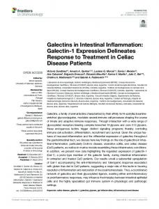
Fig. 5. Intestinal inflammation in Tir8⫺兾⫺ mice. Ten mice per group fed with DSS at the indicated time points (bottom bars). Body weight (A) and bleeding (B) were determined at different time points. Results are mean ⫾ SE from 10 mice (A) or three experiments (B). (C) Histology of control untreated, Tir8⫹兾⫹ DSS-treated, and Tir8⫺兾⫺ DSS-treated mice on day 30. (Scale bar, 100 m.)
Garlanda et al.
Student’s t test) on day 10. The difference in body weight was paralleled by the degree of intestinal bleeding (Fig. 5B). By histology, Tir8⫺兾⫺ mice showed more severe damage of intestinal mucosa with erosion and inflammatory cell recruitment (Fig. 5C). The number of mice with gross focal ulcerations was five of eight in Tir8⫺兾⫺ mice and two of nine in Tir8⫹兾⫹. Discussion The results presented here show that TIR8 has a unique pattern of expression that includes epithelial cells and DC. Previous studies have reported that TIR8 is expressed in epithelial cells and tissues but not in myelomonocytic cells (13, 14). Therefore, the finding that DC derived from monocytes or bone marrow precursors, which have a definite relationship to the myelomonocytic differentiation pathway (23), express TIR8, is unexpected. DC are heterogeneous. It remains to be established whether expression of TIR8 is shared by diverse DC populations, including, for instance, the recently characterized plasmacytoid DC (23). Given the sentinel function of DC and their localization at epithelial surfaces, the expression of TIR8 in this cell type is consistent with the view that this molecule has a regulatory role in epithelial tissues and at mucosal sites. Genetically deficient mice were generated in an effort to define nonredundant functions of Tir8. Tir8-deficient DC, but not macrophages, showed increased responsiveness to LPS and CpG oligodeoxynucleotides in terms of production of cytokines and chemokines (IL-6, CXCL10, IL-12, IL-10). LPS and CpG interact with signaling receptor complexes that include TLR4 and TLR9, respectively (4). Therefore, the finding that Tir8deficient DC show increased responsiveness to TLR agonists is consistent with its pattern of expression and its proposed function as a negative regulator of IL-1R兾TLR signaling. PNAS 兩 March 9, 2004 兩 vol. 101 兩 no. 10 兩 3525
IMMUNOLOGY
LPS injection (200 g兾kg, i.p.). In these experimental conditions we observed a 8-fold increase in myeloperoxidase activity between untreated or LPS-treated mice, with no significant differences between Tir8⫹兾⫹ and ⫺兾⫺ mice (Fig. 4C) (n ⫽ 5, one experiment performed). We studied local inflammation by analyzing leukocyte recruitment in two different models, the air pouch and thioglycollateinduced peritonitis. As shown in Fig. 4D, at 4 h after the injection of 20 or 200 ng of LPS in the air pouch, we did not observe a significant difference in leukocyte recruitment between Tir8 ⫹兾⫹ and ⫺兾⫺ mice (n ⫽ 6). Finally, Tir8 deficiency did not modify leukocyte recruitment in the peritoneal cavity at the time points analyzed (5 and 24 h and 5 days) after thioglycollate injection (data not shown). Given the expression of Tir8 in the intestinal tract and in DC, it was important to investigate the role played in vivo by Tir8 in intestinal inflammation. DSS-induced colitis is caused by a toxic effect of DSS on colon epithelial cells followed by phagocytosis by lamina propria cells and production of proinflammatory cytokines such as TNF␣, IL-6, and IL-1 (20, 21). In the chronic phase, the slow regeneration after DSS damaged the colon epithelial barrier causes further perpetuation of intestinal inflammation by bacterial products from the lumen and activation of DC and a T-cell mediated colitis (22). In a first series of three experiments, DSS-induced acute colitis was studied. Although only a nonsignificant trend in greater body weight loss in Tir8⫺兾⫺ mice was observed, Tir8-deficient mice showed increased blood loss with a score of 4 ⫾ 0 compared to 2.3 ⫾ 0.7 of control mice (data not shown). Chronic bowel inflammation was then investigated. As shown in Fig. 5A, in chronic colitis, Tir8⫺兾⫺ mice showed an increased weight loss compared to Tir8⫹兾⫹ mice, with, for instance, a body weight of 19 ⫾ 0.8 g and 21.9 ⫾ 0.4 g for Tir8⫺兾⫺ and ⫹兾⫹, respectively (P ⬍ 0.01,
Tir8⫺兾⫺ mice showed increased severity of colitis induced by DSS. Recognition of microbial moieties of the enteric flora and production of inflammatory cytokines play a key role in intestinal inflammation in experimental systems and in humans (24–27). For instance, evidence points to a key role of IL-18 and IL-1␣, which is the predominant IL-1 form expressed in epithelial cells, in intestinal inflammation (1, 5, 19, 24, 25). Therefore, the observation of increased severity of colitis in Tir8⫺兾⫺ mice is consistent with a nonredundant regulatory role of this molecule in the gastrointestinal mucosa. It is conceivable that increased production of inflammatory cytokines in response to tissue damage and exposure to microbial molecules by DC in the lamina propria and possibly by epithelial cells is responsible of a more severe colon inflammation in Tir8⫺兾⫺ mice. Tir8-deficient mice showed normal inflammatory reactions at sites other than the gastrointestinal tract, including normal peritoneal inflammation to thioglycollate and normal systemic or local inflammation in response to LPS. Wald et al. (14) reported that SIGIRR兾Tir8⫺兾⫺ mice showed increased susceptibility to systemic administration of LPS in terms of mortality. In the present study, in four separate experiments we observed no significant difference in LPS toxicity between Tir8⫺兾⫺ and ⫹兾⫹ mice. Mononuclear phagocytes and endothelial cells, which generally do not express TIR8 (13, 14), are credited with playing a central role in endotoxic shock (28, 29). The apparent discrepancy between results reported here and those of Wald et al. is likely due to the different genetic background and may reflect a differential involvement in the systemic toxicity of LPS of
cellular components other than myelomonocytic and endothelial cells. The restricted pattern of expression of TIR8 and the selectivity of the inflammatory phenotype of deficient mice are consistent with a selective regulatory role of TIR8 in epithelial tissues and mucosal surfaces. TIR8 has been shown to dampen signaling in response to activation of members of IL-1R兾TLR superfamily (13, 14). Evidence suggests that TIR8 recruited at signaling receptor complexes may act as an intracellular decoy trapping key components of the transduction cascade (TRAF6 and IRAK) (14). The increased responsiveness of DC to TLR engagement, in terms of cytokine production, and the increased severity of intestinal inflammation in Tir8⫺兾⫺ mice is consistent with the view of this molecule as a molecular trap for components of the signaling cascade. Thus, TIR8 represents a negative pathway of regulation of the IL-1R兾TLR system, with a unique pattern of expression in epithelial cells and DC, crucial for tuning inflammation in the gastrointestinal tract. Preliminary results (L. Vago, C.G., and A.M., unpublished data) indicate that TIR8 is expressed in human intestinal mucosa. It will be important to investigate its expression and involvement in the pathogenesis of human inflammatory bowel disease.
Dinarello, C. A. (1996) Blood 87, 2095–2147. Medzhitov, R. (2001) Nat. Rev. Immunol. 1, 135–145. O’Neill, L. A. (2002) Trends Immunol. 23, 296–300. Takeda, K., Kaisho, T. & Akira, S. (2003) Annu. Rev. Immunol. 21, 335–376. O’Neill, L. A. & Dinarello, C. A. (2000) Immunol. Today 21, 206–209. Mantovani, A., Locati, M., Vecchi, A., Sozzani, S. & Allavena, P. (2001) Trends Immunol. 22, 328–336. Colotta, F., Dower, S. K., Sims, J. E. & Mantovani, A. (1994) Immunol. Today 15, 562–566. Novick, D., Kim, S. H., Fantuzzi, G., Reznikov, L. L., Dinarello, C. A. & Rubinstein, M. (1999) Immunity 10, 127–136. Kobayashi, K., Hernandez, L. D., Galan, J. E., Janeway, C. A., Jr., Medzhitov, R. & Flavell, R. A. (2002) Cell 110, 191–202. Burns, K., Janssens, S., Brissoni, B., Olivos, N., Beyaert, R. & Tschopp, J. (2003) J. Exp. Med. 197, 263–268. Kinjyo, I., Hanada, T., Inagaki-Ohara, K., Mori, H., Aki, D., Ohishi, M., Yoshida, H., Kubo, M. & Yoshimura, A. (2002) Immunity 17, 583–591. Thomassen, E., Renshaw, B. R. & Sims, J. E. (1999) Cytokine 11, 389–399. Polentarutti, N., Penton Rol, G., Muzio, M., Zoja, C., Benigni, A., Tomasoni, S., Vecchi, A., Garlanda, C. & Mantovani, A. (2003) Eur. Cytokine Network 14, 1–8. Wald, D., Qin, J., Zhao, Z., Qian, Y., Naramura, M., Tian, L., Towne, J., Sims, J. E., Stark, G. R. & Li, X. (2003) Nat. Immunol. 4, 920–927. Vecchi, A., Massimiliano, L., Ramponi, S., Luini, W., Bernasconi, S., Bonecchi, R., Allavena, P., Parmentier, M., Mantovani, A. & Sozzani, S. (1999) J. Leukoc. Biol. 66, 489–494.
16. Garlanda, C., Hirsch, E., Bozza, S., Salustri, A., De Acetis, M., Nota, R., Maccagno, A., Riva, F., Bottazzi, B., Peri, G., et al. (2002) Nature 420, 182–186. 17. Goldblum, S. E., Wu, K. M. & Jay, M. (1985) J. Appl. Physiol. 59, 1978–1985. 18. Romano, M., Sironi, M., Toniatti, C., Polentarutti, N., Fruscella, P., Ghezzi, P., Faggioni, R., Luini, W., van Hinsbergh, V., Sozzani, S., et al. (1997) Immunity 6, 315–325. 19. Siegmund, B., Lehr, H. A., Fantuzzi, G. & Dinarello, C. A. (2001) Proc. Natl. Acad. Sci. USA 98, 13249–13254. 20. Suzuki, A., Hanada, T., Mitsuyama, K., Yoshida, T., Kamizono, S., Hoshino, T., Kubo, M., Yamashita, A., Okabe, M., Takeda, K., et al. (2001) J. Exp. Med. 193, 471–481. 21. Arai, Y., Takanashi, H., Kitagawa, H. & Okayasu, I. (1998) Cytokine 10, 890–896. 22. Dieleman, L. A., Palmen, M. J., Akol, H., Bloemena, E., Pena, A. S., Meuwissen, S. G. & Van Rees, E. P. (1998) Clin. Exp. Immunol. 114, 385–391. 23. Shortman, K. & Liu, Y. J. (2002) Nat. Rev. Immunol. 2, 151–161. 24. Dinarello, C. A. (2000) N. Engl. J. Med. 343, 732–734. 25. Siegmund, B., Fantuzzi, G., Rieder, F., Gamboni-Robertson, F., Lehr, H. A., Hartmann, G., Dinarello, C. A., Endres, S. & Eigler, A. (2001) Am. J. Physiol. 281, R1264–R1273. 26. Ardizzone, S. & Porro, G. B. (2002) J. Intern. Med. 252, 475–496. 27. Bouma, G. & Strober, W. (2003) Nat. Rev. Immunol. 3, 521–533. 28. Vallet, B. & Wiel, E. (2001) Crit. Care Med. 29, S36–S41. 29. Froidevaux, C., Roger, T., Martin, C., Glauser, M. P. & Calandra, T. (2001) Crit. Care Med. 29, S13–S15.
1. 2. 3. 4. 5. 6. 7. 8. 9. 10. 11. 12. 13. 14. 15.
3526 兩 www.pnas.org兾cgi兾doi兾10.1073兾pnas.0308680101
We thank Dr. Stefano Comazzi for technical assistance. This work was supported by Istituto Superiore di Sanita`, Ministero dell’Istruzione, dell’Universita e della Ricera (Fondo per Gli Investimenti in Ricerca di Base), Ministero della Salute, Associazione Italiana per la Ricerca sul Cancro, and by European Union Fifth and Sixth Framework Program QLG1-2001-02171 (to E.H.) and LSHP-CT-2003-503240 (to A.M.).
Garlanda et al.