THIEME
GLOBAL SPINE JOURNAL
Review Article
425
Intramedullary Spinal Cord Tumors: Part I— Epidemiology, Pathophysiology, and Diagnosis Christopher C. Gillis3
Patrick Shih4
1 Department of Orthopaedics and Traumatology, The University of
Hong Kong, Pokfulam, Hong Kong, SAR, China 2 The Laboratory and Clinical Research Institute for Pain, The University of Hong Kong, Pokfulam, Hong Kong, SAR, China 3 Department of Neurosurgery, Rush University Medical Center, Chicago, Illinois, United States 4 The Neurological Brain and Spine Center, Houston, Texas, United States Global Spine J 2015;5:425–435.
Abstract
Keywords
► ► ► ► ► ► ► ►
intradural intramedullary spinal cord tumors epidemiology ependymoma astrocytoma
Richard G. Fessler3
Address for correspondence Dino Samartzis, DSc, Department of Orthopaedics and Traumatology, The University of Hong Kong, 102 Pokfulam Road, Professorial Block, 5th Floor, Pokfulam, Hong Kong, SAR, China (e-mail:
[email protected]). Richard G. Fessler, MD, PhD, Department of Neurosurgery, Rush University Medical Center, Rush Professional Office Building, 1725 W. Harrison Street, Suite 855, Chicago, IL 60612, United States (e-mail:
[email protected]).
Study Design Broad narrative review. Objectives Intramedullary spinal cord tumors (IMSCT) are rare neoplasms that can potentially lead to severe neurologic deterioration, decreased function, poor quality of life, or death. As such, a better understanding of these lesions is needed. The following article, part one of a two-part series, addresses IMSCT with regards to their epidemiology, histology, pathophysiology, imaging characteristics, and clinical manifestations. Methods The authors performed an extensive review of the peer-reviewed literature addressing the aforementioned objectives. Results Numerous IMSCT exist with varying epidemiology. Each IMSCT has its own hallmark characteristics and may vary with regards to how aggressively they invade the spinal cord. These lesions are often difficult to detect and are often misdiagnosed. Furthermore, radiographically and clinically, these lesions may be difficult to distinguish from one another. Conclusions Awareness and understanding of IMSCT is imperative to facilitate an early diagnosis and plan management.
Introduction Primary spine tumors are rare neoplasms that can lead to significant patient morbidity and mortality.1,2 Intramedullary spinal cord tumors (IMSCTs) are the rarest of these neoplasms and can potentially lead to severe neurologic deterioration, decreased function, poor quality of life, or death.2 Within the IMSCT category, the most common lesions are ependymomas, astrocytomas, and hemangioblastomas, followed by other rare lesions.2,3 Each specific lesion has its own hallmark characteristics, and the tumors vary with regards to the current understanding of their etiology or associations, their imaging and clinical characteristics, and/or
received February 8, 2014 accepted after revision February 9, 2015 published online March 31, 2015
John E. O’Toole3
DOI http://dx.doi.org/ 10.1055/s-0035-1549029. ISSN 2192-5682.
how aggressively they invade the spinal cord. Due to the rarity of occurrence, these lesions are often difficult to detect and are often misdiagnosed, leading to delays in the appropriate patient care.4 Additionally, despite the hallmark characteristics and current diagnostic abilities, these lesions remain both radiographically and clinically difficult to distinguish from one another.5 Preoperative neurologic status and tumor histology are two of the most important variables affecting treatment outcome with these lesions, and thus awareness of these lesions is imperative to facilitate an early diagnosis.6–9 Early diagnosis allows early referral for treatment, which can be the critical difference in the treatment outcome. The following is a broad narrative review of the literature
© 2015 Georg Thieme Verlag KG Stuttgart · New York
This document was downloaded for personal use only. Unauthorized distribution is strictly prohibited.
Dino Samartzis1,2
Intramedullary Spinal Cord Tumors: Part I
Samartzis et al.
addressing IMSCTs with regards to their epidemiology, histology, pathophysiology, imaging characteristics, and clinical manifestations.
Epidemiology Incidence Primary spinal cord tumors are 10 times less common than their cranial counterparts, although they are histopathologically similar. Spine tumors are historically classified as (1) extradural, (2) intradural extramedullary, and (3) intradural intramedullary.1 Extradural tumors are the most common spinal tumors and are usually of metastatic origin.3,4 Intradural intramedullary lesions comprise 20 to 30% of all primary spinal cord tumors.4 The remaining 70 to 80% of primary intradural tumors are intradural extramedullary tumors.3 Intradural tumors are most likely to be found in the thoracic region, followed by the cervical and then lumbar spine. Gliomas make up 80% of all intramedullary tumors. The gliomas are further subdivided into astrocytomas (60 to 70%) and ependymomas (30 to 40%).3 Astrocytomas have a higher occurrence in children than adults, whereas ependymomas are more common in adults than children.10 Hemangioblastomas are the third most frequent IMSC tumor, comprising 2 to 15% of all intramedullary tumors.11 Metastatic intrame-
dullary tumors are rare but present in 2% of all intramedullary tumors. Autopsy series have reported that 0.9 to 2.1% of all patients with cancer exhibit intramedullary spinal cord metastases.12,13 Thus, the incidence of these tumors may be higher than initially predicted. Of the intramedullary tumors with metastatic origins, 40 to 60% and 14% arise from primary neoplasms of the lung and breast, respectively.13–15 The remaining constituents of IMSCTs include lipomas (1%) and other rare tumors that have been documented sparingly in the literature (►Table 1).
Genetic Factors Several genetic factors are associated with IMSCTs. Understanding the various genetic mutations assists in illustrating the clinical manifestations, progression, and management of these tumors. Additionally, through a brief understanding of tumor genetics, the association of spinal tumors with lesions elsewhere in the body can be appreciated. The clinical syndromes currently associated with IMSCTs include neurofibromatosis 1, 2 (NF-1, NF-2) and Von Hippel-Lindau disease (VHL).
Neurofibromatosis Neurofibromatosis is a common autosomal-dominant disorder with 100% penetrance in familial lines.16 Two subtypes have
Table 1 Types of intramedullary spinal cord tumors Tumor class
Associated tumors
Neuroepithelial tissue
Astrocytic • Astrocytoma • Glioblastoma Embryonal • Primitive neuroectodermal Ependymal • Ependymoma • Subependymoma Mixed glioma Neuronal and mixed neuronal-glial • Gangliocytoma • Ganglioglioma • Ganglioneuroblastoma Oligodendroglial • Oligodendroglioma Uncertain origin • Polar spongioblastoma
Spinal nerves
Neurofibroma Schwannoma
Nonmeningothelial, mesenchymal
Hemangioblastoma Lipoma Melanoma Sarcoma
Germ cell tumors
Germinoma Teratoma
Cysts and tumorlike lesions
Dermoid Epidermoid
Hematopoietic neoplasms
Primary central nervous system lymphoma (microglial)
Metastatic tumors and other rare neoplasms
Global Spine Journal
Vol. 5
No. 5/2015
This document was downloaded for personal use only. Unauthorized distribution is strictly prohibited.
426
Intramedullary Spinal Cord Tumors: Part I
427
clinical deterioration. The milder form is characterized by fewer tumors with a later onset and a gradual clinical progression.
Von Hippel-Lindau Disease VHL is a rare autosomal-dominant disease with 90% penetrance. Moller first suggested that VHL was a genetic disorder in 1929.90 Briefly summarized, mutation of a tumor suppressor gene located on chromosome 3p25–26 is responsible for this condition.21,22 The VHL tumor suppressor proteins form a protein complex that interacts with elongins B and C and other proteins, marking them for degradation by the cell, which inhibits hypoxia-related cell transcription factors (HIF1a, HIF2a).19,23 The VHL proteins also suppress the hypoxia-induced production of vascular endothelial growth factor, erythropoietin, and platelet-derived growth factor.19,24 Without the VHL protein present, an increased presence of these transcription factors occurs on the cellular level, eventually leading to tumor formation, and given the specifics of the transcription factors involved, these are often highly vascular lesions. VHL is characterized by widespread formation of both benign and malignant tumors throughout the body. According to Melmon and Rosen, VHL is diagnosed if the following manifest: the presence of more than one hemangioblastoma of the central nervous system (CNS), the presence of an isolated lesion associated with a visceral manifestation of the disease, or the presence of one characteristic of the disease and a family history of the disease.25 A
Table 2 Genetic basis of intramedullary spinal cord tumors Genetic disorder
Associated tumor
Neurofibromatosis-1
Acute nonlymphocytic leukemias Astrocytomas Carcinoid tumors Hematomas Hypothalamic gliomas Malignant nerve sheath tumors Meningiomas Neurofibromas Optic nerve gliomas Pheochromocytomas Primitive neuroectodermal tumors Rhabdomyosarcomas Wilms tumor
Neurofibromatosis-2
Bilateral acoustic schwannomas Ependymomas Gliomas Meningiomas Schwannomas
Von Hippel-Landau
Adrenal pheochromocytomas Central nervous system hemangioblastoma (medulla oblongata or spinal cord) Epididymal cystadenomas Lindau tumor (cerebellum hemangioblastoma) Nephritic cysts Pancreatic cysts Renal cell carcinomas Renal cysts Retinal hemangioblastomas
Global Spine Journal
Vol. 5
No. 5/2015
This document was downloaded for personal use only. Unauthorized distribution is strictly prohibited.
been established: NF-1 and NF-2. Fifty percent of patients with neurofibromatosis possess a family history of the disorder with new mutations developing in the remaining constituents. The reported prevalence of NF-1 (also known as von Reckinghausen disease or peripheral neurofibromatosis) is 1 in 3,000 to 4,000 individuals. 17 Although the incidence of IMSCT in individuals with NF-1 is speculative, it has been suggested that almost 19% of subjects diagnosed with NF-1 develop IMSCT. 18 Expressivity varies due to the heterogenetic characteristics of the disorder. The mutation is located on the long arm of chromosome 17 (17q), which encodes for neurofibromin, a negative regulator of the RAS cellular proliferation pathway. 19 Neurofibromin is a tumor suppressor gene, whereby mutation disinhibits the RAS pathway leading to increased cellular division and proliferation and eventual tumor development. 19 In the presence of NF-1, multiple neurofibromas may appear. Although many tumors are associated with NF-1, in relation to IMSCTs, astrocytomas are the most likely to develop (►Table 2). 18,20 Less prevalent than NF-1, NF-2 (also known as central neurofibromatosis) occurs in 1 in 40,000 individuals and represents 2.5% of patients with IMSCT (►Table 2).18 The location of NF-2 has been identified as a mutation of the “Merlin” gene on chromosome 22q12.19 NF-2 is largely associated with ependymomas and occasionally meningiomas (extramedullary). 18 The severe form of the condition exhibits multiple tumors with an earlier onset and a rapid
Samartzis et al.
Intramedullary Spinal Cord Tumors: Part I
Samartzis et al.
wide array of tumors is associated with this disease, and especially common are retinal hemangioblastomas (►Table 2). Hemangioblastomas of the medulla oblongata and spinal cord as well as renal cell carcinoma may ensue and lead to significant mortality in 50% of individuals with the disease. Of CNS hemangioblastomas, 80% occur in the posterior fossa and 20% appear in the spinal cord. Furthermore, 10 to 15% of cranial hemangioblastomas are associated with VHL, whereas 25% of spinal cord hemangioblastomas are associated with VHL.1,3 In association with VHL, there is an earlier age of development of hemangioblastomas than in those of sporadic origin.
hindbrain disorders, which include such theories as Gardner’s “water hammer” theory, Williams’ “suck and slosh” theory, Ball and Dayan’s theory of infiltration through perivascular (Virchow-Robbin) spaces, and Oldfield’s theory, which combines features of all of the above.26,35,37 However, tumor obstruction of the CSF dynamics and subsequent relative blockage in CSF flow at the local level may also have a role in the pathogenesis of syrinx formation by increasing the focal transmedullary pressure in addition to the two main tumor-related etiologies of hemorrhage/infarction and secretory ability.29,38
Pathophysiology Syringomyelia
Astrocytomas
The classification and terminology related to fluid-filled cavities in the spinal cord remains controversial. In this text, we will refer to the tumor-associated fluid-filled cavity within the spinal cord as syringomyelia or syrinx.26 Syrinx occurs with 25 to 58% of patients with IMSCT, and in this association, syringes appear more commonly in the lower cervical and upper thoracic region. They occur most frequently with ependymomas, followed by hemangioblastomas and cavernomas.27–31 Irrespective of tumor type, there is a correlation between the degree of cephalad extension for a tumor and the presence of a syrinx. According to Samii and Klekamp, 49% of syringes occur above the tumor level, 11% occur below tumor level, and 40% are bipolar. A small percentage involves the medulla oblongata.29 The presence of a syrinx is considered a favorable prognostic sign. Noninfiltrative tumors with distinct cleavage planes are more likely to display syringes than more diffuse, infiltrative tumors. After tumor removal is performed, the syrinx will typically dissolve. Moreover, patients presenting with the concomitant finding of syringomyelia with their tumor tend to recover from surgery more rapidly. As mentioned, the classification and etiology of syringomyelia is a current area of investigation and a continually evolving field, but it can be generally divided into three broad subgroups: (1) resulting from alteration of cerebrospinal fluid (CSF) flow dynamics related to hindbrain disorders, (2) resulting from intramedullary tissue damage secondary to hemorrhage or infarction, and (3) resulting from direct secretory ability of intramedullary tumor.26 The latter two mechanisms of syrinx formation pertain to IMSCT.27,32–37 The resultant syringes from hemorrhage and infarction can be seen occasionally in ependymomas, which often hemorrhage, and especially so in highly vascular lesions such as hemangioblastoma and cavernoma, which are the most common lesions to have associated syrinx. The secretory theory proposes that pathologic tumor vessels create transudation and the secretion of fluid from tumor cells, which thus account for syrinx development. Because the chemical composition of the tumor syrinx has an elevated protein concentration compared with CSF and syrinx fluid associated with non-IMSCT, the syringomyelia may be inherent to the tumor. These mechanisms of syrinx formation in relation to IMSCT are wholly different from those related to CSF obstruction and Global Spine Journal
Vol. 5
No. 5/2015
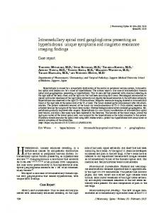
Astrocytomas are red, gray, glossy tumors that are characterized by a poorly defined plane and are generally infiltrative in nature. Astrocytomas are the second most common IMSCT in adults at 30 to 35% of tumors and the most common in children at 90% of tumors.1,10,39 Of all astrocytomas arising in the CNS, 3% occur in the spinal cord.40 They primarily occur in the cervical spine, and they often involve multiple spinal segments due to the expansive nature of these tumors (►Fig. 1). Nearly 20% of such lesions are associated with syrinx formation.29 Astrocytomas have an association with NF-1, occur predominantly in males, and rarely manifest in patients over the age of 60.41 Adults mainly exhibit highgrade lesions, whereas low-grade lesions are associated with the younger population.42 Malignant degeneration develops in 25% of adult astrocytomas.43
Ependymomas Ependymomas are soft, encapsulated, reddish gray or yellow tumors with modest vascularity that present in the third or fourth decade of life. Half of all ependymomas are located in the region spine and a slight majority are located in the cord (55%), relative to the cauda equina (45%).1 They are not sex discriminant.44,45 The majority of them are classified as benign tumors, and ependymomas have a propensity to grow slowly. Spinal ependymomas are primarily present in the cervical or cervicothoracic region.46,47 The cysts associated with these tumors are predominantly found in the superior margin of the tumor and have an increased incidence in the cervical region (►Fig. 2A).29,43,48 Approximately 65% of these tumors are associated with syrinx formation.29 Originating from ependymal cells, ependymomas are more centromedullary located as compared with astrocytomas, and they tend to blend with the cord (►Fig. 2B). They appear as a focal enlargement within the spinal cord. Ependymomas have a mean extension of three to four vertebral bodies, whereas astrocytomas average five to six segmental levels.49 Abnormalities of chromosome 22 are associated with ependymomas, and this leads to their association with NF-2.19 Various histologic subtypes of ependymomas exist. The most common is the cellular variant or the “typical” form, which is a World Health Organization (WHO) grade II tumor that is well circumscribed. Pathologic analysis reveals both true ependymal rosettes and more commonly perivascular
This document was downloaded for personal use only. Unauthorized distribution is strictly prohibited.
428
Samartzis et al.
429
This document was downloaded for personal use only. Unauthorized distribution is strictly prohibited.
Intramedullary Spinal Cord Tumors: Part I
Fig. 1 (A) Sagittal T1-weighted post–gadolinium contrast cervical magnetic resonance image (MRI) noting a heterogeneously enhancing intramedullary astrocytoma (arrow). (B) Sagittal T2-weighted cervical MRI demonstrating an intramedullary astrocytoma (arrow). (C) Axial T1weighted post–gadolinium contrast cervical MRI illustrating a cystic astrocytoma; the cystic portions are highlighted by arrows and are hypointense compared with the enhancing surrounding rim.
pseudorosettes.50 Tanycytic ependymomas are also WHO grade II, are less common overall, and are found more commonly in the spinal cord than in the brain. Histologically,
tanycytic ependymomas lack ependymal rosettes and their pseudorosettes are vaguely apparent, which can lead to misdiagnosis as pilocytic astrocytoma (WHO grade I).19 Global Spine Journal
Vol. 5
No. 5/2015
Intramedullary Spinal Cord Tumors: Part I
Samartzis et al.
Fig. 3 Sagittal T1-weighted post–gadolinium contrast lumbar magnetic resonance image illustrating the presence of a homogenously enhancing ependymoma at the levels of L1 and L2 (arrow).
Fig. 2 (A) Sagittal T1-weighted post–gadolinium contrast cervical magnetic resonance image illustrating the presence of a homogeneously enhancing ependymoma at the levels of C5 and C6 (arrow). (B) Corresponding axial view noting the centromedullary location of the ependymoma (arrow).
Myxopapillary ependymomas are WHO grade I tumors commonly found at the filum terminale, but these are considered extramedullary (►Fig. 3). Rarely anaplastic ependymomas, WHO grade III, can be encountered, which develop more rapidly than lower grades, can occasionally develop from malignant degeneration, and have an overall poor prognosis.19 Ependymomas may also be found outside the nervous system in the soft tissue, ovaries, and mediastinum. Subependymomas (WHO grade I), which may be histologically related to ependymomas, have also been reported as rare IMSCT.51
Hemangioblastoma Hemangioblastomas are small benign richly vascularized solitary neoplasms that rarely extend beyond one or two segments. Although more often located in the cerebellum, hemangioblastomas are also found in the spinal cord, predominantly located at the posterior or posterolateral region of the spinal canal (►Fig. 4). Most hemangioblastomas develop sporadically but, as previously mentioned, they are associated with VHL, especially when presenting in the spinal cord. They constitute between 2 and 8% of all IMSCTs.1 Commonly, they are located in the cervical spine but may be found in any portion of the neuroaxis. Cyst formation is evident within 50 to 70% of tumors, and the Global Spine Journal
Vol. 5
No. 5/2015
tumors have an association with syrinx.52–54 Hemangioblastomas are more commonly found in men.52–55 Although hemangioblastomas are not age discriminant, most occur during the fourth decade of life. It is possible that individuals with a hemangioblastoma can remain asymptomatic throughout life, with tumor only apparent at autopsy.55 Hemangioblastomas are composed of a dense network of vascular capillary channels containing endothelial cells, pericytes, and lipid-laden stromal cells.56 The cells of origin remain unknown, but based on the genetics of VHL and mutations found in sporadic lesions, they are likely vascular endothelial growth factor-secreting undifferentiated mesenchymal cells.1,19 Two histologic forms exist: reticular and cellular. The former is composed of irregular nuclei with conspicuous vessels. Conversely, the cellular form exhibits fewer conspicuous vessels with varying stromal cells; this form bears a similarity to astrocytomas and may be difficult to discriminate from these lesions. Hemangioblastomas are well-demarcated tumors that are amenable to resection. With complete resection, the prognosis is excellent. Pain and sensory changes are the most frequent presenting complaints. Imaging is a crucial factor in surgical planning and should be thoroughly evaluated. An associated cord widening is commonly found adjacent to the tumor, possibly attributed to edema related to increased blood flow in surrounding draining veins or within the tumor creating vascular congestion. After tumor removal, widening regresses. In addition, spinal angiography is recommended to delineate the characteristics of the spinal draining veins.57
This document was downloaded for personal use only. Unauthorized distribution is strictly prohibited.
430
Intramedullary Spinal Cord Tumors: Part I
Samartzis et al.
431
The mechanism of cord infiltration of metastatic IMSCT is ambiguous. Because the majority of metastatic IMSCTs metastasize from the lung, the tumor is thought to spread from hematogenous dissemination leading to arterial embolization.15,58,59 Other proposed theories of tumor seeding involve retrograde infiltration via the spinal cord venous system (Batson’s plexus),60 spread through perforating veins in the bone, metastatic antidromic cellular migration via nerve root to the spinal cord, CSF dissemination through intraspinal perineural sheaths, and finally, penetration of the spinal cord parenchyma via penetrating vessels within the Virchow-Robin spaces.14
Lipomas are rare congenital tumors that constitute 1% of intraspinal tumors. Commonly found in the cauda equina and conus medullaris, these tumors tend to violate the posterior cord and are usually extramedullary.61,62 Lipomas are commonly associated with spinal dysraphism and are thought to arise from premature disjunction of the cutaneous ectoderm from the neural ectoderm prior to neural tube closure, allowing mesenchymal cells to infiltrate into the neural groove.1 These tumors possess a higher water content than other intramedullary tumors and tend to attach firmly to the dura; their cellular content is indistinguishable from normal adipose tissue.62–64 These tumors are slow-growing, and they become symptomatic due to focal mass effect. They present no cleavage plane from the surrounding cord with blending of the neural and fatty tissue.1,65 As a result, complete tumor resection is extremely difficult, and some neurologic damage may be expected when attempting resection. Although little is known about these tumors, patients tend not to improve from their baseline neurologic state but the long-term prognosis is excellent with 90% progression-free survival at 16 years following total or near total excision and 35% progression-free survival at 10 years following a debulking procedure.1 Developmentally, these tumors prevent normal maturation of the surrounding neural tissue.
Other Rare Neoplasms Fig. 4 (A) Sagittal T1-weighted post–gadolinium contrast magnetic resonance image noting an avidly homogenous enhancing nodule representing a hemangioblastoma at the level of T2–T3 (arrow). (B) Corresponding axial view noting the involvement of the tumor in relation to the cord (arrow).
Intramedullary Spinal Cord Metastases The literature supports the belief that metastatic IMSCTs are rare. More recently, combining routine magnetic resonance imaging (MRI) and autopsy observation, it appears that metastatic IMSCTs may be more common than initially reported. These fast spreading tumors may escape diagnosis, as patients bearing these lesions may be asymptomatic. Although imaging is helpful in demarcating the location and extent of these tumors, the proper diagnosis is accomplished by surgical biopsy and the presence of a primary source of cancer.
Numerous additional IMSCTs exist but are extremely rare. Cavernomas are rare vascular lesions that may hemorrhage repeatedly during the lifetime of an individual, and they can have associated syringes. Intramedullary lymphomas may occur in isolation or with other central nervous system lesions. They respond to chemotherapy. Gangliogliomas are benign tumors consisting of glial and neuronal cells that are estimated to comprise 3.8% of CNS tumors found in the upper cervical cord.66,67 Oligodendrogliomas possess ill-defined borders and less extensive cord growth compared with other IMSCTs; fewer than 50 cases have been literature reported. Other IMSCT neoplasms include melanocytomas, melanomas, fibrosarcomas, peripheral neuroectodermal tumors, and amyloid angiopathies (►Table 1).1,68–70
Imaging Radiographic evaluation is necessary to determine the location and extent of tumor involvement and may help to Global Spine Journal
Vol. 5
No. 5/2015
This document was downloaded for personal use only. Unauthorized distribution is strictly prohibited.
Lipomas
Intramedullary Spinal Cord Tumors: Part I
Samartzis et al.
differentiate between lesions. A role still exists for plain radiographs in evaluation as they can illustrate bony erosions or evaluate for scoliosis. The presence of scoliosis can be indicative as advanced IMSCT can present with scoliosis even in adults.71,72 In adults, widening of the spinal canal is usually not noted except with large myxopapillary ependymoma in the conus medullaris or filum terminale. In children, widening of the spinal canal and kyphoscoliosis are more frequently encountered.71,73–75 Myelography has been utilized in the past to evaluate the cord contours; however, cord widening is sometimes difficult to appreciate and the procedure may exacerbate symptoms. Myelography has been supplanted by MRI except in rare circumstances. MRI is the preferred modality in identifying and characterizing IMSCT. Each tumor has a defined T1-, T2-, and gadolinium-enhanced T1-weighted imaging pattern. MRI may be helpful in defining the presence or absence of a cord-tumor interface, illustrating associated cysts or syringomyelia, which may support the ability to achieve a favorable resection. With modern imaging capabilities, differentiation between ependymomas, astrocytomas, and even hemangioblastomas remains difficult; however, several radiographic features may help in this distinction.76 A recently published article by Arima et al scrutinized the ability of MRI to accurately differentiate between these three most common IMSCTs and were able to achieve an 89% accuracy.76 Although this appears to be a high rate of diagnostic accuracy, it does not yet achieve a value that allows treatment based on MRI features alone. Looking at gliomas specifically (ependymomas and astrocytomas), both lesions demonstrate fusiform configuration over numerous vertebral segments, and they both appear hypo- or isointense on T1-weighted images and hyperintense on T2-weighted images. Furthermore, these two tumors enhance with contrast. The rare subependymoma shares these MRI T1- and T2-weighted features, can have associated syrinx, and is usually eccentrically located and nonenhancing; contrast enhancement is faint if present.51 Ependymomas tend to be centrally located within the cord and display symmetric expansion with diffuse heterogeneous enhancement (►Fig. 2). They tend to occupy the whole width of the cord and generally produce enhanced margins (►Fig. 2). Astrocytomas, in comparison, tend to be eccentrically positioned, may display an exophytic component, and can be nonenhancing or heterogeneously enhancing or even have an enhancing nodule (►Fig. 1).76,77 Radiographically, they do not present with well-defined borders but large satellite cysts may be visualized.78–80 Polar cysts may appear and have a low signal intensity on T1-weighted images and high signal intensity on T2-weighted images. Intradural hemorrhage may be seen in both types of glioma but are more likely in ependymoma, with the imaging characteristics dependent on the age of the hemorrhage and possible secondary development of syrinx. Hemangioblastomas are richly vascularized tumors that can have significant surrounding edema.81 They are associated with syringomyelia, can be found at any spinal level, and are most commonly sporadic. In patients with VHL, they are Global Spine Journal
Vol. 5
No. 5/2015
associated with additional visceral lesions or neuraxis hemangioblastomas. On MRI, they have mural nodules, which appear isointense on T1-weighted images and hyperintense on T2-weighted images with homogenous contrast enhancement, in contrast to the usual heterogenous contrast pattern of ependymomas and astrocytomas (►Fig. 4).76 Vascular imaging or even spinal angiography may be useful to delineate feeding vessels and illustrate associated dilated pial veins due to vascular shunting and can be helpful for consideration of preoperative embolization in these highly vascular lesions. Metastatic IMSCTs are well encapsulated and are unlikely to present with cystic change or intralesional hemorrhage.82 They most commonly present as single lesions in the thoracic cord but can be multiple or present in the cervical and lumbar spine.82 Metastatic lesions are most likely eccentrically located in the cord and expand the cord parenchyma, which was historically visible on myelography and illustrated clearly on MRI.14,82,83 MRI is the imaging tool of choice, and recently described “rim” and “flame” signs have been described to help differentiate non-CNS intramedullary metastases from primary spinal cord tumors, which occur significantly more frequently either alone or in combination in metastases than in primary cord tumors with high specificity but low sensitivity (specificity 97% for either alone and 100% when both present).84 The rim sign is defined by Rykken et al as a complete or partial rim of gadolinium enhancement, and the flame sign is defined as an ill-defined flame-shaped gadolinium-enhancing region at the superior or inferior margin of an otherwise well-defined lesion.84 Metastases are most likely to be T1 isointense and T2 hyperintense relative to the cord. The amount of edema may be out of proportion to the size of the tumor, and this is evidenced by extensive T2 hyperintensity, which can be on average 3.6-fold larger than that of the enhancing portion of the lesion.82 The estimated prevalence of spinal cord metastases from a postmortem series ranged from 0.9 to 2.1% of patients with cancer.84 Any patient with a known history of cancer and an intramedullary spinal cord lesion should have metastatic IMSCT as part of the differential diagnosis. Common IMSCTs may be difficult to differentiate from the rare tumors radiographically. Intramedullary lymphomas are rare tumors often linked to other supratentorial lesions. Oligodendrogliomas are characterized by ill-defined borders, no peritumoral edema, and slight T1-weighted image hyperintensity. Lipomas have imaging characteristics similar to fat (high T1-weighted image intensity).
Clinical Manifestations IMSCTs present with a wide array of symptoms that vary in intensity and chronicity. The clinical features of each tumor are related to the growth rate, location, and longitudinal extent of the tumor.19 In general, spinal cord lesions should be suspected when patients present with bilateral motor and sensory symptoms not involving the head and face, often with other upper motor neuron symptoms consistent with a myelopathic syndrome.1 Hydrocephalus occurs in 1 to 8% of patients with IMSCT.85,86 The hydrocephalus may result from
This document was downloaded for personal use only. Unauthorized distribution is strictly prohibited.
432
Intramedullary Spinal Cord Tumors: Part I
Conclusion IMSCTs are rare manifestations that can severely impair neurologic function and the quality of life. The proper diagnosis of IMSCT is essential to provide the best treatment and obtain the optimal outcome. The preoperative functional status, tumor histology, and degree of tumor invasion dictate postoperative results. Therefore, prompt diagnosis and intervention are essential. However, the manifestation of symptoms can be misleading and the diagnosis can be missed. Overall, various types of IMSCT exist, and the management of these tumors demand individualized scrutiny and understanding.
433
Disclosures Dino Samartzis, none Christopher C. Gillis, none Patrick Shih, none John E. O’Toole, none Richard G. Fessler, none
Acknowledgment We would like to thank the Hong Kong Theme-Based Research Scheme (T12-708/12N) for their support of this work.
References 1 Mechtler LL, Nandigam K. Spinal cord tumors: new views and
future directions. Neurol Clin 2013;31(1):241–268 2 Fisher CG, Goldschlager T, Boriani S, et al. A novel scientific model
for rare and often neglected neoplastic conditions. Evid Based Spine Care J 2013;4(2):160–162 3 Grimm S, Chamberlain MC. Adult primary spinal cord tumors. Expert Rev Neurother 2009;9(10):1487–1495 4 Duong LM, McCarthy BJ, McLendon RE, et al. Descriptive epidemi-
5
6
7
8
9
10
11
12
13
14
15
ology of malignant and nonmalignant primary spinal cord, spinal meninges, and cauda equina tumors, United States, 2004–2007. Cancer 2012;118(17):4220–4227 Setzer M, Murtagh RD, Murtagh FR, et al. Diffusion tensor imaging tractography in patients with intramedullary tumors: comparison with intraoperative findings and value for prediction of tumor resectability. J Neurosurg Spine 2010;13(3): 371–380 Ahmed R, Menezes AH, Awe OO, Torner JC. Long-term disease and neurological outcomes in patients with pediatric intramedullary spinal cord tumors. J Neurosurg Pediatr 2014;13(6):600–612 Lee SH, Chung CK, Kim CH, et al. Long-term outcomes of surgical resection with or without adjuvant radiation therapy for treatment of spinal ependymoma: a retrospective multicenter study by the Korea Spinal Oncology Research Group. Neuro-oncol 2013; 15(7):921–929 Wilson DA, Fusco DJ, Uschold TD, Spetzler RF, Chang SW. Survival and functional outcome after surgical resection of intramedullary spinal cord metastases. World Neurosurg 2012;77(2):370–374 Wong AP, Dahdaleh NS, Fessler RG, et al. Risk factors and long-term survival in adult patients with primary malignant spinal cord astrocytomas. J Neurooncol 2013;115(3):493–503 DeSousa AL, Kalsbeck JE, Mealey J Jr, Campbell RL, Hockey A. Intraspinal tumors in children. A review of 81 cases. J Neurosurg 1979;51(4):437–445 Mandigo CE, Ogden AT, Angevine PD, McCormick PC. Operative management of spinal hemangioblastoma. Neurosurgery 2009; 65(6):1166–1177 Costigan DA, Winkelman MD. Intramedullary spinal cord metastasis. A clinicopathological study of 13 cases. J Neurosurg 1985; 62(2):227–233 Chason JL, Walker FB, Landers JW. Metastatic carcinoma in the central nervous system and dorsal root ganglia. A prospective autopsy study. Cancer 1963;16:781–787 Edelson RN, Deck MDF, Posner JB. Intramedullary spinal cord metastases. Clinical and radiographic findings in nine cases. Neurology 1972;22(12):1222–1231 Jellinger K, Kothbauer P, Sunder-Plassmann E, Weiss R. Intramedullary spinal cord metastases. J Neurol 1979;220(1): 31–41
Global Spine Journal
Vol. 5
No. 5/2015
This document was downloaded for personal use only. Unauthorized distribution is strictly prohibited.
tumoral obstruction of the subarachnoid CSF flow or impaired CSF absorption induced by nondisseminated subarachnoid space tumors that increase CSF protein levels.85 Focal block of CSF flow by a tumor (or abscess formation) with increased CSF protein and presence of xanthochromia is known as Froin syndrome, which can lead to a “dry” lumbar puncture.87 The most common tumor presentation is back or neck pain. This is hypothesized to result from dural distension and irritation. The pain is of constant intensity and varies between individual patients; it is classically worsened in the recumbent position. Nerve root compression can produce weakness, spasticity, and clumsiness.88 Centrally located lesions can produce myelopathic symptoms. If the tumor extends cranially, cranial nerve involvement is possible. Paresthesias or dysesthesias can present unilaterally, often starting distally and progressing proximally before affecting the opposite side. Also, radiculopathy is implicated with lumbosacral involvement.88 Additional factors can contribute to the symptomology of IMSCT. Among them are age, degenerative changes of the spinal column, spinal canal size, medical comorbidities, and tethering structures, which may alter sensory and motor function. Tethering effects of the dentate ligament and dorsal and ventral roots occur as a response to cord distension by the tumor mass. Compromise of the corticospinal tract produces upper motor neuron deficit. Decrease in temperature sensation and pain results from spinothalamic tract encroachment. Compression of the dorsal columns can manifest with defects in proprioception and gait abnormalities. Tumors affecting the autonomic pathways produce disturbances of the sympathetic and parasympathetic system. Severe cord defects can also complicate respiratory, bowel, bladder, or sexual function.19,88 Damage to cranial nerves is also a possibility with tumors located in the upper cervical spine. The hypoglossal nerve can be subject to compression by tumors located laterally at the foramen magnum leading to ipsilateral tongue paresis and atrophy. Arising from the C1–C5 anterior horn cells, the accessory nerve travels cephalad through the foramen magnum via the subarachnoid space between the ventral and dorsal rootlets and unites with its cranial counterpart. Thus, tumors around the upper cervical spinal cord can compress the accessory nerve, leading to weakness and atrophy of the sternocleidomastoid and trapezius muscles.89
Samartzis et al.
Intramedullary Spinal Cord Tumors: Part I
Samartzis et al.
16 Mapstone TB. Neurofibromatosis and central nervous system
40 Sloof JL, Kernohan JW, MacCarthy CS. Primary Intramedullary
tumors in childhood. Neurosurg Clin N Am 1992;3(4):771–779 Riccardi VM, Eichner JE. Neurofibromatosis: Phenotype, Natural History and Pathogenesis. Baltimore, MD: Johns Hopkins University Press; 1986 Lee M, Rezai AR, Freed D, Epstein FJ. Intramedullary spinal cord tumors in neurofibromatosis. Neurosurgery 1996;38(1): 32–37 WHO Classification of Tumors of the Central Nervous System. 4th ed. Lyon, France: International Agency for Research on Cancer (IARC); 2007 Yagi T, Ohata K, Haque M, Hakuba A. Intramedullary spinal cord tumour associated with neurofibromatosis type 1. Acta Neurochir (Wien) 1997;139(11):1055–1060 Decker HJ, Neuhaus C, Jauch A, et al. Detection of a germline mutation and somatic homozygous loss of the von Hippel-Lindau tumor-suppressor gene in a family with a de novo mutation. A combined genetic study, including cytogenetics, PCR/SSCP, FISH, and CGH. Hum Genet 1996;97(6):770–776 Kley N, Whaley J, Seizinger BR. Neurofibromatosis type 2 and von Hippel-Lindau disease: from gene cloning to function. Glia 1995; 15(3):297–307 Stebbins CE, Kaelin WGJ Jr, Pavletich NP. Structure of the VHLElonginC-ElonginB complex: implications for VHL tumor suppressor function. Science 1999;284(5413):455–461
Tumors of the Spinal Cord and Filum Terminale. Philadelphia, PA: Saunders; 1964 Minehan KJ, Shaw EG, Scheithauer BW, Davis DL, Onofrio BM. Spinal cord astrocytoma: pathological and treatment considerations. J Neurosurg 1995;83(4):590–595 Houten JK, Cooper PR. Spinal cord astrocytomas: presentation, management and outcome. J Neurooncol 2000;47(3):219–224 Cooper PR. Outcome after operative treatment of intramedullary spinal cord tumors in adults: intermediate and long-term results in 51 patients. Neurosurgery 1989;25(6):855–859 Citron N, Edgar MA, Sheehy J, Thomas DGT. Intramedullary spinal cord tumours presenting as scoliosis. J Bone Joint Surg Br 1984; 66(4):513–517 Garrido E, Stein BM. Microsurgical removal of intramedullary spinal cord tumors. Surg Neurol 1977;7(4):215–219 Brotchi J, Fischer G. Spinal cord ependymomas. Neurosurg Focus 1998;4(5):e2 Hanbali F, Fourney DR, Marmor E, et al. Spinal cord ependymoma: radical surgical resection and outcome. Neurosurgery 2002;51(5): 1162–1172, discussion 1172–1174 McCormick PC, Torres R, Post KD, Stein BM. Intramedullary ependymoma of the spinal cord. J Neurosurg 1990;72(4):523–532 Balériaux DLF. Spinal cord tumors. Eur Radiol 1999;9(7):1252–1258 Sekula RF, Sandhu FA, Oliverio PJ, Henderson FC. Intramedullary tumors of the cord. Semin Spine Surg 2000;12:21–29 Wu Z, Iwanami A, Yasuda A, Mikami S, Toyama Y, Nakamura M. Intramedullary cervicothoracic subependymoma: report of three cases and review of the literature. J Orthop Sci 2014 Murota T, Symon L. Surgical management of hemangioblastoma of the spinal cord: a report of 18 cases. Neurosurgery 1989;25(5): 699–707, discussion 708 Browne TR, Adams RD, Roberson GH. Hemangioblastoma of the spinal cord. Review and report of five cases. Arch Neurol 1976; 33(6):435–441 Yasargil MG, Antic J, Laciga R, de Preux J, Fideler RW, Boone SC. The microsurgical removal of intramedullary spinal hemangioblastomas. Report of twelve cases and a review of the literature. Surg Neurol 1976;6(3):141–148 Wyburn-Mason R. The Vascular Abnormalities and Tumors of the Spinal Cord and Its Membranes. London, UK: Henry Kimpton; 1943 Richard S, Campello C, Taillandier L, Parker F, Resche F; French VHL Study Group. Haemangioblastoma of the central nervous system in von Hippel-Lindau disease. J Intern Med 1998;243(6):547–553 Nemoto Y, Inoue Y, Tashiro T, et al. Intramedullary spinal cord tumors: significance of associated hemorrhage at MR imaging. Radiology 1992;182(3):793–796 Olson ME, Chernik NL, Posner JB. Infiltration of the leptomeninges by systemic cancer. A clinical and pathologic study. Arch Neurol 1974;30(2):122–137 Price RA, Johnson WW. The central nervous system in childhood leukemia. I. The arachnoid. Cancer 1973;31(3):520–533 Batson VO. The role of the vertebral veins in metastatic processes. Ann Intern Med 1942;16:38–45 Fromm H, von Wild K. Clinical aspects, operative treatment and rehabilitation of paraplegia caused by lipomas of the spinal cordwith particular emphasis of the “intramedullary” lipomas. Paraplegia 1974;12(1):15–20 Giuffrè R. Intradural spinal lipomas. Review of the literature (99 cases) and report of an additional case. Acta Neurochir (Wien) 1966;14(1):69–95 Swanson HS, Barnett JCJ Jr. Intradural lipomas in children. Pediatrics 1962;29:911–926 Eichler ME, Dacey RG. Intramedullary spinal cord tumors. In: Bridwell KH, DeWald RL, eds. The Textbook of Spinal Surgery. Philadelphia, PA: Lippincott-Raven; 1997:2089–2102 Timmer FA, van Rooij WJ, Beute GN, Teepen JL. Intramedullary lipoma. Neuroradiology 1996;38(2):159–160
17
18
19
20
21
22
23
41
42 43
44
45 46 47
48 49 50
24 Gnarra JR, Zhou S, Merrill MJ, et al. Post-transcriptional regulation
25 26
27
28 29
30
31
32
33 34 35
36
37 38
39
of vascular endothelial growth factor mRNA by the product of the VHL tumor suppressor gene. Proc Natl Acad Sci U S A 1996;93(20): 10589–10594 Melmon KL, Rosen SW. Lindau’s disease. Review of literature and study of a large kindred. Am J Med 1964;36:595–617 Blegvad C, Grotenhuis JA, Juhler M. Syringomyelia: a practical, clinical concept for classification. Acta Neurochir (Wien) 2014; 156(11):2127–2138 Barnett HJM, Foster JB, Hudgson P. Syringomyelia. In: Walton JN, ed. Major Problems in Neurology. Philadelphia, PA: W.B. Saunders; 1973 Pullicino P, Kendall BE. Computed tomography of “cystic” intramedullary lesions. Neuroradiology 1982;23(3):117–121 Samii M, Klekamp J. Surgical results of 100 intramedullary tumors in relation to accompanying syringomyelia. Neurosurgery 1994; 35(5):865–873, discussion 873 Goy AM, Pinto RS, Raghavendra BN, Epstein FJ, Kricheff II. Intramedullary spinal cord tumors: MR imaging, with emphasis on associated cysts. Radiology 1986;161(2):381–386 Scotti G, Scialfa G, Colombo N, Landoni L. Magnetic resonance diagnosis of intramedullary tumors of the spinal cord. Neuroradiology 1987;29(2):130–135 Mehdorn HM, Stolke D. Cervical intramedullary cavernous angioma with MRI-proven haemorrhages. J Neurol 1991;238(8): 420–426 Mork SJ, Loken AC. Ependymoma: a follow-up study of 101 cases. Cancer 1977;40(2):907–915 Russell DS, Rubinstein LJ. Pathology of Tumors of the Nervous System. 5th ed. Baltimore, MD: Williams & Wilkins; 1989 Gardner WJ. Hydrodynamic mechanism of syringomyelia: its relationship to myelocele. J Neurol Neurosurg Psychiatry 1965; 28:247–259 Kiwitt JCW, Lanksch WR, Fritsch H, et al. Magnetic resonance tomography of solid spinal cord tumors with extensive secondary syringomyelia. Adv Neurosurg 1988;16:211–215 Williams B. On the pathogenesis of syringomyelia: a review. J R Soc Med 1980;73(11):798–806 Winkelman MD, Adelstein DJ, Karlins NL. Intramedullary spinal cord metastasis. Diagnostic and therapeutic considerations. Arch Neurol 1987;44(5):526–531 Epstein FJ, Farmer JP. Pediatric spinal cord tumor surgery. Neurosurg Clin N Am 1990;1(3):569–590
Global Spine Journal
Vol. 5
No. 5/2015
51
52
53
54
55 56
57
58
59 60 61
62
63 64
65
This document was downloaded for personal use only. Unauthorized distribution is strictly prohibited.
434
Intramedullary Spinal Cord Tumors: Part I
67 68 69
70
71 72
73 74 75 76
77 78
report of two rare cases and review of the literature. Neurosurgery 1997;41(6):1410–1415, discussion 1415–1416 Patel U, Pinto RS, Miller DC, et al. MR of spinal cord ganglioglioma. AJNR Am J Neuroradiol 1998;19(5):879–887 McCormick PC, Stein BM. Miscellaneous intradural pathology. Neurosurg Clin N Am 1990;1(3):687–699 Schwartz TH, Chang Y, Stein BM. Unusual intramedullary vascular lesion: report of two cases. Neurosurgery 1997;40(6): 1295–1301 Deme S, Ang L-C, Skaf G, Rowed DW. Primary intramedullary primitive neuroectodermal tumor of the spinal cord: case report and review of the literature. Neurosurgery 1997;41(6): 1417–1420 Fraser RD, Paterson DC, Simpson DA. Orthopaedic aspects of spinal tumors in children. J Bone Joint Surg Br 1977;59(2):143–151 Papagelopoulos PJ, Peterson HA, Ebersold MJ, Emmanuel PR, Choudhury SN, Quast LM. Spinal column deformity and instability after lumbar or thoracolumbar laminectomy for intraspinal tumors in children and young adults. Spine (Phila Pa 1976) 1997; 22(4):442–451 Haft H, Ransohoff J, Carter S. Spinal cord tumors in children. Pediatrics 1959;23(6):1152–1159 Lonstein JE. Post-laminectomy kyphosis. Clin Orthop Relat Res 1977;(128):93–100 Tachdjian MO, Matson DD. Orthopaedic aspects of intraspinal tumors in infants and children. J Bone Joint Surg Am 1965;47:223–248 Arima H, Hasegawa T, Togawa D, et al. Feasibility of a novel diagnostic chart of intramedullary spinal cord tumors in magnetic resonance imaging. Spinal Cord 2014;52(10):769–773 Parizel PM, Balériaux D, Rodesch G, et al. Gd-DTPA-enhanced MR imaging of spinal tumors. AJR Am J Roentgenol 1989;152(5):1087–1096 Brotchi J, Dewitte O, Levivier M, et al. A survey of 65 tumors within the spinal cord: surgical results and the importance of preoperative magnetic resonance imaging. Neurosurgery 1991;29(5): 651–656, discussion 656–657
435
79 Fine MJ, Kricheff II, Freed D, Epstein FJ. Spinal cord ependymomas:
MR imaging features. Radiology 1995;197(3):655–658 80 Kahan H, Sklar EML, Post MJ, Bruce JH. MR characteristics of
81
82
83
84
85
86
87
88
89
90
histopathologic subtypes of spinal ependymoma. AJNR Am J Neuroradiol 1996;17(1):143–150 Colombo N, Kucharczyk W, Brant-Zawadzki M, et al. Magnetic resonance imaging of spinal cord hemangioblastomas. Acta Radiol Diagn (Stockh) 1986;769:S734 Rykken JB, Diehn FE, Hunt CH, et al. Intramedullary spinal cord metastases: MRI and relevant clinical features from a 13-year institutional case series. AJNR Am J Neuroradiol 2013;34(10): 2043–2049 Choucair AK. Myelopathies in the cancer patient: incidence, presentation, diagnosis and management. Oncology (Williston Park) 1991;5(7):25–31, discussion 35–37 Rykken JB, Diehn FE, Hunt CH, et al. Rim and flame signs: postgadolinium MRI findings specific for non-CNS intramedullary spinal cord metastases. AJNR Am J Neuroradiol 2013;34(4):908–915 Mirone G, Cinalli G, Spennato P, Ruggiero C, Aliberti F. Hydrocephalus and spinal cord tumors: a review. Childs Nerv Syst 2011; 27(10):1741–1749 Cinalli G, Sainte-Rose C, Lellouch-Tubiana A, Sebag G, Renier D, Pierre-Kahn A. Hydrocephalus associated with intramedullary low-grade glioma. Illustrative cases and review of the literature. J Neurosurg 1995;83(3):480–485 Mirza S, Adams WM, Corkhill RA. Froin’s syndrome revisited, 100 years on. Pseudo-Froin’s syndrome on MRI. Clin Radiol 2008;63(5):600–604 Abul-Kasim K, Thurnher MM, McKeever P, Sundgren PC. Intradural spinal tumors: current classification and MRI features. Neuroradiology 2008;50(4):301–314 Elhammady MS, Farhat H, Aziz-Sultan MA, Morcos JJ. Isolated unilateral hypoglossal nerve palsy secondary to an atlantooccipital joint juxtafacet synovial cyst. J Neurosurg Spine 2009;10(3):234–239 Moller HU. Familial angiomatosis retinae et cerebelli. Lindau’s disease. Acta Opthalmol (Copenh) 1929;7:244–260
Global Spine Journal
Vol. 5
No. 5/2015
This document was downloaded for personal use only. Unauthorized distribution is strictly prohibited.
66 Hamburger C, Büttner A, Weis S. Ganglioglioma of the spinal cord:
Samartzis et al.