B. M. Uva et al: Ions and Water Transmembrane Transport in Nervous and Testicular Cultured Cells in Low Gravity Conditions
Bianca Maria Uva, Felice Strollo, Franco Ricci, Martina Pastorino, Grazia Tagliaferro, Maria Rita Mariani, Maria Angela Masini
Ions and Water Transmembrane Transport in Nervous and Testicular Cultured Cells in Low Gravity Conditions Aim of the present study was to investigate on the possible alterations induced by on ground modeled microgravity on ion-water transport proteins at cellular level. For the purpose we used astrocytes, C6 line, neurons (NT2 line from human teratocarcinoma) and testicular cells (germ cells, Sertoli cells, and Leydig cells; primary cultures from trypsinised prepuberal pig testes). Modeled microgravity was achieved by a desktop 3D Random Positioning Machine, cultures were kept rotating for 30’, 1h and 24h. After 30’, immunopositivity for the antibodies to Na+/K+ATPase and Na+/K+/Cl- co-transporters was greatly diminished, the plasma membrane appeared to be altered, and the mitochondria inner cristae were disrupted. Immunostaining to the antibody to the water channel aquaporin 4 was very bright. After 1h at random rotation immunostaining for the heat shock protein Hsp27 was visible, After 24h, immunostaining for the ion transport proteins was again like that of the controls, Authors Bianca Maria Uva, Martina Pastorino, Grazia Tagliafierro, Maria Angela Masini Dipartimento di Biologia, Università di Genova, Italy, Felice Strollo Unità Endocrinologia INRCA & Università di Roma La Sapienza Italy, Franco Ricci ENEA C.R. Roma Italy, Maria Rita Mariani IST Genova
Correspondence Bianca Maria Uva phone +39 0103538042 fax: +39 010 3538047 e-mail:
[email protected]
plasma membrane and the mitochondria were again normal. Immunostaining for aquaporin 4 become again similar to that of the controls. We conclude that low gravity induces only transient alterations in the cell’s transmembrane ion-water transport: the cells are able to adapt to the gravity vector changes in few hours. 1 Introduction Life on earth has evolved under the influence of gravitational force and, under the pressure of the earth gravitational field, body fluids are differently distributed between intra- and extracellular compartments (ICF and ECF), they are in constant flux and circulate under controlled hydrostatic, oncotic, colloid pressure and gravitational force. Under microgravity condition, the distribution of ICF and ECF is altered: during the initial phase of the space flight there is a rapid shift of blood to the upper part of the body [1] and after 22h of flight; hypovolemia is produced with a 17% reduction in plasma volume [2, 3]. Gravitational force may have therefore effects at tissue- and cellular-level; removal of gravitational force could result in variations of cell volume. Intracellular potassium plays an important role in controlling the cytoplasmatic ion homeostasis for maintaining cell volume and it has been demonstrated that potassium hemoconcentration changes during the initial phase of space flight [2]. Ion and water transmembrane transport must therefore be affected at cellular level, however, the direct effect of microgravity on cell volume changes and its regulation is still under investigation. During low g, the cytoskeleton disorganizes [4], the intracellular organelles and the plasma membrane appear to be damaged, the mitochondria inner cristae are disrupted [5, 6]; the plasma membrane plays an important role in transmembrane transports as it is a barrier between the ECF and the ICF and ions and water transport mechanisms are located at its level, the
© Z-Tec Publishing, Bremen Microgravity sci. technol. XVIII-3/4 (2006)
239
B. M. Uva et al: Ions and Water Transmembrane Transport in Nervous and Testicular Cultured Cells in Low Gravity Conditions inner mitochondrial membranes are the site of synthesis of ATP. Aim of the present research was to investigate if weightlessness might be directly responsible for the alterations of enzymes and other proteins involved in ion-water transmembrane movements. For the purpose we used cultured cells (astrocytes, neurons and testicular cells). Modeled microgravity was obtained by a 3D desktop Random Positioning Machine (Dutch Space). The presence of Na+/K+ATPase, Na+/K+/Cl- cotransporter, proteins of the inner mitochondrial membrane (AMA), water channel aquaporin 4 (AQP4) as well as the presence of small heat a)
b)
shock proteins Hsp27 was investigated by immunohistochemistry. Plasma membrane and mitochondria alterations at unltrasctructural level were also investigated. The results were visualised with a conventional epifluorescence microscope and with a Transmission Electron Microscope.
c)
d)
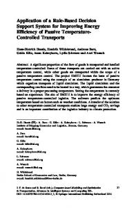
Fig. 1 a, b, c, d: a, b)Transmission electron micrographs showing mitochondria in C6 cells. a) 1xg control, 14000x; b) after 30 min at simulated microgravity, 17700x. Note the disruption of the inner cristae in b). c, d) Transmission electron microscopy micrographs showing outer plasma membrane in C6 cells. c) 1xg control, 14000x; d) after 1h at simulated microgravity, 15500x. Note the absence of the outer coat in d).
a)
b)
c)
d)
e)
Fig. 2 a,b,c.d,e: Na+/K+/ATPase immunostaining. a) 1xg C6 controls, 1700x; b,c) after 30 min at simulated microgavity b=C6 cells, 1100x; c= Leydig cell, 1300x; d,e) after 24h at simulated microgravity (d= C6 newly formed cells, 1100x; e= Leydig cell, 1100x. In b, and c, the immunostaining is low; in d, and e, the immonostaining is again strong.
a)
b)
c)
d)
e)
Fig. 3 a,b,c,d,e: a,b,c) Aquaporin 4 immunostaining in C6 cells. a) 1xg control, 2000x; b) after 30 min at simulated microgravity, 2700x; c) after 24h at simulated microgravity, 2500x. Note the intense immunostaining after 30 min-e) Heat shock proteins (Hsp 27) immunostaining in neurons (NT2 cells). d) after 1h at simulate microgravity; 1700x; e) after 24 h at simulated microgravity, 1700x.
240
Microgravity sci. technol. XVIII-3/4 (2006)
B. M. Uva et al: Ions and Water Transmembrane Transport in Nervous and Testicular Cultured Cells in Low Gravity Conditions
2 Material and Methods In our experiments astrocites (C6 cell line), neurons (NTera 2/D1 from human teratocarcinoma cells - hNT2) and testicular cells: germ cells, Sertoli cells and Leydig cells (BS PRC 57/STe cells, 2n karyotype primary cell line from trypsinized swine 1 month old testes, Zooprophylactic Institute, Brescia, Italy), were grown in DMEM (Sigma, St. Louis, Missouri) with the addition of 10% Foetal Bovine Serum, 1% gentamycin and 1% L-glutamine, streptomycin and amphotericin, at 37°C in a 5% CO2 incubator. The cells were seeded at 20,000 cells/ml using slide flasks (NUNC, flasks apposed onto a removable slide, 9.0 cm2). The flasks were positioned on the Random Positioning Machine (Dutch Space desktop three-dimensional RPM) and kept under continuous rotation at 56 Deg/sec for 30’, 1h, 24h (simulated microgravity, 10-6 g). For transmission electron microscope (TEM) analyses, cells were plated onto 25 cm3 flasks and submitted to random rotation as described before. Static controls (ground controls,1xg), hosted in an identical equipment and treated in parallel, were placed onto the supporting frame of the machine in order to get the cells face to the same vibration stress conditions. At the end of each experiment the cells were fixed with 4% paraformaldehyde, and submitted to immunohistochemical techniques using antisera to: Na+/K+ATPase (α subunit), Na+/K+/Cl- cotransporter (NKCC1, T4) (raised in mouse - these antibodies were obtained from the Developmental Studies Hybridoma Bank, Department of Biological Sciences University of Iowa, Iowa City, USA), the inner mitochondrial proteins - AMA (Medic Italy, raised in rabbit) and Hsp27 (Chemicon International, raised in mouse). The specificity of the immunostainings was verified by replacing the primary antisera with non-immune rabbit serum or PBS. The cells were observed by conventional epifluorescence microscope (Olympus). For Transmission Electron Microscopy analyses, cells were fixed with glutaraldehyde 2.5%, postfixed with osmium tetroxide 1.5%, dehydrated and embedded in epoxy resins. The ultra thin sections were stained with lead citrate and uranyl acetate. 3 Results Damages at mitochondrial level were observed in all cell types investigated after 30 min and 1h of modeled microgravity, they became clustered and often gathered at one side of the nucleus. At the TEM, disruption of the inner mitochondrial cristae (see for example in astrocytes, Fig. 1a,b) was often present and the plasma membrane of all cell types was very thin and deprived of outer coat (see for example in astrocites, Fig. 1c,d). In the astrocytes, neurons, and testicular cells, immunoreactivity for Na+/K+/ATPase (see for example in astrocytes and Leydig cells, Fig. 2a,b,c,d,e) and Na+/K+/Cl- co-transport proteins was very low after 30 min and 1h of RPM rotation, conversely the immunoreactivity for the water channels AQP 4 was enhanced Microgravity sci. technol. XVIII-3/4 (2006)
(see for example in astrocytes, Fig. 3a,b,c). Cell shrinkage was observed at 30min and 1h of modeled microgravity in the cultures. Immunoreactivity for Hsp 27 (see for example in neurons, Fig. 3d,e ) was visible in all cell type after 1h of RPM rotation and became stronger at later times. After 24h, mitochondria and plasma membrane became normal, newly formed astrocytes gathered in cluster, immunohistochemistry for ion transport proteins became normal again in all cell types (see for example in astrocytes and Leydig cells, Fig. 2d,e) and after 1h immunohistochemistry for AQP 4 was again similar to the controls (see for example in astrocytes, Fig. 3c). Immunohistochemical controls yielded negative results. 4 Discussion and conclusion Cell volume is primarily controlled by the intracellular ion homeostasis, thus ion transport across the plasma membrane is of primary importance for the regulation of cell volume [7], K+ is the dominant cation in the cytosol. Intracellular [K+] is mainly controlled by Na+/ K+/ATPase, and by Na+/K+/Cl- co-transport proteins that are a class of membrane proteins that transport Na+, K+, and Cl- ions into and out of cells in an electrically neutral manner [8], inactivation of the sodium pump and the presence of K+ leak channels causes potassium efflux from the cell with consequent volume decrease. From our results, after only 3 min at modeled microgravity in all the cell types investigated, the plasma membrane appears to be damaged, the mitochondria are abnormal, and the inner cristae are disrupted with consequent loss of energy supply, as ATP is generated in the inner cristae through the mitochondrial respiratory chain. In fact after 30 min at low g the proteins involved in ion transport (Na+/K+/ATPase and Na+/K+/Cl- co-transport proteins) appear to be downregulated. Disruption of the mitochondrial inner cristae causes also cytochrome-c release. Cytochrome-c, a proton transporter, translocated from the mitochondrial intermembrane space to the cytosol may open K+ leak channels by oxidizing the channel proteins [9]. The resultant decrease of cytosolic ions concentration mediates water efflux through the plasmalemmal water channels (aquaporins) causing cellular volume decrease. In fact after 30 min at low g, aquaporins are upregulated in all the cell studied. After 1h at simulated weightlessness immunoreactivity for heat shock proteins is evident. Cellular small stress proteins (sHsp) as Hsp 27 polypeptide are oligomeric phosphoproteins that belong to the superfamily of heat shock or stress proteins and are involved in cellular repair. Hsp 27 interferes with the release of cytochrome-c from the mitochondria [10], this may explain the recovery of the enzymatic activity observed by us in all the cells investigated. We may conclude that modeled low g may induce per se cellular volume decrease, the alteration is however only transient, after few hours nervous cells and testicular cultured cells are able to adapt and restore their ultra structure and normally express the enzymes and other proteins involved in ion water transport. 241
B. M. Uva et al: Ions and Water Transmembrane Transport in Nervous and Testicular Cultured Cells in Low Gravity Conditions
References 1 2
3
4
5
6
7
8 9
10
242
Diana, N.: Transcapillary Water Flux. The Physiologists, vol. 25, pp.365375 (1970) Leach, C.S., Alfrey C.P., Suki, W.N., Leonard, J.L., Rambaut, P.C., Inners, L.D., Smith, S.M., Lane, H.W., Krauhs, J.M.: Regulation of body fluid compartments during short-term space flight. J. Appl. Physiol., vol. 81, pp. 105-116 (1996) Alfrey, C.P., Udden, M.M., Leach-Huntoon, C., Driscoll, T., Pickett, M.H.: Control of red blood cell mass in spaceflight. J. Appl. Physiol., vol.81, pp.98-104 (1996) Uva, B.M., Masini, M.A., Sturla, M., Prato, P., Passalacqua, M., Giuliani, M., Tagliafierro, G., Strollo, F.: Clinorotation - induced weightlessness influences the cytoskeleton of glial cells in culture. Brain Res, vol. 934, pp. 132-139 (2002) Shatten H, Lewis M, Chakraburti A.: Spaceflights and clinorotation cause cytoskeleton and mitochondria changes and increases in apoptosis in cultured cells. Acta Astronautica, vol. 49, pp. 399-418. (2001) Uva, B.M, Masini, M.A., Sturla M, Buzzone, F., Giuliani, M., Tagliaferro, G,. Strollo, F. : Microgravity-induced apoptosis in cultured glial cells. Eur. J. Histochem., vol. 46, pp. 209-214. (2002) Lang, F. ,Busch, G. L., Ritter, M., Volkl, H., Waldegger, S., Gulbins, E.,Haussinger, D. Functional significance of cell volume regulatory mechanisms Physiol. Rev., vol 78, pp247-306 (1998) Haas, M. The Na+/K+/Cl- cotransporter. Am. J. Physiol., vol 267, pp C869-C885 (1994) Platoshyn, O., Zhang, S., McDaniel S.S., Yuan JX. J. : Cytochrome – c activates K+ channels before inducing apoptosis. Am. J. Physiol. Cell. Physiol., vol. 283, pp C1289-C1305 (2002) Paul, C., Manero, F., Gonin, S., Kretz-Remy C., Virot, S., Arrigo, A.P.: Hsp 27 as a Negative Regulator of Cytochrome-c Release. Mol. Cell Biol., vol, 22, pp 816-834 (2002)
Microgravity sci. technol. XVIII-3/4 (2006)