Iron Indices in Chronic Kidney Disease in the National Health and Nutritional Examination Survey 1988 –2004 Steven Fishbane,* Simcha Pollack,† Harold I. Feldman,‡ and Marshall M. Joffe§ *Winthrop University Hospital, Mineola, New York; †Department of Computer Information Systems and Decision Sciences, St. John’s University, Jamaica, New York; ‡Renal Electrolyte and Hypertension Division, Department of Medicine, and §Center for Clinical Epidemiology and Biostatistics, University of Pennsylvania School of Medicine, Philadelphia, Pennsylvania Background and objectives: Anemia is a common and early complication of nondialysis chronic kidney disease (CKD). One contributing factor is iron deficiency, which may be particularly problematic during erythropoietin replacement therapy. The aim of this study was to examine the prevalence of iron deficiency in nondialysis CKD. Design, setting, participants, & measurements: The National Health and Nutritional Examination Survey (NHANES) data for NHANES III (1988 to 1994) and subsequent NHANES 2-yr datasets, 1999 to 2000, 2001 to 2002, and 2003 to 2004 were analyzed for individuals >18 yr old. Results: It was found that low levels of iron tests [either serum ferritin < 100 ng/ml or transferrin saturation (TSAT) < 20%] were present in most patients with reduced creatinine clearance (CrCl). The percentage of low iron tests was higher among women than men, present in 57.8 to 58.8% of men and 69.9 to 72.8% of women (P < 0.001). With declining levels of CrCl, in women, TSAT levels decreased, whereas, surprisingly, serum ferritin tended to progressively increase. The percentage of anemic subjects increased progressively with declining quartiles of TSAT but was unrelated to serum ferritin quartiles. Conclusions: It was found that low levels of iron tests, following National Kidney Foundation/Kidney Disease Outcomes Quality Initiative guidelines (either serum ferritin < 100 ng/ml or TSAT < 20%) were present in most patients with reduced CrCl. Clin J Am Soc Nephrol 4: 57– 61, 2009. doi: 10.2215/CJN.01670408
ittle is known regarding iron status among patients with chronic kidney disease (CKD), except for those with stage 5 disease on hemodialysis. In hemodialysis, iron deficiency is a common problem that hinders the effectiveness of erythropoietin treatment (1). Iron deficiency in hemodialysis results primarily from excessive blood loss due to dialysis filter and line blood retention, frequent blood testing, access bleeding, and surgical blood loss (2). Most of these factors are not present, or present to a lesser degree in earlier stages of CKD. Therefore, it is unclear whether iron deficiency is as frequent a problem in these patients. The population of patients with nondialysis CKD is particularly important, however, in that it is much larger than that of hemodialysis (3). Furthermore, anemia develops relatively early in CKD (4), becoming an important cause of fatigue and other symptoms that diminish quality of life. The National Kidney Foundation’s Kidney Disease Outcome Quality Initiative (KDOQI) anemia guidelines recommend that during erythropoiesis-stimulating agent (ESA) treatment in nondialysis CKD that serum ferritin and transferrin saturation (TSAT) be maintained ⬎100 ng/ml and 20%, respectively (5).
L
Because of the lack of published data on this subject, it is not currently possible to determine what proportion of patients with nondialysis CKD have iron values below these threshold levels. For patients not undergoing ESA treatment, the KDOQI guidelines make no recommendations as to levels of iron tests that indicate sufficiency or deficiency. Because serum ferritin and TSAT are the most commonly used tests to assess iron status in CKD, it would be useful to know mean test results and prevalence of decreased levels at different stages of CKD. A helpful source of information in this regard is the National Health and Nutritional Examination Survey (NHANES). This community-based survey includes test results for a sample of individuals representative of the general U.S. population. Hsu et al. studied the NHANES III database as part of an analysis of anemia in CKD (4). The authors found low iron indices to be frequently present at all levels of reduced creatinine clearance (CrCl). The analysis, however, was limited in scope because most data were collected before the widespread use of ESA or the publication of the first KDOQI guidelines. The purpose of the analysis presented here was to fully examine the distribution of serum ferritin and TSAT among individuals with CKD studied by the NHANES program.
Received April 8, 2008. Accepted September 21, 2008.
Materials and Methods
Published online ahead of print. Publication date available at www.cjasn.org.
NHANES is a program of ongoing studies designed to assess the health of adults and children in the United States. The survey combines interviews, physical examinations, and blood sampling for laboratory
Correspondence: Dr. Steven Fishbane, 200 Old Country Road, Suite 135, Mineola, NY 11501. Phone: 516-663-2169; Fax: 516-663-2179; E-mail
[email protected] Copyright © 2009 by the American Society of Nephrology
ISSN: 1555-9041/401–0057
58
Clinical Journal of the American Society of Nephrology
testing. NHANES is a program of the National Center for Health Statistics (NCHS), part of the Centers for Disease Control and Prevention (CDC). The NHANES program began in the early 1960s and has been conducted as a series of surveys focusing on different population groups or health topics. In 1999, the survey became a continuous program, rather than one that conducted periodic large-size surveys. The survey currently examines a nationally representative sample of approximately 5000 persons each year. In the study presented here, data for NHANES III (1988 to 1994) and subsequent NHANES 2-yr datasets, 1999 to 2000, 2001 to 2002, and 2003 to 2004 were analyzed. The term-combined cohort is defined as a combination of data from each of the four included NHANES datasets. The purpose of study was to describe aspects of iron status among community patients with CKD. Renal function was estimated using data collected through the NHANES program using the Cockroft– Gault formula, [(140 ⫺ age) ⫻ weight (kg)]/72 ⫻ serum creatinine (result multiplied by 0.85 for girls/women) (6,7). Serum creatinine in NHANES III and the 1999 to 2000 dataset required correction as per Selvin et al. (8). We adjusted serum creatinine accordingly for these two datasets using the correction, if years ⫽ NHANES3 then lbxscr ⫽ .9601 ⫻ lbxscr ⫺ 0.184; if years ⫽ 1999 to 2000 then lbxscr ⫽ 1.013 ⫻ lbxscr ⫹ 0.147. The Cockroft–Gault formula was used to facilitate comparison to the earlier work of Hsu et al. (4), which also used this formula. Recalculation of our results using the four-variable Modification of Diet in Renal Disease Study Equation (MDRD) (9) revealed results that were very similar to those obtained with the Cockroft–Gault equation, with no substantive differences in classification to a specific stage of kidney disease (0.2 to 3.1%) in any of our analyses, and only very small changes in iron test categorization (0.2 to 2.9%) or hemoglobin (Hgb) quartile (0.7 to 4.0%). Stage of CKD was classified as per National Kidney Foundation (NKF) guidelines (10). Only subjects off hemodialysis and aged 18 or over were studied.
Descriptive Analyses
Clin J Am Soc Nephrol 4: 57– 61, 2009
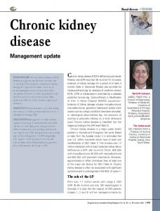
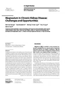
Figure 1. Percentage of subjects in each creatinine clearance (CrCl) classification for the combined cohorts (n ⫽ 34,782), chronic kidney disease (CKD) stage 5, CrCl 0 to 14.9, 0.2%; CKD stage 4, CrCl 15 to 29.9, 0.8%; CKD stage 3, CrCl 30 to 59.9, 9.9%; CrCl 60 to 90 (may be confused with CKD stage 2), 20.7%; CrCl ⬎ 90, 68.4%. iron tests in nondialysis CKD are defined by NKF-KDOQI as serum ferritin ⬍ 100 ng/ml or TSAT ⬍ 20%. We found that a high percentage of patients with CKD met these criteria. Figure 2 displays the percentage of individuals with serum ferritin ⬍ 100 ng/ml or TSAT ⬍ 20% for different ranges of CrCl corresponding to CKD stages 1 to 5. Serum creatinine-based estimating equations have their greatest precision for defining the presence of CKD when estimated CrCl (or GFR) ⬍ 60 ml/min. Therefore the data for CrCl ⬎ 60 ml/min is presented for informational purposes and should be interpreted with caution. Many or most of these patients may have normal kidney function, in which case the KDOQI iron thresholds would not be relevant. Instead we focus on patients with estimated CrCl ⬍ 60 ml/min. Among these patients, we found low iron test results
TSAT (serum iron/total iron binding capacity ⫻ 100) and serum ferritin were available in 28,207 and 26,283 of the subjects, respectively. We determined the percentage of individuals with serum ferritin ⬍ 100 ng/ml and/or TSAT ⬍ 20% at different levels of kidney function. These levels of ferritin and TSAT were chosen because they are the recommended levels for patients with nondialysis CKD of the NKF’s KDOQI anemia guidelines (11). All continuous variables are described using their means (SD). Outliers defined as more than 4 SD above or below the mean were removed. Sample weights were used in all analyses to account for the probability sampling that NHANES utilized; that is, observations in strata that are under (over) represented with respect to the U.S. population are weighed more (less) heavily. Sample weights across NHANES surveys are combined according to the procedure suggested on the NHANES website. P values ⬍ 0.05 were considered to be statistically significant. Data were analyzed using the statistical package SAS 9.1 (SAS Institute, Inc., Cary, North Carolina).
Results The combined cohort (NHANES III (1988 to 1994), 1999 to 2000, 2001 to 2002, and 2003 to 2004), represents 62,437 patients. After removing outliers and missing data we were left with a usable n of 34,782 (TSAT and ferritin results were not available for all patients). We observed clearly reduced CrCl (⬍60 ml/ min, CKD stages 3 to 5) in 6.6% of individuals (Figure 1). Low
Figure 2. The percentage of men and women with either serum ferritin ⬍ 100 ng/ml or transferrin saturation (TSAT) ⬍ 20% at different levels of CrCl for the combined National Health and Nutritional Examination Survey (NHANES) cohorts. National Kidney Foundation (NKF) CKD stages relative to CrCl are stage 5, 0 to 14.99; stage 4, 15 to 29.99; and stage 3, 30 to 59.99. Patients with CrCl 60 to 90 ml/min may have CKD stage 2. Patients with CrCl ⬎ 90 ml/min may have CKD stage 1 if other renal abnormalities are present.
Clin J Am Soc Nephrol 4: 57– 61, 2009
Iron Indices in CKD in the NHANES
59
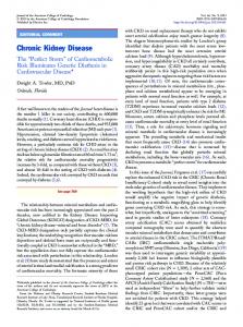
to be present in most patients. The percentage of low iron tests was higher among women than men, present in 57.8 to 58.8% of men and 69.9 to 72.8% of women (P ⬍ 0.001). There were no significant differences between races for TSAT, but Blacks had significantly higher serum ferritin levels than others with CKD (32 to 46% higher with P ⬍ 0.0001). The percentage of patients with low iron tests did not significantly differ between CKD stages 3, 4, or 5 (P ⫽ NS). Among men, there was no linear relationship between either mean serum ferritin or TSAT as a function of the level of CrCl. In contrast, among women there was a trend toward lower decreasing mean TSAT for progressively lower levels of renal function (P ⫽ 0.02) (Figure 3a). Paradoxically, among women, there was a statistically significant trend toward increasing serum ferritin for progressively lower levels of renal function (P ⬍ 0.0001) (Figure 3b). One possible explanation for a divergence between serum ferritin and TSAT would be an effect of inflammation. Although NHANES does not contain in-depth information on
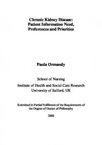
inflammatory markers, it does contain at least one marker, C-reactive protein (CRP). For men, mean CRP increased progressively from CKD stages 3 to 5, from 0.43 ⫾ 0.52, to 0.50 ⫾ 0.58, to 0.57 ⫾ 0.76, respectively (P ⫽ 0.01). In women, there was a similar trend, with mean CRP increasing through CKD stages 3 to 5 from 0.43 ⫾ 0.52, to 0.48 ⫾ 0.59, to 1.02 ⫾ 0.82, respectively (P ⫽ 0.02). The KDOQI iron threshold values of serum ferritin 100 ng/ml and TSAT 20% were generally based on expert opinion (although as discussed below they are validated to an extent by the work of Gotloib et al. (16)). In Figure 4 we present the percentage of subjects with CrCl ⬍ 60 ml/min who would be defined as iron deficient using different cutoff values for serum ferritin and TSAT. In addition, the effect of using AND compared with OR logic is displayed. With the more commonly used OR logic, the frequency of defined iron deficiency decreases progressively with lower threshold values for iron tests. One of iron’s important biologic roles is for erythropoiesis, so iron deficiency’s effect on anemia risk is highly relevant. The relationship between iron tests and probability of anemia was studied for subjects with CrCl ⬍ 60 ml/min. Using the KDOQI definition of anemia in CKD, Hgb ⬍ 13.5 g/dl in men or 12.0 g/dl in women, risk for anemia was found be generally unrelated to quartiles of mean serum ferritin. In contrast, the probability of anemia increased with progressively lower quartiles of TSAT, from 10.9% in quartile 4 to 27% in quartile 1 (Figure 5). The change in mean serum ferritin and TSAT over time was assessed for patients with moderate to severe CKD (CrCl ⬍ 60 ml/min) for successive NHANES cohorts. There was no significant change in mean serum ferritin or TSAT over the three data sets for which it could be calculated (1988 to 1994, 1999 to 2000, and 2001 to 2002).
Figure 3. Mean (a) serum ferritin and (b) TSAT as a function of CrCl for the combined NHANES cohorts (error bars are SD). The trend for both TSAT and ferritin is NS for men but for women P ⬍ 0.0001 for serum ferritin and P ⬍ 0.02 for TSAT. NKF CKD stages relative to CrCl are stage 5, 0 to 14.99; stage 4, 15 to 29.99; and stage 3, 30 to 59.99. Patients with CrCl 60 to 90 ml/min may have CKD stage 2. Patients with CrCl ⬎ 90 ml/ min may have CKD stage 1 if other renal abnormalities are present.
Figure 4. Percentage of individuals defined as iron deficient using different threshold combinations of serum ferritin and TSAT. The NKF Kidney Disease Outcomes Quality Initiatives (KDOQI) thresholds of serum ferritin 100 ng/ml and TSAT 20% are different than indices of iron deficiency in the non-CKD population in which lower thresholds are generally used. The light blue bars indicate AND logic, both test results below the specified threshold, whereas the dark blue bars indicate OR logic, with either test result being below the threshold.
60
Clinical Journal of the American Society of Nephrology
Figure 5. There was no observable trend for mean hemoglobin (Hgb) by quartiles of serum ferritin (dark bars) (P ⫽ NS). The trend for mean Hgb by quartiles of TSAT (light bars) was significant, P ⬍ 0.0001 in the combined NHANES cohorts for patients with CrCl ⬍ 60 ml/min.
Discussion There has long been a great interest in iron tests and iron status in hemodialysis patients (12,13). In contrast, far less is known regarding iron status of patients with nondialysis CKD. The NHANES surveys are an excellent source for learning about serum ferritin and TSAT in this population. The community-based focus and population-sampling techniques yield a relatively clear view of CKD in the general population. It should be noted that our results are most applicable to patients with estimated GFR ⬍ 60 ml/min in NHANES, in which CKD is most likely to be present. Hsu et al. (4,15) studied iron status in CKD in the NHANES III survey (1988 to 1994) and found iron indices suggestive of iron deficiency to be present (4) and to contribute to anemia (15) in many subjects. The purpose of our analysis was to broaden the scope to include a fuller exploration of iron indices in nondialysis CKD and to update these findings. Typical markers of iron deficiency used in CKD are serum ferritin ⬍ 100 ng/ml and TSAT ⬍ 20%. Clinicians often use these thresholds to base iron treatment decisions, and KDOQI guidelines recommend these levels in nondialysis CKD (10,11). Specifically, the KDOQI guidelines indicate that if either value is low then iron treatment is recommended. Our primary finding is that between 57.8 and 72.8% of subjects with CKD have either serum ferritin ⬍ 100 ng/ml or TSAT ⬍ 20%. This remarkably high prevalence might indicate that these indices may not be specific enough and may falsely identify too many subjects as being iron deficient. In contrast to these relatively high values of serum ferritin (100 ng/ml) and TSAT (20%) that indicate insufficient iron in hemodialysis, in the general population lower thresholds of serum ferritin (15 to 30 ng/ml) and TSAT (15%) are often used. Whether iron deficiency in CKD should be indicated by iron indices closer to those used in hemodialysis or the indices used for the general population, or even some interim set of values, is unclear. To explore this further we studied the proportion of subjects defined as iron deficient with indices set at different threshold levels using
Clin J Am Soc Nephrol 4: 57– 61, 2009
different conjunctive criteria either to maximize the sensitivity or specificity with which iron deficiency was detected. On the basis of this analysis, a more realistic prevalence for reduced iron indices occurs with serum ferritin ⬍ 50 ng/ml or TSAT ⬍ 15% (just above 30% prevalence) or serum ferritin ⬍ 100 and TSAT ⬍ 20% (approximately 20% prevalence). However, these are simply the frequencies of low iron tests, not necessarily the presence of iron deficiency. A study that included either bone marrow testing or response to treatment with iron would be required to properly assess the true prevalence of iron deficiency. In hemodialysis, repeated blood loss makes iron deficiency an almost universal problem (12). In contrast, in nondialysis CKD, dialysis-related blood loss does not occur, so iron deficiency could occur less frequently. The great prevalence of low iron indices we found may not indicate iron deficiency per se, but rather impaired iron delivery concurrent with inflammation, a complex syndrome that occurs with progressive CKD. However, it is plausible that iron deficiency might be more common than expected because in CKD the prevalence of gastrointestinal pathology with blood loss is probably increased (15), sampling of blood for laboratory testing is common, and hospitalizations and surgery for other intercurrent illness could contribute to blood loss. In addition, many patients are treated with ESA, further depleting iron stores. Evidence in support of the high prevalence of iron deficiency that we found in CKD is provided by a recent publication by Gotloib et al. (16). These investigators performed sternal bone marrow biopsies on 47 patients with CKD and Hgb ⬍ 12 g/dl. Remarkably, severe iron deficiency was found in 46 of 47 subjects. Also, the mean serum ferritin was greater than 200 ng/ml, strongly validating our results. Patients were subsequently treated with intravenous iron, with most responding with improved Hgb concentration (16). If these results are replicated by others it would serve as further confirmation of the external validity of the NHANES data we report here, indicating that iron deficiency is an extraordinarily common problem among patients with nondialysis CKD. We found a divergence of trends for serum ferritin and TSAT in relation to renal function in CKD among women, but not men. Although mean serum ferritin tends to unexpectedly increase with lower levels of CrCl in women, mean TSAT does the opposite, decreasing progressively. Because both tests are indices of iron status, it is surprising that one would indicate a greater prevalence of iron deficiency with progressive CKD whereas the other indicates a lower prevalence. The cause of this discordant behavior is unclear. One possible explanation is the effect of inflammation. It is well known that occult inflammation is commonly present in CKD and may increase in prevalence with progressive disease (17,18). Inflammation has a profound effect on iron indices. Previously, in hemodialysis, CRP, an indicator of inflammation, hwas found to be highly correlated with serum ferritin values (19). We found CRP levels to increase progressively through CKD stages 3 to 5. However, because this was true for both men and women, and because the divergence in iron indices was only present in women, it would seem unlikely that inflammation would explain the
Clin J Am Soc Nephrol 4: 57– 61, 2009
discordant behavior well. Anemia in kidney disease is a complex process that reflects an interaction of the erythropoietic processes of bone marrow with iron availability and inflammation. NHANES does not contain enough variables measuring the inflammatory state to allow for more than a superficial look at the effect of inflammation in this paper. We would encourage future studies with more targeted data collection to help sort out the interesting divergent behavior of serum ferritin and TSAT and to better understand the interaction of anemia, inflammation, and iron. One way to assess the importance of iron status in CKD is to analyze its relationship to anemia. To the extent that decreased serum ferritin or TSAT are associated with reduced levels of Hgb, the findings take on greater significance because a primary effect of iron deficiency is to blunt erythropoiesis. To this end, we found that the proportion of anemic subjects increased progressively with each lower quartile of TSAT. In contrast, the result was present but blunted with serum ferritin. The stronger relationship for TSAT compared with serum ferritin might indicate greater relevance for this test as a marker of iron status in nondialysis CKD. In summary, we have found that reduced levels of serum ferritin or TSAT are present in most patients with CKD. Whether this is indicative of a high prevalence of true iron deficiency in CKD is unclear. In addition we found highly discordant behavior for serum ferritin and TSAT with declining CrCl among women with CKD; serum ferritin increases whereas TSAT declines. We found that lower levels of TSAT were clearly associated with increased risk for anemia in CKD.
Disclosures None.
References 1. Van Wyck DB, Stivelman JC, Ruiz J, Kirlin LF, Katz MA, Ogden DA: Iron status in patients receiving erythropoietin for dialysis-associated anemia. Kidney Int 35: 712–716, 1989 2. Fishbane S, Maesaka JK: Iron management in end-stage renal disease. Am J Kidney Dis 29: 319 –333, 1997 3. Coresh J, Selvin E, Stevens LA, Manzi J, Kusek JW, Eggers P, Van Lente F, Levey AS: Prevalence of chronic kidney disease in the United States. JAMA 298: 2038 –2047, 2007 4. Hsu CY, McCulloch CE, Curhan GC: Epidemiology of anemia associated with chronic renal insufficiency among adults in the United States: Results from the Third National Health and Nutrition Examination Survey. J Am Soc Nephrol 13: 504 –510, 2002 5. KDOQI; National Kidney Foundation: II. Clinical practice guidelines and clinical practice recommendations for anemia in chronic kidney disease in adults. Am J Kidney Dis 47[5 Suppl 3]: S16 –S85, 2006
Iron Indices in CKD in the NHANES
61
6. Cockcroft DW, Gault MH: Prediction of creatinine clearance from serum creatinine. Nephron 16: 31– 41, 1976 7. Gault MH, Longerich LL, Harnett JD, Wesolowski C: Predicting glomerular function from adjusted serum creatinine. Nephron 62: 249 –256, 1992 8. Selvin E, Manzi J, Stevens LA, Van Lente F, Lacher DA, Levey AS, Coresh J: Calibration of serum creatinine in the National Health and Nutrition Examination Surveys (NHANES) 1988 –1994, 1999 –2004. Am J Kidney Dis 50: 918 –926, 2007 9. Levey AS, Greene T, Kusek J, Beck G: A simplified equation to predict glomerular filtration rate from serum creatinine [Abstract]: J Am Soc Nephrol 11: 155A, 2000 10. National Kidney Foundation: K/DOQI clinical practice guidelines for chronic kidney disease: evaluation, classification, and stratification. Am J Kidney Dis 39[2 Suppl 1]: S1–S266, 2002 11. KDOQI; National Kidney Foundation KDOQI Clinical Practice Guidelines and Clinical Practice Recommendations for Anemia in Chronic Kidney Disease. Am J Kidney Dis 47[5 Suppl 3]: S11–S145, 2006 12. Van Wyck DB, Stivelman JC, Ruiz J, Kirlin LF, Katz MA, Ogden DA: Iron status in patients receiving erythropoietin for dialysis-associated anemia. Kidney Int 35: 712–716, 1985 13. Fishbane S, Kowalski EA, Imbriano LJ, Maesaka JK: The evaluation of iron status in hemodialysis patients. J Am Soc Nephrol 7: 2654 –2657, 1996 14. Hsu CY, McCulloch CE, Curhan GC: Iron status and hemoglobin level in chronic renal insufficiency. J Am Soc Nephrol 13: 2783–2786, 2002 15. Bini EJ, Kinkhabwala A, Goldfarb DS: Predictive value of a positive fecal occult blood test increases as the severity of CKD worsens. Am J Kidney Dis 48: 580 –586, 2006 16. Gotloib L, Silverberg D, Fudin R, Shostak A: Iron deficiency is a common cause of anemia in chronic kidney disease and can often be corrected with intravenous iron. J Nephrol 19: 161–167, 2006 17. Landray MJ, Wheeler DC, Lip GY, Newman DJ, Blann AD, McGlynn FJ, Ball S, Townend JN, Baigent C: Inflammation, endothelial dysfunction, and platelet activation in patients with chronic kidney disease: The chronic renal impairment in Birmingham (CRIB) study. Am J Kidney Dis 43: 244 –253, 2004 18. Oberg BP, McMenamin E, Lucas FL, McMonagle E, Morrow J, Ikizler TA, Himmelfarb J: Increased prevalence of oxidant stress and inflammation in patients with moderate to severe chronic kidney disease. Kidney Int 65: 1009 –1016, 2004 19. Kalantar-Zadeh K, Rodriguez RA, Humphreys MH: Association between serum ferritin and measures of inflammation, nutrition and iron in haemodialysis patients. Nephrol Dial Transplant 19: 141–149, 2004 20. Weiss DJ, Krehbiel JD, Lund JE: Studies of the pathogenesis of anemia of inflammation: Mechanism of impaired erythropoiesis. Am J Vet Res 44: 1832–1835, 1983
Access to UpToDate on-line is available for additional clinical information at http://www.cjasn.org/