Elhammer,k P., Poorman, R. A., Brown, E., Maggiora, L. L., Campbell, C. M., Hoogerheide, J. G., and ... 199-213, John Wiley & Sons, New York. 74.54.63-6467.
Vol. 268,No. 17,Issue of June 15,pp. 12609-12616,1993 Printed in U.S.A.
THEJOURNAL OF BIOLOGICAL CHEMISTRY Q 1993 by TheAmerican Society for Biochemistry and Mo\ecular Biology, Inc.
Isolation and Expression of a cDNA Clone Encoding a Bovine UDP-Ga1NAc:Polypeptide N-Acetylgalactosaminyltransferase* (Received for publication, December 7, 1992, and inrevised form, March 3, 1993)
Fred L.HomaS, Tameira HpllanderQll, Donna J. Lehmant, Darrell R. ThomsenS, and Ake P. ElhammerQ(1 From the $Molecular Biology and §Biochemistry Research Units, The Upjohn Company, Kalamazoo,Michigan 49001
ogen interaction, to more general, such asproviding protection from proteolytic degradation or supplying the appropriate charge and water binding properties to mucous secretions (reviewed by Sadler (1984), Paulson (19891, and Jentoft (1990)). The initial reaction in O-linked oligosaccharide biosynthesis is the transfer of an N-acetylgalactosamine residue from the nucleotide sugar UDP-N-acetylgalactosamine to a serineorthreonine residue on the protein acceptor. This reaction, which can occur post-translationally, is catalyzed by UDP-Ga1NAc:polypeptide N-acetylgalactosaminyltransferase (GalNAc-transferase)’ (EC 2.4.1.41), an intracellular enzyme believedto be localized in the secretory pathway. The exact location(s)of GalNAc-transferase is still controversial. It has been reported that the initial addition of N acetylgalactosamine to the acceptor protein can take place early (even cotranslationally) in the rough endoplasmic reticulum. Other authors have suggested that this reaction is a post-translational eventoccurring in later endoplasmic reticulum compartments and/or in the cis region of the Golgi complex. Finally, evidence has also been presented for model a in which transfer of N-acetylgalactosamine to Ser/Thr may occur in several compartmentsin the secretory pathway, including compartments later than the Golgicomplex (reviewed by Schachter and Brockhausen (1992)). Elongation and terminationof O-linked oligosaccharides is accomplished by sequential addition of individual monosaccharides by specific transferases (Roseman, 1970); current data suggest that these reactions are localized primarily in the Golgi apparatus (Schachter and Brockhausen, 1992). The acceptor (peptide) specificity of GalNAc-transferase is also poorly understood. Studies using intact protein acceptors as well as peptides containing sequences derived from known acceptors have yielded little conclusive information (Hagopian et al., 1971; Hill et al., 1977; Gooley et al., 1991; Young et al., 1979; Briand et al., 1981; Hughes et al., 1988; Wang et O-Glycosidically linked (mucin-type) oligosaccharides have al., 1992), although some progress in this area has recently been reported on a number of different types of glycoproteins been reported (Elhammer et al., 1993). (reviewed by Sadler (1984)). These structures have been asMilk (and colostrum) contains a number of glycosyltranssigned a diverse array of functions, ranging from quite specific, ferase activities (e.g. Prieels et al. (1975),Paulson et al. (1977), such as being involved in cell-cell recognition and host-path- Bushway and Keenan (1979), and Parodi et al. (1984)). Previous work has shown that bovine colostrum contains what * The costs of publication of this article were defrayed in part by appears to be a soluble form of GalNAc-transferase (Elhamthe payment of page charges. This article must therefore be hereby marked “advertisement” in accordance with 18 U.S.C. Section 1734 mer and Kornfeld, 1986). This report also described a procedure for the purification of this molecule to homogeneity. In solely to indicate this fact. The nucleotide sequence(s)reported in thispaperhas been submitted the currentstudy, we have used this procedure for purification to the GenBankTM/EMBL Data Bank with accessionnumber(s) of GalNAc-transferase in amounts sufficient for NHZ-termiL87780. nal sequencing. The information obtained was used to isolate
NHs-terminal amino acid sequence obtained from a UDP-GalNAc:polypeptide N-acetylgalactosaminyltransferase (GalNAc-transferase) isolated from bovine colostrum was used for the construction of synthetic oligonucleotide primers. Subsequent polymerase chain reaction and library screenings of a bovine intestine cDNA library produced seven positive clones. The conlargest clone had a 2294-base pair insert that tained an open reading frame coding for a protein a predicted polypepcomposed of 559 amino acids with tide molecular mass of 64,173Da. The cloned molecule has no significant sequence homology to previously reported cloned glycosyltransferases, but appears to have a similar domain structure. It is a type I1 membrane protein with a 23-amino acid putative transmembrane regionstarting 8 amino acids from theNH2 terminus. The transmembrane segment of the molecule is immediately followed by a sequence rich in proline residues. The molecule contains three consensus sequences for N-linked glycosylation and five predicted sites for O-glycosylation. Northern blot analysis of poly(A+) mRNA isolated from Madin-Darby bovine kidney cells, bovine mammary tissue, and eight human tissues demonstrated the expression of two transcripts differing in size by -1 kilobase. The cloned DNA was expressed in insect cells using a baculovirus vector. Thisresultedin an almost 100-foldincreasein GalNAc-transferase activity in lysates prepared from cells infected with viruscontaining the GalNAc-transferase gene compared to cells infected with virus containing DNA coding for an unrelatedmolecule or uninfected cells. Immunoprecipitation from lysates prepared from infected cells labeled in vivo with [ssS] methionine showed a large increase in the recoveryof an -67-kDa protein.
n Summer Intern. Present address: University of Michigan, Ann Arbor, MI 48109. (1 To whom correspondence should be addressed Biochemistry Research Unit, 7240-209-622, The Upjohn Co., 301 Henrietta St., Kalamazoo, MI 49001. Tel.: 616-384-9399; Fax: 616-385-7373.
The abbreviations are used are: GalNAc-transferase, UDPGa1NAc:polypeptide N-acetylgalactosaminyltransferase; PCR, polymerase chain reaction; PAGE, polyacrylamide gel electrophoresis; HPLC, high performance liquid chromatography; bp, base pair(s).
12609
12610
cDNA Cloning of GalNAc-transferase
a cDNA clone encoding a full-length (membrane-bound) transferase that, upon expression in the insect cell line Sf9, resulted in the synthesis of a fully active enzyme. The availability of the cloned molecule should greatly facilitate studies both on the subcellular localization and acceptor specificity as well as other characteristicsof this enzyme.
labeled with [ C X - ~ ~ P I ~by A Tnick P translation (Goldin et al., 1981). The cDNA library (containing 2.5 x lo6 independent clones) was screened by plaque hybridization using the above-described labeled DNA fragment as a probe. Seven positive plaques were obtained from the primary screen, and each isolate was plaque-purified three times. DNA was isolated from each of the seven X clones and digested with EcoRI or analyzed by PCR using oligonucleotides F and G as primers (see Fig. 2B). The size of the X g t l O inserts was determined on 1% agarose gels. EXPERIMENTALPROCEDURES DNA Sequence Analysis-The inserts in pCR1000-931, pCR1000MateriaL+"c~-~~P]dATP (300 Ci/mmol), UDP-N-[1-3H]acetylga- 600, pCR1000-91B, and pCR1000-52A weresequenced by the dideoxy lactosamine (8.3 Ci/mmol), and NalZ5I(15.2 mCi/pg) were purchased chaintermination method (Sanger et al., 1977) using Sequenase from Amersham COT. [ ( u - ~ ~ P I ~ A was T Pfrom Du Pont-New England Version 2.0 with [ C Y - ~ ~ P I ~ A Double-stranded TP. DNA sequencing was Nuclear, and [35S]methioninewas from ICN (Tran3%1abel, 1 mCi/ done with 20-mer oligonucleotide primers, synthesized according to ml). Bovine colostrum was obtained from a local farmer. UDP-N- the sequence of the cDNA insert. The sequencing strategy is shown acetylgalactosamine, UDP, phenylmethylsulfonyl fluoride, chymosta- in Fig. 2C. Sequence analysis was performed using the sequence tin, leupeptin, antipain,pepstatin,aprotinin, bovine submaxillary analysis software package of the University of Wisconsin Genetics mucin, Nonidet P-40, Triton X-100, taurodeoxycholate, Sephadex G- Computer Group (Devereux et al., 1984). 100 superfine, and rabbitanti-chicken IgG antibodies were from RNA Isolation and Northern Hybridization-Total RNA and Sigma. DEAE-Sephacel, Sepharose 6B, and protein A-Sepharose were poly(A+) RNA were prepared from bovine mammary tissue and from from Pharmacia LKB Biotechnology Inc. IODO-GEN was from Madin-Darby bovine kidney cells using the Invitrogen Fastrack kit Pierce Chemical Co. Peptide N-glycosidase F was from Oxford Gly- following the manufacturer's procedure. Two pgof poly(A+) RNA cosystems. The GeneAmp kit (for PCR), was obtained from Perkin- were denatured by glyoxylation, and Northern blot analysis was Elmer Cetus Instruments. A bovine small intestine cDNA library performed as previously described (Homaet al., 1986). Ahuman cloned in a X g t l O vector was purchased from Clontech (BL1010a). multiple tissue Northern blot was purchased from Clontech (7760-1). The TA cloning vector pCRlOOO was from Invitrogen. Sequenase The blots were prehybridized in 50% formamide, 5 X SSC, 1 X Version 2.0 was from U. S. Biochemical Corp. The baculoGold trans- Denhardt's solution, 1%SDS, 100 pg/ml denatured salmon testis fection kit was from PharMingen. Restriction enzymes and all other DNA a t 42'C for 2 h and then hybridized overnight a t 42 "C with reagents were from standard sources. the "P-labeled 600-bp insert isolated from pCR1000-600. Filters were washed three times for 15 min in 0.1 X SSC, 0.1% SDS at 55 "C. Isolation of UDP-Ga1NAc:Polypeptide N-AcetylgalactosaminylExpression of pCR1000-9IB Insert in Sf9Cells-pCR1000-91B was transferase-The first four steps inthe purification of the transferase were identical to theprocedure described by Elhammer and Kornfeld digested with SstII and Hind111 (both enzymes cut only in pCRlOOO (1986), except that the samples loaded on the affinity columns were sequences that flank the insert) (see Fig. 2C), and these sites were adjusted to 1 mM ATP (in addition to the reported buffer, salt, and blunted using T4 DNA polymerase. BamHI linkers were then ligated UDP concentrations) to compensate for an apparently higher pyro- onto the blunted ends, and the resulting sample was ligated into the phosphatase activity(ies) in the colostrum used in the current work. BamHI site of the baculovirus expression vector pAC373 (Summers Equilibration, loading, washing, and elution buffer volumes were and Smith, 1986). The resulting isolates were screened for proper adjusted (scaled up) for the larger columns used. Assays, sample orientation of the GalNAc-transferase open reading frame with reconcentration procedures, column elution patterns, etc. were as de- spect tothe baculovirus polyhedrin promoter, to yieldpAC373scribed by Elhammer and Kornfeld (1986). The conditions for the GalNAcT. Cotransfection of Sf9 cells with pAC373-GalNAcT and fifth step in the purification, size-exclusion chromatography, were linearized baculovirus DNA from the PharMingen baculoGold transaltered in an effort to improve the poor recoveries obtained in this fection kit was performed using calcium phosphate precipitation step in the previous purification (Elhammer and Kornfeld, 1986). (Summers and Smith, 1986). The baculovirus DNA provided in the The column used was Sephadex G-100 superfine (20-50-pm bead size PharMingen transfection kit contains a lethal mutation that can be 1.5 X 100 cm) equilibrated in 25 mM imidazole, pH 7.2,300 mM NaCl, corrected by homologous recombination with sequences contained in 0.1% taurodeoxycholate, 10% glycerol. The column was run at a the pAC373 vector. Therefore, following transfection, only recombinant viruses willgrow on Sf9 cells. Transfections were done in pressure of -30 cm of HzO, which resulted in a flow of -2.3 ml/h; and fractions (100 total) were collected a t 40-min intervals. Other, duplicate, and theresulting virus samples are referred to asGalNAcT less difficult to work with matrixes were tested for this step, but none 2-1A and GalNAcT 2-1B. Preparation of Chicken Anti-GalNAc-transferase Antibodies-A yielded a comparable resolution. The recoveries from this step using chicken was injected intramuscularly axillary with 100 pg of purified the conditions described above typically ranged from 80 to 90%. NHz-terminal sequencing of the purified molecule was done by enzyme (with Freund's complete adjuvant). One month later, the automated Edman degradation in an Applied Biosystems Sequencer chicken was boosted subcutaneously with another 50 pg of antigen (Model 470) fitted with an on-line HPLC analyzer (Model 120A) for (with Freund's incomplete adjuvant); asecond intramuscular booster, phenylthiohydantoins. Quantitation of the latterwas afforded by the 50 pg of enzyme axillary, was administered after an additional 21 Nelson Analytical Turbochrom chromatography data system con- days. Test bleeds were done 2 weeks after each booster. After the second test bleed (which, upon analysis, was found to contain antinected in parallel with the recorder to the output from the HPLC GalNAc-transferase antibodies), eggswere collected each day and system. used as a source for antibodies. IgG was isolated from egg yolk as Assays for UDP-Ga1NAc:Polypeptide N-Acetylgalactosaminyltransferase Activity-Activity assays during the purification were done as described by Jensenius et al. (1981). Immunoprecipitations-Immunoprecipitation of the in vivo [35Ss] described by Elhammer and Kornfeld (1986). Assays for activity in lysates from Sf9 cells were carried out as described by Thomsen et al. methionine-labeled enzyme was done from crude cell lysates. Infected cells were labeled between 24 and 48 h postinfection with 50 pCi/ml (1990). PCR-Oligonucleotide primers were prepared with a Applied Bio- [35S]methioninein medium that containedone-tenth the normal systems DNA synthesizer (Model 380B). PCR was carried out in 0.1 methionine concentration. Approximately 1.5 X lo6 labeled infected ml of solution containing 50 mM KCI, 10 mM Tris-HC1, pH 8.3, 1.5 cells were dissolved in 670 pl of phosphate-buffered saline containing mM MgCl,, a 0.2mM concentration of each of the four dNTPs, a 1 0.5%Triton X-100,0.5% taurodeoxycholate, 0.05% SDS, 0.1 Trypsin inhibitory units/ml aprotinin,and 10 pg/ml each leupeptin, antipain, p~ concentration of each oligonucleotide, either 5 pl of the bovine intestine cDNA library or 10 ng of plasmid or X DNA, and 2.5 units chymostatin, and pepstatin. Following incubation on ice for >2 h, of Taq polymerase. The reaction was covered with 0.1 ml of mineral any undissolved debris was sedimented at 10,000 x g for 20 min, and oil and subjected to atemperature step cycle. When degenerate the supernatant was collected. Immunoprecipitation was carried out chicken IgG) of chicken antioligonucleotides were used, the steps were 94 "C (1 min), 37"C (2 by the addition of 4 pl (-20pgof min), and 72 "C (3 min) for a total of 35 cycles. For nondegenerate GalNAc-transferase antibodies; purified IgG isolated from egg yolk oligonucleotides, the steps were94 "C (1 rnin), 55 "C (2 rnin), and was used for all immunoprecipitation experiments. Following incubation a t 4 "C overnight, the antigen-antibody complexes were iso72 "C (3 min) for a total of 25 cycles. Screening of u t 1 0 Bovine Small IntestinecDNA Library-A bovine lated by overnight adsorption to 22 p1 (volume of sedimented gel) of small intestine cDNA library cloned into a X g t l O vector was used. A protein A-Sepharose coated with rabbit anti-chicken IgG antibodies. subcloned 600-bp PCR product (insert of plasmid pCR1000-600)was The coated protein A-Sepharose was prepared by incubating 330 pl
cDNA Cloning of GalNAc-transferase of sedimented protein A-Sepharose with 2.3 mg of rabbit anti-chicken IgG antibodies (an affinity-purified IgG fraction) in 1 ml of phosphate-buffered saline overnight; the coated protein A-Sepharose was washed three times with1ml of phosphate-buffered saline containing 0.5% Triton X-100, 0.5% taurodeoxycholate, 0.05% SDS. Following adsorption of the antigen, the immunosorbent was sedimented by centrifugation and washed extensively essentially as described by Dunphy et al. (1985). The washed antigen-antibody-immunosorbent complexes were suspendedin 50 pl of SDS-PAGE sample buffer (Laemmli, 1970) and heated for 5 min in a boiling water bath to release the bound antigen.Following sedimentation of the protein ASepharose, the antigen-containing supernatantswere aspirated and loaded on SDS-polyacrylamidegels. SDS-PAGE and fluorography of the dried gels were done as described previously (Davis et al., 1986). RESULTS
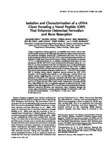
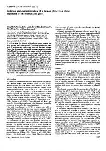
Isolation of GalNAc-transferase from Bovine Colostrum and NH2-terminul Sequencing-The purified GalNAc-transferase preparation contained only one polypeptide, with a molecular mass of-70 kDa, detectable with silver staining (Fig. L4). NH2-terminal sequencing of this material yielded the 34 amino acids shown in Fig. 2A. Aportion of the purified preparation was labeled in vitro with ''I and separated by SDS-PAGE before and afterdigestion with peptideN-glycosidase F. Fig. 1B shows that this treatmentresulted in an -6kDa shift in the apparentmolecular mass of the protein. Isolation and Characterization of cDNAClones Encoding Bovine GalNAc-transferase-Three degenerate oligonucleotides were synthesized based on the partial NHZ-terminal amino acid sequence of the purified bovine colostrum enzyme (Fig. 2 A ) . The cDNA encoding the GalNAc-transferase gene was cloned using the following approach. Oligonucleotides A and C were used as opposing primers in a PCR. A bovine small intestine cDNA library cloned in a X g t l O vector was used as the template for the reaction. On the basis of the amino acid sequence, the predicted size of the amplified PCR product should be 93 bp. The products of the PCR were analyzed by Southern blot analysis using oligonucleotide B as a probe (Fig. 2 A ) . Although the PCR yielded a number of ethidium bromide staining bands, only a single band of -90 A -
"200
B -=" "200
- 116 - 92.5 (I)
-
66.2
-45
- 116 - 92.5 0I
m'
-
66.2
- 6
FIG.1. Separation of bovine colostrum GalNAc-transferase by SDS-PAGE. A , purified bovine colostrum enzyme separated by SDS-PAGE on a 10% polyacrylamide gel and visualized by silver staining; B, in vitro '251-labeled purified bovine colostrum enzyme visualized by autoradiography. Left lane, products fromdigestion with peptide N-glycosidase F; right lane, enzyme incubated as for peptide glycosidase. The migration N-glycosidase F digestion, but without the of molecular weight markers is indicated to the right.
12611
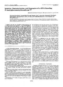
bp hybridized to the probe. This fragment was gel-purified and cloned into the TA cloning vector pCRlOOO to yield plasmid pCR1000-931. Determination of the DNA sequence (Fig. 3) of the pCR1000-931 insert revealed that the deduced amino sequence perfectly matched amino acids 4-34 in the NHz-terminal sequence of the purified transferase (compare PCR-amplify and toclone the with Fig. 2 A ) . In an attempt to GalNAc-transferase gene from the bovine X g t l O library, oligonucleotides D-G (Fig. 2, A and B) were synthesized. Oligonucleotides D and E were derived from the sequence of the pCR1000-931insert, andoligonucleotides F and G areprimers that directly flank either side of the EcoRI cloning site of X g t l O (Fig. 2B). PCRs were run using the bovine cDNA library as template with oligonucleotides D and F or oligonucleotides D and G as primers. The resulting PCR products were analyzed by Southern blot analysis using oligonucleotide E as a probe. No hybridizing bands were seen in PCRs when oligonucleotides D and F were used, but the oligonucleotide D G combination yielded a single hybridizing fragment of -600 bp. This fragment was gel-purified and cloned into the TA cloning vector to yield pCR1000-600,and thesequence of this insert was determined (Fig. 3). The 621-bp insert contained a 207-amino acid open reading frame, with the first 23 amino acids of that open reading frame being a perfect match to amino acids 12-34 of the purified protein (Fig. 2 A ) . Assuming that the621-bp fragment contained aportion of the GalNActransferase gene, this fragment was used as a probe to screen the bovine cDNA library, and seven positive plaques were obtained. The sizes of the X inserts were analyzed on agarose gelsfollowing restriction digestion with EcoRI or by PCR using oligonucleotides F and G as primers. Five of the seven isolates were found to contain inserts of600 bp or smaller, while the two remaining isolates contained inserts of -1600 and 2300 bp. The two larger inserts were PCR-amplified and cloned (using oligonucleotides F and G as primers) into the TA cloning vector to yield pCR1000-52A (1600-bpinsert) and pCR1000-91B (2300-bp insert). Nucleotide Sequence and Predicted Amino Acid SequenceThe complete nucleotide sequences of the inserts in pCR100052A (1582 bp) and pCR1000-91B (2294 bp) were determined using the sequencing strategy outlined in Fig. 2C. As can be seen in Fig. 3, the first ATG codon of the sequence obtained from the 91B clone is present at nucleotide 53. The translated sequence from the ATG codon predicts a polypeptide of 559 amino acids with a predicted M , of 64,173, which is in good agreement with the M , for the purified bovine GalNAc-transferase protein (Fig. LA). The sequence of the 52A clone demonstrated that it is a truncated version of the 91B clone in that thesequence of this clone starts atnucleotide 162 and ends at nucleotide 1744 of the larger 91B clone. The 52A insert covers nearly all of the open reading frame sequences (missing codons for the first37 amino acids) found in the 91B clone. The nucleotide sequence of the 52A clone is identical to that of the 91B clone, with the exception that nucleotide 358 is a G residue in the 52A clone instead of an A residue. This base change is in the wobble position (AGA to AGG) of codon 102, so it does not alter the arginine at that position. The 3"untranslated region of the 91B clone is562bpin length and contains a consensus polyadenylation signal (nucleotides 2176-2182) and a track of 25 A residues at the end of the clone (Fig. 3), indicating that the 91B clone contains all the 3'-terminal sequences of the GalNAc-transferase mRNA. Analysis of Primary Structure of Bovine GalNAc-transferase-Comparisons of the nucleotide and predicted amino acid sequences of the cloned molecule with the sequences in the
+
12612
cDNA Cloning of GalNAc-transferase A.
6.
C.
52A
SStII 01 B
+ + +
-
500
EcoRI
1000
+
___)
G
1500
2000
___)
f-"
___)
.f".
-
___)
w
___)
+ f"
___)
" + +
4 " FIG. 2. NHderminal sequence of bovine colostrum GalNAc-transferase, sequences of oligonucleotide primers, restriction map for cDNA clones (pCR1000-91B and pCR1000-52A) containing GalNAc-transferase, and sequencing strategy. A, NH,terminal amino acid sequence (34amino acids) obtained from purified bovine colostrum GalNAc-transferase. The oligonucleotide (oligonucleotides A-E) sequences of the primers and probes usedin PCRs and Southern blot analysis are shown below the amino acid sequence. The degeneracy of oligonucleotides A-C is 512, 64,and 64,respectively. B, nucleotide sequence of the region surrounding the EcoRI cloning site of the X g t l O vector. Oligonucleotides F and G were synthesized and used in PCRs with the bovine small intestine cDNA library cloned in A g t l O (see text). C, restriction map of cDNA clones pCR1000-91B and pCR1000-52A. The protein coding region of the GalNAc-transferase protein is represented by an open box, the noncoding regions by a straight solid line, and vector sequences by a solid box. The arrows beneath the 91B clone and above the 52A clone indicate the direction and extentof sequencing of the clones.
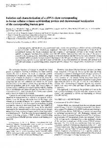
GenBank data base and the SWISSPROT protein sequence data base yielded no significant similarities. Inspection of the predicted amino acid sequence shows that thecloned molecule has the characteristics of a type I1 membrane protein. A KyteDoolittle (1982) hydropathicity analysis of the molecule resulted in apredicted transmembrane domain between residues 9 and 28 (Fig. 4A);secondary structure analysis suggests that this domain of the molecule has an a-helical conformation (Gamier etal., 1978). Furthermore, the predicted amino acid sequence contains three sites for N-linked glycosylation (asparagines 95,141, and 552) as well as four predicted (Elhammer et al., 1993) sites for 0-linked glycosylation (serine 119 and threonines 117, 118, and 288) (Fig. 4B). Northern Blot Analysis-Expression of the GalNAc-transferase mRNA from Madin-Darby bovine kidney cells, bovine mammary tissue, and various human tissues was analyzed by Northern blot analysis using the 600-bp insert of pCR1000600 as a hybridization probe. As shown in Fig. 5 , a t least two different sized GalNAc-transferase mRNAs were detected
from all the samples. The sizes of the bovine messages were -4.1 and 3.2 kb, while all the human tissues expressed messages of 4.8 and 3.9 kb. In addition, a thirdmRNA of -1.5 kb was detected in the skeletal muscle sample. Expression of GalNAc-transferase-The putative GalNActransferase coding region was cloned intoa baculovirus expression vector. Expression of this construct in Sf9 cells resulted in an almost 100-fold increase (compared to cells expressing an unrelatedprotein or to uninfected cells) in intracellular GalNAc-transferase activity (Table I).Increased transferase activity (approximately four times) was also detected in the culture medium from the infected cells. It is likely that this represents the intact or a proteolytic fragment(s) (compare below) of the molecule leaking from dead or dying cells; the totalactivity in the medium was never >4% of the totalactivity in the cells. The baculovirus-expressed protein was examined further by immunoprecipitation and SDS-PAGE analysis. Baculovirus-infected cells were labeled from 24 to 48 h postinfection
cDNA Cloning of GalNAc-transferase
12613 115 21
TTG GGAACIAACCCTGAAGTTAGRT~GGATTACTTTCATTTGACTTAAAGTGCC ATG AGA AAA TIT GCA TAC TGCAAG GTG GTC CTA GCC ACC TCC 7TG ATT TGG GTA CTC GAT net A ~ QLYS P!E la Tyr ~ y LYS s a1 Val Leu A l a Thr ser Leu Ile TIP Val Leu Leu ASP CTA A m TTC CTG CTG CTT TAC TTCAGT GAA TGC AAC AAA TGT GAT GAAM A AAA GAG AGA GGA CTT CCTGCT GGG GAT GTT n e ~phe ~ e uLeu ~ e ~u y rPhelser ~ l Cys u Asn Lys Cys Asp Glu Lys LysG l u Arg Gly Leu Pro Ala Gly Asp Val Leu +""""""""" GM GGT CCT GGA GAA ATG CGG AAA CCA GTC GTC ATT CCT AAA GAG GAT CAA CAR AAG ATG AAA GAG ATG TTT AAA ATC RAT Glu Gly Pro Gly Glu Met Gly Lys Pro Val Val Ile Pro Lys G l u Asp Gln Glu Lys Her Lys Giu Mer Phe Lys Ile Asn """"""""""""-""I GAG ATC AT? GCA CTC AAC AGA TCT CTA CCA GAT GTTAGA TTA GAR GGG TGT ARR ACA AAG GTG TAT CCA GAT AAC CTT Glu Mer Ile Ala Leukyn-ArT cr1;eu PKO Asp Val Arg Leu Glu Gly C y s Lys Th: i y s Val Tyr Pro Asp Asn Leu Prc
4
217 55
GAG CCA GTACAA AAG CCT CAT Glu Pro Val Gln Lys Pro H i s
319 89
CAG TTC AAT TTA ATG GCA AGT Gln ?he Asn Leu Mer Ala Ser ACA CCT ACC AGT G E GTG ATT GTT Thr Thr Ser Val Vai Ile Val
421 123
GAAATT GTT CTA GTA GAT GAT Ile Val Leu Val Asp Asp Ala
GCC 5 2 1 157
GTG AAA M A TTA AAA GTA CCC GTT CAC GTC ATT CGA ATG GAG CAG CGT TCT GCATTG ATC AGA Lys Lys Leu Lys Val p r o Val His Vai Iie Arg Mef. G l u G i n ArQ S e r Gly Leu :le Arg
€25
GCG CAC TGT GAG TGC ACA GTG GGG TGG CTG GAG CCT CTC TTA GCC AGG GCT AGG TTA M A GGT GCT GCTGPG TCT AAA GGC CAA GTG ATC ACC TTT TTA GAC Ala Arg Leu Lys Gly Ala Ala Val Ser Lys Gly Glc Val Ile Thr Fhc Leu ASP Ala His Cys Glu CYs Thr Val Gly Trp Leu Glu Pro Leu Leu Ala Afg
727 225
TAT GGC GGG TTC G1y Gly ?he I
829 259
" "
TTC CAC AAT GAG GCT AGC TCGACA CTT CTG CGA ACT GTC Phe His Asn Glu Ala Trp Ser Thr Leu Leu Arg Thr Val ACT
GAA AGA GAC TTT TTA AAA AGA CCT CTA GAG
ACT
CATAGC GTC ATTAAT CGC TCA CCA AGG CAC ATG CTA GAA Ser Val I l e l ~ n A 311 r ~ Pro A r g His Met Leu Glu
E15
TAC
Ser Glu Arg Asp Phe Leu Lys Arg F r o Leu Glu Ser Tyr \'a:
e..
Glu
ATC ATA G?.T GTG A T AGT GAT GAC ACT TTC GAG TAC ATG GCA GGT TCT GAC ATC ACC ATC AAA CAT GAC A X AAG ACA GPG GTC TGT CCC :le Lys H l s Asp Arg Lys Tt: Val Val Cys F r o lie I l e Asp Val I l e Ser Asp Asp Thr ?he Glu Tyr M e L Ala Gly Se: ASP Her Thr
TYK
CAR AGA GAA ATG GAC AGA AGG AAA GGT GAT CGC ACT CTT CCT GTG AGA ACA CCT ACA ATG GCA GGA AAC ?GG AAG CTC AAC TTT CG'Z TGG TAT CCT GTT CCC As" T r p Lys Leu Asr. ?he AIS T r p Tyr Pro Val Pro G l n Arg Gll; Met Asp Arg A r g Lys Gly Asp Arg Thr Leu Pro Val A r g Thr P:oTh: Mec Ala Gly
19!
931 293
0
GCC CTT TTT TCA ATA GAC AGA GAT TAC TTT CAG G A A Gly Leu Phe Ser Ile A ~ CA r g ASP Tyr Phe ~ l GIG n
A X GGA
ACA TAT GAT GCT GGX ATG GAT ATTTGG GGA GGA GAA AAC CTA GAA ATT TCC TTT AGG ATT 'Wr Asn Ala ~ ! ynet A S P Ile Trp Gly Gly GluA m Leu Glu lie ser Phe Arq Ilr
le ~ l Thr y
TGG CAG TGT GGA GGA ACT TTG GAG ATT GTT ACT TGCTCA CAT GTT GGA CAT GTG TTT CGG AAA GCT ACA CCC TACACG TTT CCA GGA GGC ACG GGG CAG ATTrp Gln C y s G:y C:y ?: Leu Glu Ile Val Thr Cys Se: P i s Val Gly H i s Val Fhe Arg Lys Ala Thr Pro TYr Thr Phe Pro Gly Giy Thr Gly G l n Ile
1033 327 1135
36:
ATA ATT TCT CCA CGT ACA GTT ?.AC GTA GAT TATGGA GAT ATC ART AAA RAT AAC ACA CGA CTT GCAGAR GTA TGG ATG GAT GAA TTC AAG AAT TTC TTC TAT :le Asn Lys A 6 3 AS> Arg Ars leu Ala G l u Val . ; : T Mer h . 6 ~G l u P h e Lys Asn Phe Fhe > z Ile Ile Ser ?ro G I). Vai Thr Lys \'a1 Asp ?yr Cly As;
1237
ATA TCA TCA ASA K T GGT CTA AGG CAC AAA CTC CAA TGC AGA CCA TTC TCT TGG TAC CTA GAG AAT ATT TAT CCTGAT TC'I CAC AT: CCT CGT CAC TAT TTC I;e Ser Ser R r g Leu Gly Leu Arg His Lys Leu Gln Cys A : G Pro ?ne S e r T:€ T y r Leli C I u Asn ile Tyr F r o A s p SQr Gin I l t P r o A r g H I S Tyr >ne
1739
TC? TTG GGA GAS AYA CGA XAT GTC GAA ACA AAT CAS TST CTA GAT M C AT:; GCT AGA XAA GAG A A T GAR AAA CTT GGA ATT TTT AAC TGT C?.; GGT ATG GGA Ser Leu Gly Glu ile Arg Asn Va: G l u Thr Asn Glr. s k c Asp Asn Met Ala P.:g Lys Glu Asc Glu iys Val Gly Ile Phe ASC Cys His Gly Mer Gly
1421 4ii
GGT AAT CAG GTT TTC "CT TAC ACT GCC AAC AAA GAA ATT AGA A C A GAT GAC CTT TGC TTG GAT GTC TCCAAA CTT RATG,GC CCA G I C ACA A?G CTC AAA TCC Gly Asn G l n V1; ?he Ser T y : T h r Ala Asn Lys Giu Ile Arg: T h r Asp Asp L?u Cys Leu Asp Val Ser Lys Leu As2 Gly P r o \'dl Tk,: M ~ Leu L Lys Q s
1543 497
CAC CAC CIA AAA GGC AAC CAG CTT TGG GAG TAT GAC CCG STG AAG TTG ACC CTC CAG CAT GTG ARC ACT AAC CAG TGC CTG GAC A?.& GCC ACA GAC GAG GAC H1S His Leu Lys Gly Asn Gln Le'd Trp G l u ?\ir Asp Pro Va! Lys Leu Tnr Lec Sln H I S Vai Afin Ser >.sn Gln Cys Leu Asp LyP Ala Th: Asp Glu As3
1645 531
AGC CGA TCC CAG CAG TGG CTT CTT CGG LAC-GE L C S C T T CCA GA> ATA TTC TGA GACCA?ATTTACAAAAAAA AGC CAG GTG CCC P.GC XTC AGA GAC TGC AGC GGA Se: Gln Val P r o Ser lie A r g As3 i y s Ser Gly Ser Arg Ser Gln Gin T r p Leu Leu ArglAsn -",J vai Thr LOL: P:o GIL lie Phe End
1751 559
GGAAARCGTAAGGhCTGACTCGGGCTACCTCAGCATACATTTCTGCCA~hTTCTTAAGTAGCAAAMAACGAAAAGTGCTTTCCTTCTGiAGGATGTAAGG'iTTATCAGCCATTAXAACTTTATAGACTGCCCTCG
1886
395
420
i Ao 2~ C 1 CTTCCACTAGCTGTGAACCA~CC7GTCCTG~CCCAGGACGTGC~CTA~~TAGThGCGAGA~G~GCACACTGX~GTTTACMGATTGAAAGP.GTCCGTCATCAAGAATCCTCGTAAAGAATACTCAGACTG AGT~GCGA.4CT~TGCT7TCCAGAGAGCTGCGCCTTTTATGGT~TGC~TGCACAGCAGTGAGTTCCGCTGACTGTGCTGTCATAATGAAGAGACGTCTAAGATTTTTTTTCTGATTAGAAC~GTAGCCAGTA~A 2l5€ T ~ A R R T A C T G A T A T A A ~ C A A C T C r , R 4 C C A G A T T C A G A A T C ~ T G A A h A C A T T T T T A C A A T T T A R R A A A h A ~ A A A A ~ T A T A T ~ A A A C A C G G T T T A A A G G A A T T ~ A A ~ A A A A A , ~ A A A A A A A A A ~ A A X A2294 AAAAA
FIG.3. Amino acid sequence of cloned GalNAc-transferaseinferred from nucleotide sequence of cDNA clones 9 l B and 52A. The proposed transmembrane sequence is boxed. Potential sites for N-linked glycosylation are indicated by the dashed boxes, and predicted sites for 0-linked glycosylation are marked with dots under the appropriate amino acids. The NH2terminus of the soluble bovine GalNActransferase (determined by NHp-teminal sequencing) is indicated by the arrow. The consensus poly(A+) sequence (AATAAA) is boxed, and the sequences of the 93-bp insert of pCR1000-931 and the 621-bp insert of pCR1000-600 are indicated by the dashed and solid underlines, respectively. The numbering of the nucleotide (upper)or amino acid sequence (lower) is indicated to the right of the sequence. with [36S]methionine,and GalNAc-transferase was immunoprecipitated from lysates andculturemediaof the labeled cells using a chicken polyclonal antibody raised against the purified bovine colostrum enzyme. The results from separation of the precipitated materialon SDS-PAGE are shown in Fig. 6. Two closely spaced proteins with molecular masses of -67 and 63.5 kDa were detected in both lysates and media from cells infected with viruses containing the GalNAc-transferasegene (Fig. 6). This is close to the molecular mass predicted for the cloned protein andis also comparable to the molecular mass of the purified bovine colostrum enzyme (Fig. 1). The endogenously expressed enzymeis only barely detectable in this experiment, but appearsto have a similar molecular mass (Fig. 6, lanes 5 and 6); phosphor screen autoradiography of the gel showed that -75 times more radioactivity wasincorporatedin the clonedenzymecompared to the endogenous one (data not shown). The additional lower molecular weight bands seen on the gel probably represent non-
specificallyprecipitatedmaterial since the samebandsare found in the control samples (lanes 5 and 6). DISCUSSION
The cloning of bovine GalNAc-transferase usingthe strategy described above yielded two cDNA clones. The sequences of the overlapping region of these two clones were identical, with the exception of a single base difference (which does not change the amino acid) in the codon for amino acid 102 of the GalNAc-transferase coding region.Possible explanations for this basedifferenceare as follows. 1) Since the phage inserts were subcloned into plasmid vectors following PCR, the base difference may be a PCR artifact; and 2) the two clones isolated may represent two different genes. The second possibility is supported bythe detection of two different sized GalNAc-transferase messages on Northern blots of mRNA isolated from bovine mammary tissue (see below). In either case, the fact that the cloned moleculeexhibits full enzymatic
cDNA Cloning of GalNAc-transferase
12614
TABLE I
jr?A
,
Expression of GalNAc-transferase i n Sf9 cells The cloned GalNAc-transferase DNA was expressed in Sf9 cells from a recombinant baculovirus. Cells were harvested 48 h postinfection and lysed in a detergent-containing buffer. Following sedimentation of undissolved material, the cleared lysates were assayed for GalNAc-transferase activity as outlined under "Experimental Procedures." Lysates from uninfected cells or from cells infected with either a baculovirus containing an unrelated gene (human cytomegalovirus DNA polymerase (CMW Pol-1) or two separatebaculovirus isolates of theGalNAc-transferasegene(GalNAcT 2-1A and GalNAcT2-1B) were assayed. Relative Construct Specific activity rate
I
units/mg protein
r>.,I
..,.
: " - L J 4 b ? 4 . r - L -,".-
7 ,
-,
J4.,":I
,.++,cy0
-
"a
Residue number
FIG. 4. Predicted transmembranedomain and 0-linked glycosylation sites for cloned GalNAc-transferase. The aminoacid sequence of the cloned molecule was analyzed for putative transmembrane segment(s) as described by Kyte and Doolittle (1982) ( A ) and for 0-linkedglycosylation sites as outlined by Elhammer e t al. (1993) (B). 3
4
5
6
7
8
1
A
B 93 75
1
2
3
'
1 0.8 87 74
-=5
4
u -
6
-~
-200
" c
.
2
0.16 0.13 13.9 11.9
Uninfected cells CMWPol-1 GalNAcT 2-1A GalNAcT 2-1B
- 116
-
92.5
-
66.2 c?
-
-
7.5 4.4
2.4
-
1.4
-
-45
B X
93
-
2.41.4
-
FIG. 6. Immunoprecipitationof in uiuo [S6S]methionine-labeled GalNAc-transferase expressed in baculovirus-infected Sf9 cells. The cloned GalNAc-transferase DNA was expressed in Sf9 cells using a baculovirus vector. The infected cells were switched to culture medium containing [?3]methionine 24 hpost-infection and harvested after another 24 h. The cells were lysed in a detergentcontaining buffer, and the labeled transferase was immunoprecipitated from thecell lysates and thecorresponding culture media. The washed immunoprecipitates were separated by SDS-PAGE on a 10% polyacrylamide gel. Lanes I, 3, and 5 contain radioactivity precipiactivity when expressed from a baculovirus vector indicates tated from cell lysates of cellsinfectedwith virus containing the GalNAcT 2-1A, GalNAcT 2-1B, and cytomegalovirus that the isolated sequence represents an authentic GalNAc- constructs DNA polymerase, respectively.Lanes 2,4, and 6 contain radioactivity transferase. immunoprecipitated from the corresponding culture media. The two The cloned GalNAc-transferase is atype I1 membrane molecular mass forms of the immunoprecipitated protein are indiprotein with the same general domain structure as that of cated by the arrowheads. The migration of molecular weight markers other cloned glycosyltransferases (Paulson and Colley, 1989). is indicated to theright.
FIG.5. Northern blot analysis. T w o pg of poly(A+) mRNA isolated from bovine mammary tissue, Madin-Darby bovine kidney cells, and eight different human tissues were probed with the "Plabeled 600-bp insert isolated frompCR1000-600 (see Fig. 3). A , lanes I and 2 contain mRNA from Madin-Darby bovine kidney cells and bovine mammary tissue, respectively. B, lanes 1-8 contain mRNA isolated from human heart, brain, placenta,lung, liver, skeletal muscle, kidney, and pancreas, respectively.
Also similar is the insignificant sequence homology to other glycosyltransferases as well as to other previously reported sequences. The amino acid sequence contains threeconsensus sequences for N-linked glycosylation. The results from peptide N-glycosidase digestion of the purified in vitro lz5I-1abeled protein suggest that at least two of these are utilized on the bovine enzyme; an -6-kDa shift in molecular mass was observed by SDS-PAGE following this treatment (Fig. 1B). Furthermore, in uiuo labeling experiments using mouse and rat cells have shown that GalNAc-transferase is capable of incorporating both radioactive N-acetylglucosamine and N-
acetylgalactosamine into protein-linked oligosaccharide structures (data notshown). The latter observation suggests that atleast one of the predicted sites for 0-glycosylation also is utilized. The amino acid sequence predicted for the larger clone isolated in this work provides a plausible explanation for the water solubility of the bovine colostrum GalNAc-transferase. This enzyme apparently lacks the NH2-terminal 40 amino acids of the membrane-bound molecule, a segment that includes both the cytoplasmic and membrane-spanning do-
12615
cDNA Cloning of GalNAc-transferase mains. The Kyte-Doolittle (1982) hydropathicity plot from the cloned enzyme (Fig. 4A)shows only one sequence segment (residues 9-28) with a high membrane-spanning probability. It is not clear, at present, if the soluble bovine colostrum enzyme is the result of proteolytic cleavage of a membranebound molecule or if it represents aborn fide secretory protein. Soluble, enzymatically active forms of a 61-4-galactosyltransferase and an a2-6-sialyltransferase have been reported, both of which appear to be the result of proteolytic cleavage of membrane-bound proteins(Paulson and Colley, 1989). In addition, the translation products from the different mRNA species related to both these molecules appear in most tissues to be membrane-bound molecules (Joziasse, 1992). By analogy, it appears likely that the two mRNAs observed in our Northern blotting experiments (Fig. 5 ) both code for membrane-bound enzymes and thus that the soluble bovine colostrum enzyme (again in analogy with Dl-4-galactosyltransferase and a2-6-sialyltransferase) must represent a proteolytic fragment of a membrane-bound enzyme. On the other hand, work reported on the different transcriptssynthesized for rat kidney a2-6-sialyltransferase suggests that some of these molecules contain start codons that, when translated, would yield proteins without cytoplasmic and membrane-anchoring domains (Svensson et al., 1990; Wang et al., 1990; Wen et al., 1992). Clearly, further experiments are needed to positively answer this question. The larger sizes of the two GalNAc-transferase messages (compared to the cloned DNA) are presumably related to untranslated sequences larger than those recovered in the isolated clones in the 5'- and/or 3'-end of the native molecules. Messenger RNA molecules from previously characterized cloned glycosyltransferases frequently contain extensive 5'- and 3"untranslated sequences (e.g. Weinstein et al. (1987), Larsen et al. (1989), Russo et al. (1990), Scocca and Krag (1990), Sarkar et al. (1991), and Nagata et al. (1992)). There is also a precedence for more than one mRNA species for at least four glycosyltransferases; the genes coding for these enzymes appear to be under control of more than one promoter (reviewed by Joziasse (1992)). In addition, a GalNActransferase has been described that catalyzes the synthesis of a specific oncofetal epitope on fibronectin (Matsuura et al., 1988, 1989). This observation is consistent with the existence of more than one form of the enzyme. Expression of the cloned sequence in Sf9 cells resulted in a large increase in intracellular transferaseactivity, thus establishing the identity of the cloned sequence with a GalNAc-toSer/Thr transferase. The marked increase in a protein(s)with a molecular mass similar to that predicted from the amino acid sequence furthersupports this conclusion (Fig. 6). A closer examination of the protein(s) immunoprecipitated in this experiment reveals two closely spaced polypeptide bands with an -3.5-kDa difference in molecular mass. The exact identity of these two proteins is not known at present; they may represent differentglycoforms of the enzyme, or, perhaps more likely, the lower molecular mass form may be a proteolytic fragment, similar to the enzyme purified from bovine colostrum. The latter possibility is supported by two observations. 1)The mass difference between the two molecules is roughly equal to thatof the sequence (40 amino acids) missing in bovine colostrum enzyme; and 2) while the immunoprecipitates from cell lysates containpredominantly the higher molecular mass form of the enzyme, the culture medium appears to be enriched in the lower mass form. High-speed centrifugation of the culture medium failed to sediment more than -30% of the enzymatic activity (data not shown). The smaller molecular mass of the insect cell-produced molecule
compared to thepredicted mass of a membrane-bound bovine enzyme (the molecular mass of the soluble colostrum enzyme plus -4 kDa for the transmembrane and cytoplasmic domains) may bethe result of differences in glycosylation of the two molecules. Insect cells typically synthesize truncated, non-sialylated N - and 0-linked oligosaccharides (e.g. Hsieh and Robbins (1984), Doming0 and Throwbridge (1988), Kuroda et al. (1990), Thomsen et al. (1990), Wathen et al. (19911, and Chen and Bahl(l991)); this results in a reduced molecular mass of insect cell-produced glycoproteins on SDS-PAGE. The identity of higher molecular mass bands (-120-180 kDa) on the gel is not clear. We have observed these bands previously in immunoprecipitates (by our anti-GalNAc-transferase antibody) from in vivo labeled mammalian and insect cells? They may represent nonspecifically precipitated material, another protein(s) containing an epitope(s) recognized by our polyclonal antibody, or aggregated GalNAc-transferase. The latter possibility appears less likely, however, since immunoprecipitates of purified GalNAc-transferase contain only one protein band. The fact that theintensity and molecular mass of these bands appear to vary (while the GalNAc-transferase band remains constant) between different experiments and cell types suggests that theyrepresent nonspecifically adsorbed contaminants (Fig. 6, compare lanes 1-4 and lanes 5 and 6). The efficient production of the cloned molecule using the baculovirus expression system should facilitate preparation of GalNAc-transferase in amountssufficient for more detailed biochemical and enzymatic studies as well as for preparation of immunological reagents for localization studies. Acknowledgments-We thank Heidi Zurcher-Neely and Robert Heinrikson (The Upjohn Co.) for assistance with NH2-terminal sequencing. REFERENCES Briand, J. P., Andrews, S. P., Jr., Cahill, E., Conway, N. A,, and Young, J. D. (1981) J. Biol. Chem. 2 5 6 , 12205-12207 Bushway, A. A., and Keenan, T. W. (1979) Biochim. Biophys. Acta 5 7 2 , 146rn
.
LOL
Chen, W., and Bahl, 0. P. (1991) J. Biol. Chem. 2 6 6 , 6246-6251 Davis, c. G., Elhammer, A. P., Russel, D. W., Schneider, W. J., Kornfeld, S., Brown, M. S., and Goldstein J. L. (1986) J. BioL C h e m 2 6 1 , 2828-2838 f Res. 12,387Devereux, J., Haeherh, P., and Srnlthies, 0. (1984) N ~ l e i Acids 395 Domingo, D. L., and Throwbridge, I. S. (1988) J. Bioi. C k m . 2 6 3 , 1338613392 Dunphy, w. G., Brands, R., and Rothman, J. E. (1985) Cell 4 0 , 463-472 Elhammer, and Kornfeld, S. (1986) J. Biol. Chem. 261,5249-5255 Elhammer,k P., Poorman, R. A., Brown, E., Maggiora, L. L., Campbell, C. M., Hoogerheide, J. G., and Kezdy, F. J. (1993) J. B i d . Chem. 2 6 8 , 1002910038 Gamier, J., Osguthorpe, D. J., and Robson, B. (1978) J . Mol. Biol. 1 2 0 97-120 Goldin, A. L., Sandri-Goldin, R. M., Levine, M., and Glorioso, J. C. ( h 1 ) J . Virol. 38, 50-58 Gooley, A. A., Classon, B. J., Marschalek, R., and Williams, K. L. (1991) Biochern. Biophys. Res. Cornmun. 1 7 8 , 1194-1201 Ha OPian, A., Westall, F.c.,Whitehead, J. S., and Eylar, E. H. (1971) J . Biol. &ern. 246.2519-2523 Hill, H. D., Jr., Schwyzer, M., Steinman, H. M., and Hill, R. L. (1977) J . Bid. Chem. 262,3799-3804 Homa, F. L., Otal, T. M., Glorioso, J. C., and Levine, M. (1986) Mol. Cell. Biol. R. 36.52-Rfififi -__Hsieh, P., and Robhins, P. W. (1984) J. Biol. Chem. 259, 2375-2382 Hughes, R. C., Bradbury, A. F., and Smyth, D. G. (1988)Carbohydr. Res. 1 7 8 , 259-269 Jensenius, J.-C., Andersen, I., Hau, J., Crone, M.,and Koch, C. (1981) J. Imrnunol. Methods 46,63;68 Jentoft, N. (1990) Trends Blochem. Sei. 1 5 , 291-294 Joziasse, D. H. (1992) Glycobiology 2,271-277 Kuroda, K., Geyer, H., Geyer, R., Doerfler, W., and Klenk, H.-D. (1990) Virology 174,418-429 Kyte, J., and Doolittle, R. F. (1982) J. Mol. Biol. 1 5 7 , 105-322 Laemmli. U. K. (1970) Nature 227.6XO-fi% , ". Larsen, R . D., Rajan, P., Ruff, M. M., Kukowska-Latallo J. Curnmings, R. D., and Lowe, J. B. (1989) Proc. Natl. Acad. Sci. U. S. A. 86,'8227-8231 Matsuura, H., Takio, K, Titani, K., Greene, T., Levery, S. B., Salyan, M. E. K., and Hakamon, S.-I.(1988) J. Btol. Chem. 263,3314-3322 Matsuura, H., Greene, T., and Hakamori, S.-i. (1989) J. Biol. Chem. 264, 10472-11147fi Nagata, Y., Yarnashiro, S., Yodoi, J., Lloyd, K. O., Shiku, H., and Furukawa, ~~
-.
111-
~~~
~~
* A. P. Elhammer, unpublished observations.
12616
cDNA Cloning of GalNAc-transferase
K. (1992)J. BioL Chem 267,12082-12089 Parqdi, A. J., Blank, E. W., Peterson, J., and Ceriani, R. (1984)MOLCeU.
Blochem. 68.157-163 Paulson, J. C. (1989)Trends Biaehem. Sei. 14, 272-275 Paulson, J. C., and Colley, K. J. (1989)J. BioL Chem 264, 17615-17618,and references therein Paulson, J. C., Beranek, W.E., and Hill, R. L. (1977)J. Biol Chem. 262, 2356-2362 Prieels, J.-P., Maes, E., Dolmans, M.,andLeonis, J. (1975)J.Biochern. (Tokyo) 60,525-531 Roseman, S.(1970)Chem. Phys. Lipids 6.270-280 Ruaso, R. N., Shaper, N. L., and Shaper,J. H.(1990)J.Biol. Chern. 266,33243331
Sadler, J. E. (1984)in Biology of Carbohydrates (Ginsburg, V., and Robbins, P. W., eds) Vol. 2,pp. 199-213,John Wiley & Sons, New York Sanger, F.,Nicklen, S., and Coulaon, A. R. (1977)Proc. Natl. A c d . Sci. U. S. A. 74.54.63-6467 . -,- -. - - .- .
Sarkar, M., Hull, E., Nishikawa, Y., Simpson, R. J., Moritz, R. L., Dunn, R., and Schachter, H. (1991)Proc. A d . Natl. Sci U. S. A. 88.234-238 Schachter, H., and Brockhausen, I. (1992)in Glycoconjugates (Allen, H. J., and
Kisailus, E. C., e&) pp. 263-332,Marcel Dekker, Inc., New York Scocca J. R. and Kr S. S. (1990)J. BloL Chem. 266,20621-20625 Summ&, M:D., and%nith, G. E. (1986)A Manunl of Methods for Baculouirus Vectors and Insect Cell Culture Procedures, Texas Agricultural Experiment Station, College Station T X Svensson, E. C., Soreghd, B., and Paulson, J. C. (1990)J. BioL C h m 266, 20863-20868 Thomsen, D. R., Post, L. E., and Elhammer, A. P. (1990)J. CeU. Biochern. 43, 67-19 Wang, X., O’Hanlon, T. P., Young, R. F., and Lau, J. T. Y. (1990)Glycobiobgy 1,25-31 Wan Y., Abernethy, J. L., Eekbart, A. E., and Hill, R. L. (1992)J. Biol. Chem. 26~,12706-12716 Wathen, M., Aeed, P. A., and Ethammer, A. P. (1991)Biochemistry 30,28632868 Weinstein, J., Lee, E. U., McEntee, K., Lai, P.-H., and Paulson, J. C. (1987)J. Bid. Chem. 262,17735-17743 Wen, D. X., Svensson, E. C., and Paulson, J. C. (1992)J. BioL Chem 267, 2512-2518 Yo,ung, J. D., Tmchiya, D., Sandlii, D. E., and Holroyde, M.J. (1979)Biochernmtry 18,4444-4448