Rufino et al., [12] reported that the biosurfactant produced by Candida lipolytica ..... [16] DK Santos; RD Rufino; JM Luna; VA Santos; AA Salgueiro; LA Sarubbo.
Available online www.jocpr.com
Journal of Chemical and Pharmaceutical Research, 2016, 8(7):357-367
Research Article
ISSN : 0975-7384 CODEN(USA) : JCPRC5
Isolation and partial Structural & Functional Characterization of Glycolipid Biosurfactant producing Pichia sorbitophila WG1 from Rotten Grapes Vidur Bhatia and *Baljeet Singh Saharan Department of Microbiology, Kurukshetra University, Kurukshetra, India _____________________________________________________________________________________________ ABSTRACT Surfactants are amphiphilic compounds containing the hydrophobic and hydrophilic moieties which reduce surface tension between water and hydrocarbons. In the present investigation, a Glycolipid Biosurfactant producing yeast was isolated from rotten grapes. The isolate was selected on the basis of reduction in surface tension, oil displacement measurements and emulsification index. On the basis of 18S rRNA gene amplification and sequencing chosen isolate was identified as Pichia sorbitophila WG1. The biosurfactant produced was structurally characterized by FTIR, NMR and GC-MS. It was found to be a glycolipid stable over a vast range of pH and temperature. Further, the biosurfactant showed significant broad spectrum antimicrobial activity against various Gram positive and Gram negative pathogens. The cytotoxicity test confirmed its non toxicity, when tested against Triticum aestivum and Brassica nigra. Keywords: Biosurfactant, Glycolipid, Pichia sorbitophila, Characterization, Antimicrobial & Phytotoxicity assessment. _____________________________________________________________________________________________ INTRODUCTION Microbial surfactants are amphipathic, surface-active agents that accumulates at interfaces reducing interfacial tension, surface tension and prominent to the establishment of accumulated micellular structures in aqueous phase [1]. Microbial surfactants are a group of microorganism’s derived products for environmental, biotechnological and commercial applications [2,3,1]. Biosurfactants (BS) are commonly used in industrial formulations, agricultural products, feed and food, cosmetic formulations and pharmaceutical preparations [3]. The unique profits of biosurfactant permit their consumption in a limitless number of industrial preparations. There is rising consumer compression for industrials formulations that are further natural, ecological, and environmentally viable, comprising foods and health care products. Industries have reacted by trying to recognize sustainable replacements to synthetic or chemical ingredients within the industrial formulations. Commonly bacteria are capable to produce biosurfactants, but in reasonably less quantity [4,1]. On the other hand, Yeasts usually produce biosurfactants in large amount as compared to the bacteria [5] because of its rigid cell walls. To make biosurfactants economically viable, it is vital to select microorganisms capable of producing high amount of biosurfactant [6].Yeasts have appeared as a note worthy group with considerable biological significance and suitable applications. The major benefits of using yeasts for industrial viability is their generally regarded as safe (GRAS) status [7]. In the present study, we had considered potential of various yeast isolates to produce biosurfactants and studied its structural and functional properties.
357
Vidur Bhatia and Baljeet Singh Saharan J. Chem. Pharm. Res., 2016, 8(7):357-367 ______________________________________________________________________________ EXPERIMENTAL SECTION 2.1 Screening for Biosurfactant production Pure culture of the yeast strains were used to screen for the BS production by various methods i.e. hemolytic activity, oil displacement test, drop collapsing test and emulsification assay. All the experiments were performed in triplicates. 2.2 Taxonomic identification 2.2.1 Colony PCR of 18S rRNA gene Genomic DNA of the isolate was extracted and amplified for 18S rRNA gene with colony PCR for taxonomic identification. Concisely, master mix with primer ITS2: GGTCCGTGTTTCAAGACGG, 1.2 UDNA polymerase in 50 µl PCR reaction mixtures. About1 small colony was picked up with sterilized loop and transferred to the PCR tube as DNA templates. The thermocycler PCR consisted of one cycle of 95 °C for 10 min, 53 °C for 2 min, 72 °C for 2 min, and 35 cycles of 94 °C for 20 s, 57 °C for 45 s, 72 °C for 1 min, then incubation at 72 °C for 5 min, and a final incubation at 4°C. Briefly, 10 µl of each amplified mixture and the molecular marker were separated on agarose gel electrophoresis to confirm the size of the amplified gene. The 18S rRNA gene sequence of the isolate was matched with other yeast sequences by using NCBI mega BLAST for their pair wise individualities. 2.3 Measurement of Surface tension The surface tension of the culture supernatant (obtained by centrifuging the cultures at 5000g for 20 min.) was measured using a Sigma 700 digital surface tensiometer (KSV Instruments Ltd.-Finland) working on the principle of the Du Nuoy ring method. 2.4 Production and purification of biosurfactant The biosurfactant production was carried out in Erlenmeyer flasks containing 150 ml of Minimal media with 20 g/L of soybean oil, incubation at 28 °C in an orbital shaker operated at 200 rpm for 144 h. After incubation, the yeast cells were separated from the growth medium by centrifugation at 5000 rpm for 20 min at 4 °C. The cell free suspension (CFS) was used to isolate the biosurfactant by ethyl acetate solvent extraction method. The extracted biosurfactant was dialyzed beside demineralized water at 4°C in dialysis membrane (molecular weight cutoff 8,00010,000Dalton) and freeze dried for further application. 2.5 Structural characterization of biosurfactant 2.5.1 Thin layer chromatography (TLC) The composition of the biosurfactant produced was resolute by TLC followed by post chromatographic detection [8]. The polysaccharides moieties were stained with Syldatk reagents (anisaldehyde: sulphuric acid: glacial acetic acid 0.5:1:50) and separated against the chloroform and methanol (60:30). Plates were heated at 90 °C for 5 min and observed for the brown spot corresponding to the sugar present in biosurfactant. 2.5.2 Fourier transform infrared spectroscopy (FTIR) Composition of biosurfactant produced was revealed using FTIR spectroscopy by scanning it in the range of 4000400 cm-1 at a resolution of 4 cm−1 (Model-ABB). 2.5.3 Nuclear magnetic resonance (NMR) spectroscopy The 1-5 mg biosurfactant sample was dissolved in 100% CDCl3 and 1HNMR analysis was carried out using a Bruker Av II-400 spectrometer. Proton NMR chemical shifts was stated in ppm relative to the solvent shift as chemical standard. 2.5.4 Gas chromatography and mass spectroscopy (GC-MS) The biosurfactant sample was analyzed for fatty acid composition on gas chromatograph-mass spectroscopy fitted with VF-5MS column. Briefly, the preliminary column temperature was set at 100 °C for 1 min, and then ramped at the rate 30 °C min-1 to 270 °C, and finally held at 270 °C for 10 min. The temperature ranges of the transfer line, ion trap and quadruple were 280, 230, and 160 °C, respectively. The inlet temperature was 270 °C, and a 50 µl sample of biosurfactant was injected. The flow rate of the carrier gas (helium) was 1.2 ml min−1.
358
Vidur Bhatia and Baljeet Singh Saharan J. Chem. Pharm. Res., 2016, 8(7):357-367 ______________________________________________________________________________ 2.6 Ionic property of biosurfactant The ionic property partially purified biosurfactant was determined by using agar well diffusion method [9]. Briefly, 3 uniformly spaced wells were made on a soft agar (1 %) plate; central well was filled with 10 µl of BS. Either side of wells was filled with anionic compound (20 mmol-1 SDS (Sodium dodecyl sulfate) and cationic compound (20 mmol-1 Cetyl Trimethyl Ammonium Bromide (CTAB). Plates were incubated at 25 °C for 24 h and observed for the precipitation lines. 2.7 CMC determination The critical micelle concentration (CMC) of isolated biosurfactant was determined by measuring the surface tension of its different dilutions in distilled water up to a constant value. All the measurements were recorded in triplicates. The CMC was obtained by plotting surface tension against surfactant concentration and expressed as mg/ml. 2.8 Stability study of biosurfactant Partially purified biosurfactant (at a conc. of its CMC) was used to elucidate its stability at different temperature and pH range. 2.8.1 Effect of pH To elucidate the pH stability of biosurfactant, the sample was adjusted to different pH values (4-10) with 1M NaOH and 1M HCl and surface tension was measured. 2.8.2 Effect of temperature To check the stability of biosurfactant at different temperature 10 ml of biosurfactant suspension was exposed to a constant temperature of 10, 20, 30, 40, 50, 60, 70, 80, 90, 100 and 110 °C for 30 min, cooled to room temperature and then the surface tension was measured. 2.9 Antimicrobial assay The antimicrobial activity of the biosurfactant was tested against various pathogenic and non-pathogenic strains by serial dilution technique in 96 well flat bottom plastic tissue culture plates. Briefly, 125 µl of sterile, 2X culture medium was placed into the first column of the 96 wells micro-plate and 125 µl of sterile 1X culture broth in the remaining wells. Further, 125 µl of biosurfactant solution in PBS (25 mg ml-1) were added to the first column of the microplate; this results in a BS concentration of 12.5 mg ml-1; in sequence, 125 µl were transferred to the successive wells. All the wells (except negative control) were inoculated with 2.5 µl of an overnight pathogenic strain. Plates were incubated for 48 h at 37 ºC. After incubation, the absorbance at 600 nm was recorded for each well. The growth inhibition percentages at different BS concentrations for each pathogenic strain were calculated as: % Growth Inhibition = [1-(AC/AO)] X 100 Where Ac represents the absorbance of the well with BS concentration c and Ao represents the absorbance of the control well (without BS). 2.10 Phytotoxicity assay The Phytotoxicity assay of isolated biosurfactant was determined in a static seed germination and root elongation of the Brassica Nigra and Triticum aestivum slightly modified from Tiquia et al., 2008.Solutions of BS were prepared with distilled water at concentration of 1 mg/ml. 25 pre sterilized seeds were inoculated in each petri plate with 10 ml of test solution at 27 °C. After five days of incubation in the dark, seed germination, root elongation (> 5mm) and the germination index were recorded as follows: Relative seed germination (%) = (number of seeds germinated in the extract/number of seeds germinated in the control) ×100. Relative root length (%) = (mean root length in the extract/mean root length in the control) × 100 Germination index = [(% of seed germination) × (% of root growth)]/100%. Vigor index (VI) = [seedling length (cm) × germination percentage]
359
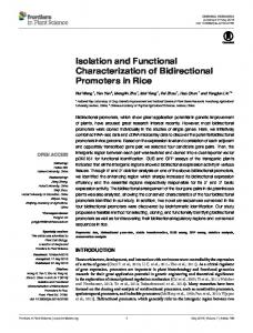
Vidur Bhatia and Baljeet Singh Saharan J. Chem. Pharm. Res., 2016, 8(7):357-367 ______________________________________________________________________________ RESULTS AND DISCUSSION 3.1. Biosurfactant screening Various yeasts isolated from rotten fruits were screened for production of biosurfactants. The screening of biosurfactant producing yeast was a two phase process. Primarily, the isolates were screened for their ability to produce biosurfactant using drop collapse method. The flattened drop, of cell free suspension (CFS) comprising biosurfactant, over the oil coated surface confirmed the presence of biosurfactant. Efficient emulsification property is critical for significant biosurfactant and its further applications [10]. In case of isolate WG1 the maximal % emulsification activity with kerosene oil after 24 hours was found to be (E24=53.57%). Further, the effectiveness of any microbial surfactant is determined by its ability to reduce the surface tension of production medium [11]. The biosurfactant produced by isolateWG1 showed a significant reduction in surface tension of CFS from 72 to 39.2 mN/m. Various yeast strains have been reported as biosurfactant producer on the basis of their ability to reduce surface tension of production media. Rufino et al., [12] reported that the biosurfactant produced by Candida lipolytica UCP 0988 reduced the water surface tension from 70 to 25.3 mN/m; similarly, while working with Candida sphaerica UCP0995, Luna et al., [13] found that the biosurfactant was able to reduce the surface tension of the medium to 25 mN/m. The flattened drop collapse, effective emulsification index and significant reduction in surface tension finally confirmed the biosurfactant production by the isolate WG1 (Fig. 1).
A
C
B
D
Fig. 1: Different observation of isolate WG1 A) colony morphology on YPD media; B) Cell morphology under 100X light microscope; C) Emulsification activity with kerosene oil and D) oil displacement assay
3.2 Taxonomic identification Isolate WG1 was taxonomically identified by 18S rRNA sequencing, for this genomic DNA was directly amplified by colony PCR using universal 18S rRNA primers. Taxonomic affiliation of the isolates was retrieved from GenBank. The BLASTn algorithm was used to determine the most related sequence relatives in the GenBank database. The BLASTn search for 18S rRNA partial gene sequence of the yeast isolate WG1 isolated from rotten white grapes showed 99% identity with Pichia sorbitophila sp. from NCBI database which have been submitted to NCBI Genbank under the Accession No. KX060549. Its phylogenetic tree (Fig.2) was constructed using the MEGA 5 (Molecular Evolutionary Genetics Analysis) software (v.5.05).
360
Vidur Bhatia and Baljeet Singh Saharan J. Chem. Pharm. Res., 2016, 8(7):357-367 ______________________________________________________________________________
Fig.2: Phylogenetic relatedness of Pichia sorbitophila strain WG1 based on 18S rRNA gene sequence
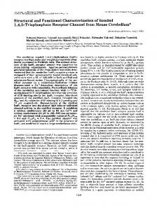
3.3 Surface tension measurement The biosurfactants produced by isolates WG1 showed a significant reduction in surface tension of cell free supernatant from 64 mNm-1(surface tension of YPD broth) to 39.2mNm-1. The reduction in surface tension is very significant as compared to the surface tension of the production medium. 3.4 Production and purification of biosurfactant The shake flask production of yeast biosurfactant was conceded out by inoculating with 1% 18 h grown pre culture at 37 °C for 72 h on shaking conditions. The surface tension of the cell free supernatant was found to be reduced to 39.2 mN/m from 72 mN/m, hyper surface tension reduction was detected at the late exponential phase of cell growth. Reduction in surface tension during the logarithmic and stationary phase has been reported and confirmed the production of biosurfactant [14]. Biosurfactant from CFS was extracted with equal amount of ethyl acetate. The yield of biosurfactants obtained by the ethyl acetate extraction method from 144 h culture of selected yeast isolate was 5.8 g/L. Furthermore, crude biosurfactant derived from yeast isolate was partially purified using column chromatography (Silica gel; 60-120 mesh size). 3.5 Characterization of biosurfactant Partially purified biosurfactant was initially described by TLC followed by post chromatographic detection. After silica plate developed, plates were sprayed with Syldatk reagent which developed a dark red spot specified the incidence of sugar moieties. The separation achieved with TLC development confirmed the presence of the glycolipid kind of biosurfactant. Commonly glycolipids kinds of biosurfactants are rich in carbohydrates in mixture with fatty acids [15]. 3.5.1 FTIR analysis The molecular arrangement and functional groups were determined by FTIR spectrum which revealed that the major fraction of biosurfactant was lipid and polysaccharide fractions. The molecular arrangement of biosurfactant showed that most noticeable adsorption bands were located at 3024 cm−1, 1713 cm−1 (C=O stretching in carbonyl groups), 1219 cm−1 (C-O stretching bands) and 748 cm−1 (CH2 group) (Fig. 3) which importantly established the fact that biosurfactants was a glycolipid kind of biosurfactant [3]. Assessment of the spectra showed that the biosurfactant is stringently comparable to glycolipid reported earlier from different yeast strains [14,16].
361
Vidur Bhatia and Baljeet Singh Saharan J. Chem. Pharm. Res., 2016, 8(7):357-367 ______________________________________________________________________________
Fig. 3: FTIR spectrum of biosurfactant produced by the isolate WG1
Fig. 4: NMR spectroscopy of biosurfactant produced by the isolate WG1
362
Vidur Bhatia and Baljeet Singh Saharan J. Chem. Pharm. Res., 2016, 8(7):357-367 ______________________________________________________________________________ 3.5.2 NMR spectroscopy The structural organization of protons in yeast derived biosurfactant has been carried out by 1H NMR spectroscopy. The chemical shifts and spatial arrangements were depicted that the biosurfactant has the spatial arrangements strictly similar to glycolipid (Fig. 4). Different spectral peaks were detected in NMR because of the presence of polysaccharides and fatty acids fractions. Characteristic spectra peaks of NMR were also reported in case of glycolipids [17,18,19]. 3.5.3 GC-MS analysis of fatty acids of methyl esters (FAME) The fatty acid composition of yeast derived biosurfactant by isolate WG1 was examined by GC-MS and obtained spectral peaks were equated with the data available in library. Biosurfactant obtained from the isolate WG1 was found to be a glycolipid mainly composed of long chain fatty acids with polysaccharides fractions (Fig. 5). Major fatty acid was found as Hexadecanoic acid on the basis of probability of similarity from standard data library. Different authors have also observed that yeast surface active agents mainly composed of hexadecanoic acid as a major fatty acid composition [20,21,22]. Biosurfactant produced by the Cryptococcus humicola JCM1461 has been reported for biosurfactants containing hexadecanoic acid as a major fatty acid [23].
Fig. 5: GC-MS analysis of biosurfactant produced by the isolate WG1
3.6 Ionic charge of biosurfactant In agar double diffusion test, samples of all the isolated biosurfactant formed white precipitation line between the well containing BS sample and the well containing cationic compound barium chloride. This confirmed that the biosurfactant isolated from Pichia sorbitophila WG1 was anionic in nature. 3.7 Critical micelle concentration (CMC) of biosurfactant The CMC of the produced biosurfactant was determined by preparing different dilutions in distilled water at pH 7. The CMC of the purified biosurfactant from Pichia sorbitophila WG1 was found to be approximately 2.25 mg ml-1 (Fig. 6).
363
Vidur Bhatia and Baljeet Singh Saharan J. Chem. Pharm. Res., 2016, 8(7):357-367 ______________________________________________________________________________ 3
Biosurfactant conc. (mg/ml)
2.5
2
1.5
1
0.5
0 72
68
61
54
48
42
39.2
39.2
39.2
39.2
Surface tension (mN/m) . Fig. 6: CMC of biosurfactant isolated from Pichia sorbitophila WG1
Surface tension (mN/m)
43 42 41 40 39 38 4
5
6
7
8
9
10
pH . Fig. 7: Effect of pH on the surface tension of the crude biosurfactant isolated from Pichia sorbitophila WG1
3.8 Stability study of biosurfactant 3.8.1 Effect of pH Biosurfactant isolated from Pichia sorbitophila WG1 shows stability over wide range of pH from 4.0 to 10.0 with maximum reduction in surface tension (39.1 mN/m) at pH 8.0. Almost same readings (39.2 mN/m) was also obtained at pH 6.0 and pH 7.0 which increased to 40.4 and 40.8 at pH 9.0, and pH 10.0 respectively. There was a slight increase in surface tension values at pH 4.0 and pH 5.0 as well, but shows stability of biosurfactant. Therefore, we can say that, the surface tension activity of BS obtained from Pichia sorbitophila WG1 was stable from pH 4.0
364
Vidur Bhatia and Baljeet Singh Saharan J. Chem. Pharm. Res., 2016, 8(7):357-367 ______________________________________________________________________________ (42 mN/m) to pH 10.0 (40.8 mN/m) with minimal variation in surface tension and showed highest stability (minimum surface tension i.e.39.2 mN/m) at pH 8.0 as shown in Fig.7. 3.8.2 Effect of temperature The surface tension of crude biosurfactant obtained from Pichia sorbitophila WG1 showed non-significant change at different tested temperatures, which was slightly higher at low temperatures i.e. at 10 °C and 20 °C but showed stability (39.2- 41.3 mN/m) at higher temperatures (30-110 °C)(Fig. 8). 46
Surface tensio (mN/m)
45 44 43 42 41 40 39 38 10
30
50
70
90
110
Temperature . Fig. 8: Effect of temperature on the surface tension of the crude biosurfactant isolated from Pichia sorbitophila WG1
3.9 Antimicrobial activity The biosurfactant of Pichia sorbitophila WG1 showed 76% inhibition of Escherichia coli, 72% inhibition of Pseudomonas aeruginosa, 71%inhibition of Salmonella typhi, 95% inhibition of Staphylococcus aureus, 97% inhibition of Staphylococcus epidermidis and96% inhibition of Bacillus cereus. The biosurfactant did not show an effective antimicrobial activity against the Shigella strains studied. It inhibited only 47.1% of the growth of Shigella flexneri. On the other hand, it showed almost complete inhibition of growth (more than 99%) of Listeria monocytogenes and Listeria innocua (Fig.9). In general, Gram positive pathogens showed higher reduction in growth as compared to the Gram negative pathogens.
365
Vidur Bhatia and Baljeet Singh Saharan J. Chem. Pharm. Res., 2016, 8(7):357-367 ______________________________________________________________________________ 100 90
Inhibition (%)
80 70 60 50 40 30 20 10 0
Pathogens
.
Fig. 9: Growth inhibition percentages of different microbes with the biosurfactant isolated from Pichia sorbitophila WG1
3.10 Phytotoxicity analysis Germination test was used to measure the phytotoxicity of the isolated biosurfactant. For this, a series of experiments were conducted to study different parameters like seed germination, root elongation, vigor index and germination index against the seeds of Brassica nigra L. and Triticum aestivum L. Table1: Effect of Pichia sorbitophila WG1 biosurfactant on different parameters of Triticum aestivum and Brassica nigra Name of tested crop Triticum aestivum Brassica nigra
Seed germination(%) 100±0.2 100±0.2
Root elongation (%) 116.7±0.3 112±0.3
Germination Index (%) 116.7±0.5 112±0.23
Vigor index 1450±35.35 1250±125
In the present study, 100 % seed germination suggests that the isolated biosurfactant doesn’t exhibit any inhibitory effect on two tested crops. Also, the germination index (GI) value was more than 110 %, indicating any absence of toxic effect on seed germination and root elongation on the tested seeds and thus our product can be considered as non-phytotoxic compound. CONCLUSION Biosurfactants are widely used as compared to chemical surfactants due to their unique features like low toxicity, biodegradable nature, easy & ecofriendly production and more effectiveness etc. Many reports are available in literature regarding production of biosurfactants from bacterial species, but due to their pathogenic nature and low production rate, most of the byproducts are unsuitable to be used. Yeast, being a GRAS (generally regarded as safe) status microorganism, produces higher quantity of biosurfactants as compared to bacterial species. The biosurfactant produced by Pichia sorbitophila WG1 in the present study was structurally characterized as glycolipid containing hexadecanoic acid chain which showed stability over a wide range of pH & temperature and also own some other properties like excellent surface tension reducing ability, CMC, antimicrobial and proved to be nontoxic. All these properties of isolated biosurfactant opens new future prospects for its use in a wide variety of effective industrial, environmental and biotechnology applications such as production of biocontrol agent etc. Acknowledgement The infrastructure and financial aid provided by the Kurukshetra University, Kurukshetra is highly acknowledged to carry out present research work.
366
Vidur Bhatia and Baljeet Singh Saharan J. Chem. Pharm. Res., 2016, 8(7):357-367 ______________________________________________________________________________ REFERENCES [1] BS Saharan; RK Sahu; D Sharma. Genet. Eng. Biotechnol. J., 2011(1), 1-14. [2] JD Desai; IM Banat. Microbiol. Mol. Biol. reviews, (1997), 61(1), 47-64. [3] IM Banat; A Franzetti; I Gandolfi; G Bestetti; G Martinotti; L Fracchia; TJ Smyth; R Marchant. Appl. Microbiol. Biotechnol., (2010), 87(2), 427-444. [4] HS Kim; BD Yoon; CH Lee; HH Suh; HM Oh; T Katsuragi; Y Tani. J. Ferment. Bioeng., (1997), 84(1), 41-46. [5] JD Desai. J. Sci. Ind. Res., (1987), 46(10), 440-449. [6] JD Desai; AJ Desai. Surfactant Sci. Ser., (1993), 65-65. [7] GC Fontes; NM Ramos; PFF Amaral; M Nele; MAZ Coelho. Braz. J. Chem. Eng., (2012), 29(3), 483-494. [8] C Syldatk; S Lang; U Matulovic; F Wagner. Zeitschrift für Naturforschung C, (1985), 40(1-2), 61-67. [9] T Meylheuc; CJ Van Oss; M Bellon‐Fontaine. N. J. Appl. Microbiol., (2001), 91(5), 822-832. [10] IM Banat; RS Makkar; SS Cameotra. Appl. Microbiol. Biotechnol., (2000), 53(5), 495-508. [11] CN Mulligan. Environ. Pollut., (2005), 133(2), 183-198. [12] RD Rufino; JM Luna; LA Sarubbo; LRM Rodrigues; JAC Teixeira GM Campos-Takaki. Colloids Surf. B: Biointerfaces, (2011), 84(1), 1-5. [13] JM Luna; RD Rufino; LA Sarubbo; GM Campos-Takaki. Colloids Surf., B: Biointerfaces, (2013), 102, 202209. [14] JMD Luna; L Sarubbo; GMD Campos-Takaki. Brazilian Archives of Biology and Technology, (2009), 52(4), 785-793. [15] S Lang. Curr. Opin. Colloid Interface Sci., (2002), 7(1), 12-20. [16] DK Santos; RD Rufino; JM Luna; VA Santos; AA Salgueiro; LA Sarubbo. J. Pet. Sci. Eng., (2013), 105, 43-50. [17] M Henkel; MM Müller; JH Kügler; RB Lovaglio; J Contiero; C Syldatk, R Hausmann. Process Biochem., (2012), 47(8), 1207-1219. [18] T Morita; T Fukuoka; T Imura; N Hirose; D Kitamoto. Biosci. Biotechnol. Biochem., (2012), 76(9), 1788-1791. [19] TB Lotfabad; S Tayyebi; R Roostaazad. World Appl. Sci. J., (2013), 22(6), 809-816. [20] T Kulakovskaya; A Shashkov; E Kulakovskaya; W Golubev; A Zinin; Y Tsvetkov; A Grachev; N Nifantiev. J. Oleo Sci., (2009), 58(3), 133-140. [21] IA Ribeiro; C Faustino; PS Guerreiro; RF Frade; MR Bronze; MF Castro; MH Ribeiro. J. Mol. Recognition, (2015), 28(3), 155-165. [22] M Konishi; M Fujita; Y Ishibane; Y Shimizu; Y Tsukiyama; M Ishida. Biosci. Biotechnol. Biochem. (2016), 17. [23] T Morita, Y Ishibashi, T Fukuoka, T Imura, H Sakai, M Abe, D Kitamoto. Biosci. Biotechnol. Biochem. (2011), 75(8), 1597-1599.
367