THE JOURNAL OF BIOLOGICAL CHEMISTRY © 1997 by The American Society for Biochemistry and Molecular Biology, Inc.
Vol. 272, No. 37, Issue of September 12, pp. 23042–23049, 1997 Printed in U.S.A.
Isolation, Expression, and Regulation of the pgr11 Gene Encoding Glutathione Reductase Absolutely Required for the Growth of Schizosaccharomyces pombe* (Received for publication, January 6, 1997, and in revised form, July 7, 1997)
Joon Lee‡§, Ian W. Dawes¶, and Jung-Hye Roe‡i From the ‡Department of Microbiology, College of Natural Sciences, and Research Center for Molecular Microbiology, Seoul National University, Seoul 151-742, Korea and ¶School of Biochemistry and Molecular Genetics, University of New South Wales, Sydney, New South Wales 2052, Australia
The pgr11 gene encoding glutathione reductase (GR, EC 1.6.4.2) was isolated from Schizosaccharomyces pombe using a polymerase chain reaction fragment as a probe. The gene consists of two exons and an intron of 55 nucleotides, encoding a polypeptide of 465 amino acids (50,238 Da) with conserved residues characteristic of GR. The transcriptional start site was localized at 239 nucleotides upstream from the ATG initiation codon. The level of transcript as well as the GR enzyme activity increased more than 11-fold when the cloned pgr11 gene was expressed on a multicopy plasmid. This overexpression conferred on S. pombe cells more resistance against menadione, a redox cycling agent, but not against H2O2. The level of pgr11 transcripts increased by treatment with oxidants such as menadione, cumene hydroperoxide, and diamide. It also increased by treatment with high osmolarity, heat shock, or at the stationary growth phase. The deletion of the pap11 gene encoding an AP-1 homolog in S. pombe caused reduction in the pgr11 gene expression. Furthermore, Dpap1 cells lost the inducibility of pgr11 gene expression by the above stresses, implying that Pap1 is involved in general stress-inducible gene expression. When the pgr11 gene was disrupted, the haploid spores were not viable. Repression of nmt1 promoter-driven pgr11 expression by thiamine caused cessation of growth, which was rescued by the episomal pgr11 gene. These results indicate that GR activity, which efficiently reduces GSSG, is essentially required for the growth of S. pombe, unlike in Saccharomyces cerevisiae or Escherichia coli. Glutathione (L-g-glutamyl-L-cysteinylglycine, GSH) is one of the most prevalent reducing thiols in eukaryotic cells, which functions in many biological processes such as protein and DNA synthesis, transport, modulation of enzyme activity, and metabolism of various xenobiotics and free radicals (1, 2). GSH is oxidized nonenzymatically through the reaction with disulfides, metals, and reactive oxygen species, or through peroxi* This work was supported by the Korea Science and Engineering Foundation (KOSEF) through the Research Center for Molecular Microbiology at Seoul National University and by a grant from the Department of Industry, Trade and Regional Development, Australia (to I. W. D.). The costs of publication of this article were defrayed in part by the payment of page charges. This article must therefore be hereby marked “advertisement” in accordance with 18 U.S.C. Section 1734 solely to indicate this fact. The nucleotide sequence(s) reported in this paper has been submitted to the GenBankTM/EBI Data Bank with accession number(s) U63845. § Recipient of Research Promotion Grant for Young Researchers from the Korea Research Foundation. i To whom correspondence should be addressed: Tel.: 82 2 880 6706; Fax: 82 2 888 4911; E-mail:
[email protected].
dase-mediated reaction, yielding oxidized glutathione (GSSG). GSSG is known to be very toxic to biological molecules due to its high reactivity with free sulfhydryl groups. The intracellular ratio of GSH to GSSG, therefore, is usually kept high in most eukaryotic cells examined (3). The reduction of GSSG to GSH is efficiently mediated by glutathione reductase (GR),1 using NADPH specifically as a reducing equivalent (4). GR is a member of the pyridine nucleotide-disulfide oxidoreductase family of flavoenzymes, which includes thioredoxin reductase, lipoamide dehydrogenase, mercury reductase, and trypanothione reductase (5). In addition to glutathione, several peptide thiols such as thioredoxin (Trx) and glutaredoxin (Grx), which contain redoxactive dithiol groups, provide reducing environment to the cell as electron (hydrogen) donors (6 – 8). They provide hydrogens to specific reductases that reduce ribonucleotide for DNA synthesis, phosphoadenosine phosphosulfate for generating cysteine, and methionine sulfoxide to repair oxidized protein (6, 9, 10). They also provide hydrogen to glutathione peroxidase (11). Trx is reduced by Trx reductase using NADPH. Grx is reduced by GSH using its own thiol-disulfide exchange activity. Recently a regulatory network of Trx, Grx, and GSH for the efficient regulation of the level of ribonucleotide reductase has been proposed in Escherichia coli, in which GSH, Trx, and Grx all compensate for each other (12). In prokaryotes the role of GSH is not well characterized. An E. coli mutant (gshA2 deficient in g-glutamylcysteine synthetase) contains less than 0.4% of the wild-type GSH but is as resistant as the wild type against treatments with oxidants, heat, or g-ray (13). Furthermore, glutathione reductase-deficient (gor2) mutants maintained the highly reduced glutathione pool, suggesting that GSSG can be reduced independently of GR (14). Either Grx or Trx system can carry out this function (15). In addition lipoate can function as a hydrogen donor of Grx in the absence of GSH (12). The gor2 mutant, however, exhibited sensitivity to hydrogen peroxide and diamide, a thiol oxidizing agent, suggesting that GR protects cells from oxidative damage. Recent work in E. coli demonstrates that GSH defends cells against attack by chlorine compounds (16). In Saccharomyces cerevisiae, GSH is critically required for aerobic survival as well as protection against oxidative stress, as judged from the phenotype of mutants deficient of GSH synthesis (17–19). The adaptation to hydrogen peroxide was suppressed by depleting GSH (20). The GLR1 gene encoding GR in S. cerevisiae is required in defending cells against oxi1 The abbreviations used are: GR, glutathione reductase; Trx, thioredoxin; Grx, glutaredoxin; MD, menadione; STRE, stress-response element; PCR, polymerase chain reaction; IPTG, isopropyl-1-thio-b-D-galactopyranoside; bp, base pair(s); kb, kilobase pair(s); EMM, Edinburgh minimal medium.
23042
This paper is available on line at http://www.jbc.org
Glutathione Reductase Gene Expression in S. pombe
23043
TABLE I S. pombe strains used in this study Strain
972 ED665 ED666 ED667 JLD JLD1 JL36 JL38 TP4–5A TP108–3C
Genotype (denotation) 2
h wild type h2 ade6-M210 leu1–32 ura4-D18 h1 ade6-M210 leu-32 ura4-D18 h2 ade6-M216 leu1–32 ura4-D18 h1/h2 ade6-M210/ade6-M216 leu1–32/leu1–32 ura4-D18/ura4-D18 (ED666 cross ED667) h1/h2 ade6-M210/ade6-M216 leu1–32/leu1–32 ura4-D18/ura4-D18 pgr11/pgr1::ura41 h2 ade6-M210 leu1–32 ura4-D18/ura41 Dpgr1/nmt-pgr1 h2 ade6-M210 leu1–32 ura4-D18/ura41 h2 ade6-M210 leu1 ura4 h2 leu1 ura4 pap1::ura41
dative stress judging from the marked sensitivity of the glr1 disruptant to hydrogen peroxide and paraquat (21, 22). However, the gene is not essentially required for normal aerobic growth. The dispensibility of GR seems to be due to the presence of Trx since glr1 trx1 trx2 triple mutant is not viable, suggesting the functional overlap of GR and Trx in S. cerevisiae (23). The GLR1 gene of S. cerevisiae is induced by hydrogen peroxide in a manner dependent on yAP-1, a yeast homolog of mammalian AP-1 (22). Schizosaccharomyces pombe displays different physiology from S. cerevisiae and is probably more closely related to higher eukaryotes (24). S. pombe cells adapt to oxidative stress accompanied by the induction of several antioxidant enzymes (25). The induction pattern of these enzymes is quite different from that of S. cerevisiae. For example, hydrogen peroxide induce catalase and peroxidase whereas menadione (MD), a redox cycling agent which can generate superoxide radical (O2. ), could induce not only catalase and peroxidase but superoxide dismutase, GR, and glucose-6-phosphate dehydrogenase. Furthermore, S. pombe cells pretreated with MD are adapted for subsequent shock of hydrogen peroxide, but not vice versa, implying different effects of these oxidants in cellular induction of defense mechanisms (25). Under the same laboratory condition, S. pombe cells are more resistant to hydrogen peroxide and MD than is S. cerevisiae. Part of this resistance can be ascribed to higher levels of catalase and GSH/GSSG ratio. As a first step to examine the role of GSH in the physiology of S. pombe, we isolated the pgr11 gene encoding GR in S. pombe and hereby report its characterization. The regulation of its expression was examined under different stressful conditions, and the involvement of Pap1, a S. pombe homolog of mammalian AP-1 (26), was investigated. Surprisingly, we found that GR is essentially required for the aerobic growth of S. pombe, and the pgr11 gene is regulated by Pap1 for induction upon various stressful conditions. EXPERIMENTAL PROCEDURES
Cells, Media, and Chemicals—S. pombe strains used in this study were listed in Table I. For routine growth YES (0.5% w/v yeast extract, 3% w/v glucose, 250 mg/liter supplements) or YEPD (1% w/v yeast extract, 2% w/v glucose, 2% w/v peptone) medium were used. Cells containing plasmid or disrupted gene were cultured in EMM with appropriate supplements (27). Cells were grown at 30 °C with shaking to A595 of 0.15– 0.2, 2.0, or more than 5.0 (72 h after inoculation) for early exponential, late exponential, or stationary phase cultures, respectively. E. coli strain DH5a was used for most plasmid construction and preparation. GSH, menadione sodium bisulfite, diamide, cumene hydroperoxide were purchased from Sigma, and 2-vinylpyridine from Aldrich. Construction of S. pombe Genomic Library—The genomic DNA from S. pombe wild type strain (972 h2) was prepared according to Moreno et al. (27). It was partially digested with restriction endonuclease BamHI, and DNA fragments of 19 –23 kb in size were fractionated by glycerol gradient. These were ligated into phage lEMBL3 DNA cut with BamHI and propagated as recommended (Stratagene). PCR and Cloning of the pgr11 Gene—The degenerate oligonucleotide primers corresponding to residues 187–192 (VGAGYI) and 326 –333
Source or reference
Laboratory Laboratory Laboratory Laboratory This work This work This work This work 26 26
collection collection collection collection
(TPVAIAAG) of GLR1 gene of S. cerevisiae were synthesized; 59-GTHGGTGCTGGTTAYATT-39 and 59-ACCAGCAGCTAAWGCRACRGGRGT-39, respectively, where Y 5 C, T; W 5 A, T; R 5 A, G; and H 5 A, C, T. PCR amplification was done for 30 cycles under the following condition; denaturation at 94 °C for 1 min, annealing at 42 °C for 2 min, and extension at 72 °C for 1 min. The amplified DNA fragment of 441 bp was cloned into pTZ18R at HincII site to generate pSPGR05. The insert DNA fragment was used as a probe to screen the l genomic library according to the standard method (28). Six positive clones were selected and the common region of a 4.3-kb PstI fragment was identified and cloned into pTZ18R at the PstI site yielding pSPGR10 and pSPGR20 with inserts in both orientations. The sequencing of the cloned DNA was done by dideoxy chain termination method either manually or by an automatic sequencer (ALFexpress, Pharmacia). Primer Extension Analysis—Total RNA was prepared as described by Schmitt et al. (29) and primer extension analysis was performed according to Grimm et al. (30). A primer (GRPRIM1; 59-TGAATTCGTTAGACTTTGCCATTG-39) corresponding to a region about 200 nucleotides upstream from the initiation ATG codon was synthesized and radioactively labeled with [g-32P]ATP by T4 polynucleotide kinase. Total RNAs (20 –30 mg) from nontreated or MD-treated cells were analyzed. GR Enzyme Assay, Activity Staining, and the Measurement of Intracellular Glutathiones—Cell free extracts were prepared as described previously (25). GR activity was measured according to Smith et al. (31) by monitoring the reduction of 5,59-dithiobis(2-nitrobenzoic acid) to thiobis(2-nitrobenzoic acid) by GSH, which is produced by GR. The activity staining for GR was performed on native polyacrylamid gel detecting yellow color caused by the reduction of 5,59-dithiobis(2-nitrobenzoic acid) in the presence of NADPH and GSSG (32). The amount of total and oxidized glutathione were determined using GR and 2-vinylpyridine as described by Griffith (33). Overexpression of the pgr11 Gene Product—PstI fragment containing the pgr11 gene was cloned into the multicopy shuttle plasmid pUR19N (34) at PstI site. The resulting plasmid, pURGR01, was introduced into S. pombe cells by lithium acetate/polyethylene glycol method (27) and selected for Ura1 phenotype. Northern Blot Analysis—When cells were grown to the appropriate growth phases external stresses such as H2O2, cumene hydroperoxide, MD, diamide, NaCl, and high temperature were administered for 1 h. The same amount (30 – 40 mg) of RNAs prepared from these cells were separated on agarose gel containing formaldehyde and was transferred onto Hybond-N filter (Amersham Corp.). Hybridization was performed with the PCR product as a probe by standard method (28). The blot was autoradiographed on x-ray film and was quantified with Molecular Imager (Bio-Rad). Disruption of the pgr11 Gene—pSPGR20 plasmid was cut with KpnI and a 5.3-kb fragment containing the pgr11 gene was liberated and self-ligated, resulting in pSPGR40. A 1.8-kb ura41 cassette (30) was inserted into pSPGR40 by replacing the HincII/Tth111I fragment of the pgr11 ORF (see Fig. 1). The resulting pGR-ura4 was digested with KpnI and PstI, and the 3.1-kb fragment containing ura41 cassette was used to transform diploid JLD strain to achieve gene replacement according to Moreno et al. (27). Stable Ura1 prototrophs were selected. The correct integration of the ura41 cassette was verified either by PCR with two specific primers from pgr11 and ura41 sequences, or by genomic Southern hybridization. Tetrads were analyzed on YES plate. Construction of Conditionally Regulated nmt-pgr1 Allele—The Nterminal portion of the pgr11 cDNA was synthesized by reverse transcription-PCR using primers mGRN1, 59-ACTCATATGGCACCTATTTCAAAGG-39, where NdeI site is underlined; and RTC, 59-GCGACGAGCTCAGCAATATTCCAT-39. The amplified DNA fragment of about 200 bp was subcloned to the HincII site of pTZ18R creating pTmGR1.
23044
Glutathione Reductase Gene Expression in S. pombe
FIG. 1. The physical map and the nucleotide sequence of pgr11. A, the physical map of 4.3-kb PstI fragment containing the pgr11 gene is shown. The primers for PCR amplification and its product (441 bp) are drawn on top. The exonal sequences of pgr11 were indicated by thick lines with an arrowhead. A 1.8-kb ura41 cassette and the region it replaced in pgr11 for its disruption is shown (see “Experimental Procedures”). The restriction endonuclease sites are: B, BglII; E, EcoRI; H, HindIII; Hc, HincII; K, KpnI; Na, NarI; Nd, NdeI; Nh, NheI; P, PstI; and T, Tth111I. B, the nucleotide sequences of 2684-bp KpnI/PstI fragment containing the pgr11 gene were shown with its deduced amino acid sequences. The major transcriptional start site (11) is indicated with an arrow and a minor start site is marked with a triangle. The putative TATA box is indicated in bold face, doubly underlined. The intron sequences are indicated in italics. The conserved regions in the intron junction as well as the branch site observed in S. pombe (39) are underlined. Two regions corresponding to the PCR primers are marked by arrows. Four domains required for the GR activity are highlighted in bold; residues 9 –39 for FAD binding, 42–55 for the active site, 180 –217 for NADPH binding, and 440 – 465 for the assembly domain (40). The full sequence of the 4334-bp PstI fragment was registered in GenBankTM/EMBL/DDBJ data base under the accession no. U63845. The 1.7-kb NarI/BamHI fragment containing the C-terminal remainder of the pgr11 gene was obtained from the pSPGR01 and was fused to the N-terminal portion cut with NarI/BamHI in pTmGR1. The resulting plasmid pTmGR10 harbors promoterless cDNA of pgr11. The thiaminerepressible promoter of nmt11 gene (35) was fused to pgr11 cDNA and was integrated into S. pombe chromosome in the following way. The ars1 region was removed from pREP82 (36) by EcoRI digest to generate pRIP82. A 642-bp NdeI fragment was cut out from pTmGR10 and inserted into the NdeI site downstream of the nmt1 promoter in pRIP82. The resulting plasmid, pRIP-GR01, was linearlized at the unique NheI site inside the pgr11 gene and used to transform ED665 cells (see Fig. 1). Among stable Ura1 transformants, the JL36 clone was
picked and verified for the correct integration of nmt-pgr1 by Southern hybridization. Its isogenic control, JL38, was constructed by transforming ED665 with pRIP82 vector (see Table I). The phenotype of JL38 was identical with its parent ED665 except Ura1 prototrophy. Rescue of GR Deficiency by Cloned pgr11—The PstI/BamHI fragment containing nmt1 region in pREP1 plasmid (35) was replaced with the 4.3-kb PstI/BamHI fragment containing the entire pgr11 gene. The resulting pGR02 plasmid DNA was introduced into JL36. The Ura1 and Leu1 transformants were selected and streaked on EMM plate with or without 10 mM thiamine and allowed to grow at 30 °C. Sequence Analyses and Data Base Entry—The translation of DNA sequence, homology search with BLAST, motifs, and PUBLISH were
Glutathione Reductase Gene Expression in S. pombe
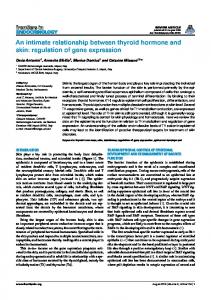
FIG. 2. Determination of the transcriptional start site by primer extension. The 59 end of the 24-nucleotide primer was labeled with 32P and hybridized with RNAs (30 mg) from cells untreated (lane 1) or treated with 0.2 mM MD (lane 2), followed by extension with mouse mammary tumor virus reverse transcriptase. The same primer was used for sequencing the pgr11 gene cloned in double stranded plasmid. The extended products were separated on 7 M urea, 5% polyacrylamide gel. The major transcriptional start site was localized on the A residue 239 nucleotides upstream from the initiation codon. A minor site was found on another A residue 3 nucleotides further upstream (large and small arrowheads, respectively). done using the GCG Wisconsin Package (version 8.1) installed at RCMM (Research Center for Molecular Microbiology, Seoul National University). The nucleotide sequences of the pgr11 gene and its flanking region (4334-bp PstI fragment) was deposited in GenBankTM/ EMBL/DDBJ under the accession no. U63845. RESULTS
Isolation and Characterization of the pgr11 Gene Encoding GR from S. pombe—From the predicted amino acid sequences of GR genes of human (37), E. coli (38), and S. cerevisiae (21), several highly conserved regions were identified. The primer pair corresponding to residues 187–192 (VGAGYI) and 326 – 333 (TPVAIAAG) of GLR1 gene of S. cerevisiae amplified a single species of PCR product of the expected size (441 bp) from the chromosomal DNA of S. pombe strain 972. This fragment was cloned and sequenced. The deduced amino acid sequence revealed about 50% identity with those of GR from E. coli and S. cerevisiae. It hybridized with a specific restriction fragment from the genome, indicating that there is only one copy of GR gene (pgr11) in S. pombe (data not shown). The same probe was used to screen the genomic phage library. From positive phage clones, the common 4.3-kb PstI fragment hybridizing with the probe was subcloned and sequenced (Fig. 1). It contained the pgr11 gene with an intron of 55 nucleotides in size near the N terminus of the open reading frame as verified by reverse transcription-PCR and sequencing of the cDNA (data not shown). The canonical 59 donor (GUAAGU), 39 acceptor ((U/ A)UAG), and branch site (YURAY) sequences were found in this intron (39). We deduced the primary structure of GR, containing 465 amino acid residues with the molecular mass of 50,238 Da (Fig. 1B). The amino acid sequence is highly homologous to other known GR sequences; 56.8% identity (76.3% similarity) with S. cerevisiae, 49% identity (68.8% similarity) with E. coli, and 50.3% identity (71.4% similarity) with human. It contained a motif of active site for pyridine nucleotide disulfide oxidoreductase class I, from residues 43 to 53 (GGTCVNYGCVP), as commonly observed in FAD flavoproteins such as GR, thioredoxin reductase (EC 1.6.4.5), trypanothione reductase (EC 1.6.4.8), lipoamide dehydrogenase (EC 1.8.1.4), and mercury reductase (EC 1.16.1.1) (5). Other conserved sequences known to be essential for binding FAD and NADPH and subunit assembly were also found (40) (Fig. 1B). To locate the transcriptional start site, a primer correspond-
23045
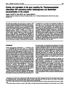
FIG. 3. Overexpression of GR on multicopy plasmids in S. pombe. A, Northern blot analysis of pgr11 mRNA. Total RNAs were isolated from S. pombe (ED665) cells containing either pUR19N parental vector (lane 1) or pURGR01 plasmid containing the pgr11 gene (lane 2). pgr11-specific transcripts were detected by using the 441-bp PCR fragment as a hybridization probe. The size of molecular mass markers is indicated on the left. B, quantitation of pgr11 transcripts and GR activity. The pgr11-specific radioactivity in panel A was quantitated by Molecular Imager. The enzyme activities in cell extracts obtained from the control (lane 1) and GR overexpressing cells (lane 2) were measured as described under “Experimental Procedures.”
ing to the region about 200 nucleotides upstream from the ATG codon was extended. The most prevalent transcript was initiated from the A residue located at 239 nucleotides upstream from the ATG codon (Fig. 2). A minor start site was mapped at another A residue 3 nucleotides upstream of the major one. A putative TATA box (TAATTAAA) was located 48 nucleotides upstream from the major start site consistent with other known positions of TATA box in S. pombe (41) (see Fig. 1B). When cells were treated with 0.2 mM MD for 1 h, transcripts from these start sites increased, suggesting that pgr11 transcription is induced by MD (Fig. 2, lane 2). Overexpression of GR and Its Effect on the Resistance against Oxidative Stress—To confirm that the cloned gene encodes active GR, we expressed its gene product in E. coli. The cDNA of pgr11 gene was placed under the control of T7 promoter in pET3a plasmid. E. coli BL21(DE3)pLysS cells were transformed with this plasmid and was treated with IPTG. The GR activity in cell extracts was examined in the native polyacrylamide gel electrophoresis by activity staining as described under “Experimental Procedures.” IPTG induced a distinct GR activity band in E. coli which comigrates with the GR activity band from S. pombe cell extracts (data not shown). We then overexpressed GR in S. pombe. The 4.3-kb PstI fragment containing the entire pgr11 gene was cloned into the multicopy plasmid pUR19N, creating pURGR01. We estimated the amount of pgr11 mRNA by Northern hybridization analysis and its gene product GR by enzyme activity assay from cells containing either pUR19N or pURGR01. As shown in Fig. 3A, pgr11-specific transcript of about 1.5 kb was detected. This specific transcript increased 11-fold in cells containing pURGR01. GR activity increased by 15-fold compared with the control cells (Fig. 3B). The increased activity was confirmed by activity staining in native electrophoretic gel (data not shown). We then measured the content of glutathiones and the ratio GSH/GSSG. The result presented in Table II demonstrates that the overproduced enzyme caused marked reduction of GSSG without changing the total amount of glutathiones. These results confirm that the cloned pgr11 gene indeed encodes active GR enzyme which efficiently reduces GSSG in vivo. We tested whether the increased level of GR or GSH/GSSG
23046
Glutathione Reductase Gene Expression in S. pombe TABLE II GR activity, amounts of GSH and GSSG from S. pombe of different genetic backgrounds
Strain (plasmid/treat)
Characteristics
GRa
Total glutathioneb 21
nmolzmg
units mg
ED665 (pUR19N) ED665 (pURGR01) JLD JLD1 JL36 (2thiamine)d JL36 (1thiamine)d
Control GR overproducer pgr11/pgr11 pgr11/pgr1::ura41 Dpgr1/nmt-pgr1 Dpgr1/nmt-pgr1
3.8 6 0.9 58.7 6 4.4 14.7 6 1.5 4.9 6 0.4 7.4 6 1.2 NDe
22.3 6 0.6 25.2 6 2.3 33.4 6 2.1 23.5 6 0.4 12.1 6 0.8 81.7 6 1.5
GSSGb
GSH/GSSG
21
0.27 6 0.02 ,,0.01c 0.31 6 0.06 0.64 6 0.02 0.57 6 0.08 39.5 6 1.8
81 ..252c 106 35 19.3 0.1
a One unit of GR was defined as the activity required for the reduction of 1 mmol of 5,59-dithiobis(2-nitrobenzoic acid)/min. Data are from three independent measurements. b The amount of total glutathione (GSH 1 2 GSSG) and GSSG was determined according to Griffith (33) as described in the text. c GGSG was not detected by this assay method whose sensitivity limit is 1022 nmol of glutathione (33). d These cultures were allowed to grow until no more increase in cell density was observed in the presence or absence of thiamine. e Not detected.
FIG. 4. Effect of overproduced GR on the cell survival against oxidative stress. Sensitivity of S. pombe cells containing parental vector (pUR19N; ●) or plasmid pURGR01 with pgr11 gene (f) against MD (panel A) or H2O2 (panel B) was measured. Exponentially growing cells were treated with each oxidant at various concentrations for 1 h. Cells were diluted and plated on YES plates in triplicate. One representative set of data is shown.
ratio contributes to the survival under oxidative stress conditions. For this purpose exponentially growing cells containing either parental vector (pUR19N) or the cloned pgr11 gene in pUR19N (pURGR01) were treated with different concentrations of H2O2 or MD for 1 h and the remaining viable cells were counted on YES plates. As shown in Fig. 4A, the increased level of GR conferred more resistance against MD. However, it did not provide cells with more protection against H2O2 (Fig. 4B). This is consistent with our previous observation that GR is more induced by MD than by H2O2, suggesting that the contribution of GR may be more critical in protection against MD toxicity than against H2O2 (25). pgr11 Expression Is Induced by Various Stresses Such as Oxidants, High Salt, High Temperature, and Starvation—We previously analyzed the changes in GR activity in S. pombe upon oxidant treatment and growth phase variation (25). At early exponential phase (A595 of 0.15– 0.2), treatment with 0.2 mM H2O2 for 1 h did not change its activity considerably (20% increase). MD, however, induced GR activity up to 3-fold. When cells were grown to the stationary phase GR activity increased more than 5-fold. To examine whether this activity variation is the result of transcript regulation, we performed Northern blot analysis of total RNAs from cells treated with several external stresses. In exponentially growing cells treated with either 0.2 or 2 mM H2O2 the level of pgr11 transcript did not change considerably (Fig. 5, lanes 2 and 3). Cumene hydroperoxide, however, increased the pgr11 transcript up to 2.8-fold (Fig. 5, lane 4). MD increased the level of pgr11 RNA by 3.3-fold (Fig. 5, lane 5). Similar extent of induction was observed in the primer extension analysis as well (Fig. 2). The fold increase in RNA level correlates well with the increase in enzyme activity as previously observed (25). Diamide, which oxidizes free thiol groups, also increased the level of pgr11 transcript by 2.5-fold (Fig. 5, lane 6). To test whether pgr11 expression is also induced by other
FIG. 5. Differential induction of pgr11 by various oxidants. Northern analysis of pgr11 mRNA. Exponentially growing cells were either untreated (lane 1) or treated with 0.2 mM H2O2 (lane 2), 2 mM H2O2 (lane 3), 0.1 mM cumene hydroperoxide (CHP, lane 4), 0.2 mM MD (lane 5), and 1.5 mM diamide (lane 6) for 1 h. Total RNAs were prepared and analyzed as described in the text. The blots were quantitated by Molecular Imager, and the average signals from three to five separate experiments are expressed as relative values.
stresses, we treated cells with high osmolarity, high temperature, or grew cells for longer period. Addition of NaCl to 200 mM, temperature upshift from 30 °C to 40 °C, and prolonged culture (A595 of 2.0 for late exponential, and A595 of 5.0 for stationary phase) all increased the expression of pgr11 by more than 3-fold (Fig. 6). These suggest that pgr11 is another stressregulated gene and there could be a common pathway which triggers the expression of GR in response to different stimuli.
Glutathione Reductase Gene Expression in S. pombe
23047
FIG. 6. Induction of pgr11 expression under conditions of high osmolarity, high temperature, and starvation. RNAs were prepared from cells at early exponential growth phase without any treatment (A595 of about 0.2, lane 1), treated with 0.2 M NaCl (lane 2), heat-shocked at 40 °C for 1 h (lane 3), at late exponential phase (A595 of about 2.0, lane 4) or stationary phase (A595 of about 5.0, lane 5). The hybridization analysis and quantitation were done as described in Fig. 5.
Pap1 Positively Regulates the pgr11 Expression—We identified two putative AP-1 binding sites in the upstream of the pgr11 promoter, TGAATCA (at 2192 ; 2186, match underlined) (42) and ATTAGGAAG (at 2160 ; 2152, match underlined) (26). To find out whether Pap1 regulates pgr11 expression, we measured the level of pgr11 RNA in different pap1 genetic backgrounds. The RNAs from the pap11 strain (TP45A) and its isogenic Dpap1 strain (TP108-3C) were analyzed by Northern blot analysis (Fig. 7A). In the Dpap1 strain pgr11 mRNA decreased about 2-fold. When the multicopy plasmid containing functional pap11 gene (pST1) was introduced into TP108-3C (26), the pgr11 transcript increased about 2-fold due to the increase in pap11 expression. The level of pap11 transcript increased about 4-fold in this cell (data not shown). In the Dpap1 strain GR activity decreased to about one-third of the wild type level, correlating with the decrease in pgr11 transcripts. In pap11 overproducing strain, however, GR activity was only half of the wild type level, suggesting that there may be a counteractive mechanism to balance the GR activity posttranscriptionally at high Pap1 concentration. The glutathione redox status paralleled the GR activity in the cell (Fig. 7B). We next investigated whether Pap1 is involved in the induction of the pgr11 gene expression in response to different external stresses. In Dpap1 cells, the induction of the pgr11 gene by oxidative stress, high temperature, high osmolarity, or starvation was almost abolished (Fig. 7C). The effect of Pap1 is most pronounced in cells under starved condition, where no significant amount of pgr11 transcript was detected. These results demonstrate that Pap1 acts as a positive regulatory factor for pgr11 expression and at the same time relays the stress signal from the external environment to induce pgr11 gene expression. Whether Pap1 affects pgr11 expression directly by binding to the putative Pap1 binding site requires further investigation. pgr11 Is an Essential Gene for the Aerobic Growth of S. pombe—To understand the role of GR in S. pombe, we constructed Dpgr1 disruptant. The disruption probe was made by replacing the 1.2-kb HincII/Tth111I fragment covering most of the pgr11 ORF with the 1.8-kb ura41 gene cassette (see Fig. 1A
FIG. 7. Positive regulation of pgr11 by Pap1. A, Pap1-dependent expression of pgr11 gene. RNAs were prepared from pap11 wild type cells (TP4-5A, lane 1), its isogenic Dpap1 cells (TP108-3C, lane 2), and Dpap1 cells containing pap11 gene on multicopy plasmid (TP1083C(pST1), lane 3). 30 mg of each RNA sample were analyzed for pgr11 mRNA as described in Fig. 5. B, the relative amount of pgr11 transcript as well as GR activity and the ratio of GSH/GSSG. Cell extracts to measure GR activity and GSH/GSSG were prepared from the same cells used for RNA preparation as described in panel A. C, dependence of the stress-induction of the pgr11 gene on Pap1. RNAs were prepared from Dpap1 (TP108-3C) cells at early exponential phase without any treatment (lane 1), treated for 1 h with 0.2 mM menadione (lane 2), 0.1 mM cumene hydroperoxide (lane 3), heat shock at 40 °C (lane 4), or 200 mM NaCl (lane 5), as well as from cells at late exponential phase (lane 6). 40 mg of each RNA sample were analyzed for pgr11 mRNA by Northern blotting.
and “Experimental Procedures”) and introduced into a diploid strain (JLD; h1/h2 ura4-D18/ura4-D18). We isolated Ura1 transformants and obtained pgr11/pgr1::ura41 heterozygous strain JLD1 (Table I). We confirmed the single copy disruption by both PCR and Southern hybridization (data not shown). GR activity and GSH/GSSG ratio in cell free extracts of JLD1 was measured (Table II). GR activity was reduced to about a third compared with the pgr11/pgr11 wild type strain (JLD). The ratio of GSH/GSSG was also reduced correspondingly. These again confirm that only one functional copy of the pgr11 gene in JLD1 strain was disrupted and the level of GR and GSH/GSSG ratio correlates tightly. Six asci were subjected to tetrad analysis and each spore was grown on YES plate in the air for 4 days at 30 °C. We detected only two spores from each ascus growing into colonies which were all Ura2 (data not shown). On microscopic observation of nonviable colonies, we were able to observe that spores had germinated and undergone several divisions before ceasing growth. This indicates that the pgr11 gene is essentially required for proliferation of S. pombe cells. Supplementation of YES plates with 1 mM GSH or 0.1 mM
23048
Glutathione Reductase Gene Expression in S. pombe
FIG. 8. Requirement of pgr11 gene for normal aerobic growth of S. pombe. The JL36 strain in which the expression of pgr11 is driven by thiamine-repressible nmt1 promoter was constructed as described in the text (Table I). It was streaked on EMM plate with (right) or without (left) 10 mM thiamine. A control strain (JL38) and JL36 transformant containing the wild type pgr11 gene on multicopy plasmid (pGR02) was streaked in parallel.
N-acetyl-L-cysteine did not promote the growth of the disruptant, suggesting that the growth inhibition is not caused by the deficiency in reduced GSH but by the accumulation of GSSG (data not shown). The growth defect of Dpgr1 strain was also observed by the repression of pgr11 gene expression. We constructed a conditionally regulated pgr1 allele in strain JL36 where the pgr11 promoter was replaced with the thiamine-repressible nmt1 promoter (see Table I and “Experimental Procedures”). The repression of pgr11 transcription by thiamine in JL36 strain was verified by both Northern analysis and enzyme assays. The amount of pgr11 mRNA decreased to undetectable level within 1 h following treatment with 10 mM thiamine (data not shown). JL36 did not grow on an EMM plate containing 10 mM of thiamine, whereas the control strain JL38 grew normally (Fig. 8). This growth defect was overcome by introducing episomal copies of the pgr11 gene (pGR02) into JL36, confirming that GR deficiency is the sole cause for the growth defect in JL36 (Fig. 8). The relationship between cell proliferation and GSH/GSSG ratio was examined using this mutant strain. JL36 cultured in the presence of 10 mM thiamine ceased growth at A595 of about 0.5 in liquid EMM medium, whereas in the absence of thiamine it grew until it reached the A595 of about 6.0. Cell-free extracts from each culture were measured for GR activity and GSH/ GSSG ratio (Table II). The expression of nmt driven pgr11 gene in JL36 in the absence of thiamine produced enough GR to keep GSSG sufficiently low to ensure high GSH/GSSG ratio. Thiamine-treated culture, however, exhibited no detectable GR activity at all. The majority of the total glutathione existed as oxidized forms (GSSG) lowering the GSH/GSSG ratio to near 0.1, despite the significant increase in total glutathiones. These results clearly demonstrate that active GR is in constant need for S. pombe cells to proliferate, and the efficient reduction of GSSG is almost entirely mediated by GR. DISCUSSION
The expression of the pgr11 gene encoding GR in S. pombe is induced by various oxidants, heat shock, high osmolarity, and
starvation. We observed that MD induced the expression of pgr11 gene, whereas H2O2 did not. In comparison, GR of S. cerevisiae is reported to be induced by H2O2 at the transcript level (22). The induction of pgr11 by cumene hydroperoxide and diamide to a level comparable with MD suggests that oxidants in general can trigger the expression of pgr11. The inert response toward H2O2 cannot result from the action of relatively high level of catalase found in this organism for the following reasons: 1) addition of 0.2 mM H2O2 was sufficient to induce adaptive response against further H2O2 treatment at lethal dose, and 2) the 10-fold higher concentration (2 mM) of H2O2 was not effective for inducing pgr11 expression, either (Fig. 5). These suggest that H2O2 does not produce the signal to induce pgr11 gene expression, whereas other oxidants do. Whatever the internal signal and its transduction pathway is, the observation that the induction of pgr11 gene expression by various external stresses is mediated by Pap1 suggests that different external stimuli may merge into a common path in the Pap1-dependent transcription of the pgr11 gene. Similar example of induction by multiple external stresses are found in catalase gene expression in S. pombe and S. cerevisiae. Catalase is induced by H2O2, MD, high osmolarity, UV, and starvation (25, 43). Catalase T (encoded by CTT1) of S. cerevisiae has been shown to be regulated by various stresses via stressresponse element (STRE) (44, 45). Although the role of catalase in such stressful conditions is not fully understood, the presence of CTT1 is required for cell viability under severe osmotic stress and heat shock (45). In S. cerevisiae, it has also been suggested that heat-induced cell death is mediated by reactive oxygen species as observed in oxidative stress conditions (46). Therefore, different external stimuli may share the common signal inside the cell, thus co-activate the same transcriptional activator. The activation of mammalian AP-1 by various external stimuli has been documented to proceed via redox-regulated reaction (47, 48). In this respect, it is tempting to speculate that the Pap1-dependent induction of pgr11 by oxidants and possibly by various other stresses may involve reactive oxygen species in the internal signaling pathway. In S. cerevisiae, the osmotic stress response is mediated by STRE through the signaling of Hog1p, a mitogen-activated protein kinase found in S. cerevisiae. Although STRE mediates induction by multiple stresses partly through the binding of Msn2p, the Hog1p signaling path is activated only by osmotic stress (45, 49). The Hog1p homolog in S. pombe has been identified as Sty1/Spc1 (50). It transmits the signal from the external stimuli such as osmotic, oxidative, and heat stresses as well as UV irradiation to the transcription factor Atf1, which then activates transcription of such genes encoding glycerol-3-phosphate dehydrogenase and catalase (51, 52). A putative Atf1 binding sequence was found in the upstream of the pgr11 gene. However it remains to be investigated whether Atf1 also regulates the pgr11 expression in response to various stimuli as Pap1 did in this study. An interesting implication of Sty1-Atf1 signal cascade is that this pathway may be distinctively different from the one involving Pap1, since purified Sty1 did not phosphorylate Pap1 (51). Our results imply that there could be an alternative pathway in S. pombe, which senses oxidative stress and/or other stress signals and transduces the signals through Pap1 to the pgr11 gene expression. The involvement of Pap1 in defending S. pombe cells against oxidative stress has been partly confirmed by the hypersensitive phenotype of Dpap1 cells against H2O2 and MD.2 A parallel phenomenon has been observed in S. cerevisiae in which yAP-1 confers resistance against various kinds of chemicals, including
2
J.-H. Roe and J. Lee, unpublished results.
Glutathione Reductase Gene Expression in S. pombe oxidants (53). It has been shown that yAP-1 regulates several oxidative defense enzymes such as thioredoxin (encoded by TRX2) (54), GR (encoded by GLR1) (22), as well as g-glutamylcysteine synthetase (encoded by GSH1) (18) via AP-1 responsive element present in their promoters. It also plays a role in regulating the induction of H2O2 adaptive response (55) and is responsible for the stationary phase induction of GR gene in S. cerevisiae (56), just as Pap1 is responsible for the stationary phase induction of pgr11 gene. The growth defect of Dpgr1 strain is not complemented by adding free thiol compounds such as GSH or N-Acetyl-L-cysteine to the media, suggesting that the lowered amount of GSH is not the cause of growth arrest. This is consistent with the observation that the limited supply of GSH is not so much detrimental in this yeast, as mutants containing lowered level of GSH has been isolated (57). GR depletion in JL36 by thiamine led to the increased accumulation of total glutathiones, possibly by triggering GSH synthesis system as observed in glr1 mutant of S. cerevisiae (23). However, in the absence of GR GSSG was not reduced efficiently and accumulated as oxidized form (Table II). Therefore the results suggest that the accumulation of oxidized GSSG rather than limited supply of GSH is the cause of growth inhibition. Furthermore it can be concluded that S. pombe cells are in constant need for GR activity to reduce (remove) GSSG, to ensure normal proliferation. The indispensibility of GR in S. pombe growth is a unique phenomenon, suggesting that the alternative pathways to remove GSSG such as Trx, Trx reductase, Grx, or the lipoamide and lipoamide dehydrogenase system as proposed in E. coli and S. cerevisiae may not function efficiently to reduce GSSG in S. pombe. Acknowledgments—We express our deep gratitude to Dr. T. Toda (Imperial Cancer Research Fund, UK) for providing Dpap1 strain and the plasmid containing pap11 gene, and to Prof. S.-D. Park (Seoul National University, Korea) for pUR19N and pREP plasmids. Appreciations are also given to Dr. L. P. Collinson (Heart and Lung Research Institute, Australia) for helpful comments and discussions, Dr. Y.-S. Suh (Samsung Biomedical Research Institute, Korea) and S.-J. Hong (Seoul National University) for guidance in obtaining Dpgr1 disruptant. REFERENCES 1. Meister, A., and Anderson, M. E. (1983) Annu. Rev. Biochem. 52, 711–760 2. Meister, A. (1988) J. Biol. Chem. 263, 17205–17208 3. Halliwell, B., and Gutteridge, J. M. C. (1989) Free Radicals in Biology and Medicine, 2nd Ed., Oxford University Press, Oxford 4. Carlberg, I, and Mannervik, B. (1975) J. Biol. Chem. 250, 5475–5480 5. Lo´pez-Barea, J., Ba´rcena, J. A., Florindo, J., Garcı´a-Alfonso, C., Lo´pez-Ruis, A., Martı´nez-Galisteo, E., and Peinado, J. (1990) in Glutathione: Metabolism and Physiological Functions (Vina, J., ed) pp. 105–116, CRC Press, London 6. Holmgren, A. (1989) J. Biol. Chem. 264, 13963–13966 ˚ slund, F. (1995) Methods Enzymol. 252, 283–292 7. Holmgren, A., and A 8. Holmgren, A., and Bjo¨rnstedt, M. (1995) Methods Enzymol. 252, 199 –208 9. Holmgren, A. (1985) Annu. Rev. Biochem. 54, 237–271 ˚ slund, F., Ehn, B., Miranda-Vizuete, A., Pueyo, A., and Holmgren, A. (1994) 10. A Proc. Natl. Acad. Sci. U. S. A. 91, 9813–9817 ˚ kesson, B., and Holmgren, A. (1994) 11. Bjo¨rnstedt, M., Xue, J., Huang, W., A
23049
J. Biol. Chem. 269, 29382–29384 12. Miranda-Vizuete, A., Rodriguez-Ariza, A., Toribio, F., Holmgren, A., Lo´pezBarea, J., and Pueyo, C. (1996) J. Biol. Chem. 271, 19099 –19103 13. Greenberg, J. T., and Demple, B. (1986) J. Bacteriol. 168, 1026 –1029 14. Tuggle, C. K., and Fuchs, J. A. (1985) J. Bacteriol. 162, 448 – 450 15. Russel, M., and A. Holmgren (1988) Proc. Natl. Acad. Sci. U. S. A. 85, 990 –994 16. Chesney, J. A., Eaton, J. W., and Mahoney, J. R., Jr. (1996) J. Bacteriol. 178, 2131–2135 17. Kistler, M., Maier, K., and Eckardt-Schupp, F. (1990) Mutagenesis 5, 39 – 44 18. Wu, A.-L., and Moye-Rowley, W. S. (1994) Mol. Cell. Biol. 14, 5832–5839 19. Grant, C. M., MacIver, F. H., and Dawes, I. W. (1996) Curr. Genet. 29, 511–515 20. Izawa, S., Inoue, Y., and Kimura, A. (1995) FEBS Lett. 368, 73–76 21. Collinson, L. P., and Dawes, I. W. (1995) Gene (Amst.) 156, 123–127 22. Grant, C. M., Collinson, L. P., Roe, J.-H., and Dawes, I. W. (1996) Mol. Microbiol. 21, 171–179 23. Muller, E. G. (1996) Mol. Biol. Cell. 7, 1805–1813 24. Nasim, A., Young, P., and Johnson, B. F. (1989) Molecular Biology of the Fission Yeast, Academic Press, San Diego, CA 25. Lee, J., Dawes, I. W., and Roe, J.-H. (1995) Microbiology 141, 3127–3132 26. Toda, T., Shimanuki, M., and Yanagida, M. (1991) Genes Dev. 5, 60 –73 27. Moreno, S., Klar, A., and Nurse, P. (1991) Methods Enzymol. 194, 795– 823 28. Sambrook, J., Fritsch, E. F., and Maniatis, T. (1989) Molecular Cloning: A Laboratory Manual, 2nd Ed., Cold Spring Harbor Laboratory, Cold Spring Harbor, NY 29. Schmitt, M. E., Brown, T. A., and Trumpower, B. L. (1990) Nucleic Acids Res. 18, 3091–3092 30. Grimm, C., Kohli, J., Murray, J., and Maundrell, K. (1988) Mol. Gen. Genet. 215, 81– 86 31. Smith, I. K., Vierheller, T. L., and Thorne, C. A. (1988) Anal. Biochem. 175, 408 – 413 32. Manchenko, G. P. (1994) Handbook of Detection of Enzymes on Electrophoretic Gels, pp. 82– 83, CRC Press, Boca Raton, FL 33. Griffith, O. W. (1980) Anal. Biochem. 106, 207–212 34. Barbet, N., Muriel, W. J., and Carr, A. M. (1992) Gene (Amst.) 114, 59 – 66 35. Maundrell, K. (1990) J. Biol. Chem. 265, 10857–10864 36. Basi, G., Schmid, E., and Maundrell, K. (1993) Gene (Amst.) 123, 131–136 37. Tutic, M., Lu, X., Shirmer, H., and Werner, D. (1990) Eur. J. Biochem. 188, 523–528 38. Greer, S., and Perham, R. N. (1986) Biochem. J. 25, 2736 –2742 39. Zhang, M. Q., and Marr, T. G. (1994) Nucleic Acids Res. 22, 1750 –1759 40. Carothers, D. J., Pons, G., and Patel, M. S. (1989) Arch. Biochem. Biophys. 268, 409 – 425 41. Russell, P. (1989) in Molecular Biology of the Fission Yeast (Nasim, A., Young, P., and Johnson, B. F., eds) pp. 244 –271, Academic Press, San Diego, CA 42. Moye-Rowley, W. S., Harshman, K. D., and Parker, C. S. (1989) Genes Dev. 3, 283–292 43. Nakagawa, C. W., Mutoh, N., and Hayashi, Y. (1995) J. Biochem. 118, 109 –116.44 44. Marchler, G., Schu¨ller, C., Adam, G., and Ruis, H. (1993) EMBO J. 12, 1997–2003 45. Schu¨ller, C., Brewster, J. L., Alexander, M. R., Gustin, M. C., and Ruis, H. (1994) EMBO J. 13, 4382– 4389 46. Davidson, J. F., Whyte, B., Bissinger, P. H., and Schiestle, R. H. (1996) Proc. Natl. Acad. Sci. U. S. A. 93, 5116 –5121 47. Pahl, H. L., and Baeuerle, P. A. (1994) BioEssays 16, 497–502 48. Schulze-Osthoff, K., Los, M., and Baeuerle, P. A. (1995) Biochem. Pharmacol. 50, 735–741 49. Schmitt, A. P., and McEntee, K. (1996) Proc. Natl. Acad. Sci. U. S. A. 93, 5777–5782 50. Millar, J. B. A., V. Buck, and M. G. Wilkinson. (1995) Genes Dev. 9, 2117–2130 51. Wilkinson, M. G., M. Samuels, T. Takeda, W. M. Toon, J.-C. Shieh, T. Toda, J. B. A. Millar, and N. Jones. (1996) Genes Dev. 10, 2289 –2301 52. Shiozaki, K., and P. Russell. (1996) Genes Dev. 10, 2276 –2288 53. Schnell, N., Krems, B., and Entian, K.-D. (1992) Curr. Genet. 21, 269 –273 54. Kuge, S., and Jones, N. (1994) EMBO J. 13, 655– 664 55. Stephen, D. W., Rivers, S. L., and Jamieson, D. J. (1995) Mol. Microbiol. 16, 415– 423 56. Grant, C. M., MacIver, F. H., and Dawes, I. W. (1996) Mol. Microbiol. 22, 739 –746 57. Mutoh, N., and Hayashi, Y. (1988) Biochem. Biophys. Res. Commun. 151, 32–39