M.Rubin for help in preparing the mouse testis cDNA library, George Grills for photographic assistance .... Baxter,J.D. (1983) DNA, 2, 329-335. Cleveland,D.W. ...
The EMBO Journal vol.5 no.6 pp. 1229 - 1235, 1986
Isolation of a mouse cDNA coding for a developmentally regulated, testis-specific transcript containing homeo box homology
D.J.Wolgemuthl'2, E.Engelmyer2, R.N.Duggal2, E.Gizang-Ginsberg1, G.L.Mutter2'3, C.Ponzetto2, C.Viviano1 and Z.F.Zakeri2
-
'Department of Genetics and Development, 2The Center for Reproductive Sciences, and 3Department of Pathology, Columbia University College of Physicians and Surgeons, 630 West 168th Street, New York, NY 10032, USA Communicated by W.J.Gehring
A clone, pHBT-1, containing sequences homologous to Drosophila homeo boxes has been isolated from a mouse testis cDNA library. The sequence is 80% homologous at the DNA level and 88% homologous at the amino acid level to the homeo box sequence of the Antennapedia gene of Drosophila. Sequences flanking the 3' end of the homeo box are highly diverged from other murine homeo box-containing genes characterized to date. RNA blot hybridization analysis of mouse testis poly(A)+ RNA revealed transcripts of 1.4 kb in length. Within the limits of sensitivity of detection of Northern blot analysis, no transcripts were seen in any of the adult somatic tissues examined. Other tissues that contain stem cells, namely those of the hemopoietic system, also lacked detectable amounts of HBT-1 transcripts. HBT-1 transcripts were limited to male germ cell-containing tissues, since RNAs from juvenile and adult ovaries did not contain detectable amounts of the 1.4-kb transcripts. Expression of the HBT-1 gene was not detected in embryonic testes, nor in testes of neonatal animals which contain germ cells up to the Type B stage of spermatogonial development. A role for the expression of the HBT-1 gene in the meiotic stages of male germ cell differentiation is postulated. Key words: homeo box sequences/mouse testis mRNAs/spermatogenesis -
Introduction Germ cells undergo one of the most complex differentiation processes of any cell within the body. The production of mature gametes is achieved through unique developmental programs in the male and female, resulting in strikingly dissimilar cells. Genes involved in this process would be predicted to exhibit tissue- and developmental-specificity of expression. These developmentally regulated genes can be divided into two categories: those that encode various enzymes and structural components of germ cells, and those whose products play a key regulatory role in the commitment of cells to a given pathway of differentiation, perhaps by activating genes responsible for the differentiated phenotype. The identification of regulatory genes has been elusive, not only in germ cell differentiation but also in other developing systems. The homeotic genes expressed during larval development of Drosophila are an example of regulatory genes. Homeotic genes affect the morphogenetic fate of cells within segments of the body (Lewis, 1978). Recent molecular analysis of the Antennapedia and Bithorax complexes (McGinnis et al., 1984a, b; Scott and © IRL Press Limited, Oxford, England
Weiner, 1984) revealed that some homeotic genes contain a region of homology of 180 bp, termed the 'homeo box' (reviewed in Gehring, 1985). Homeo box sequences have also been identified in the homeotic-like segmentation genes fushi tarazu (Kuroiwa et al., 1984; Laughon and Scott, 1984; Hafen et al., 1984) and engrailed (Fjose et al., 1985; Poole et al., 1985) and the maternal-effect gene caudal (Mlodzik et al., 1985). Under conditions of reduced hybridization stringency, the genomes of diverse higher eukaryotic organisms, including frog, chicken, mouse, and human, were found to contain sequences with homology to the Antennapedia (Antp) homeo box region (McGinnis et al., 1984b; Shepherd et al., 1984; Carrasco et al., 1984; Levine et al., 1984). The significance of the high level of conservation of the homeo box region, and the functions of the products encoded by homeo box-containing genes, remain to be determined. Recent observations of the expression of homeo box-containing genes during oogenesis in lower vertebrates (Muller et al., 1984) and in mammalian embryonal carcinoma cell lines of germ cell origin (Colberg-Poley et al., 1985a) prompted us to consider if genes containing homeo box sequences would be expressed during mammalian germ cell development. To test this hypothesis, a mouse testis cDNA library was screened with cloned homeobox sequences from the Anmp gene under conditions of reduced stringency (McGinnis et al., 1984a, b, c). We isolated a cDNA clone, named pHBT- 1, which appears to represent a new homeo box-containing gene expressed only in the testis. The pattern of expression of the HBT- 1 gene during testicular development suggests a role in the meiotic stages of male germ cell differentiation.
Results Isolation of a homeo box-containing cDNA clone from a testis cDNA library Approximately 5 x 10⁵ plaques from an amplified mouse testis cDNA library in XgtlO were screened using a cloned insert containing the homeo box region of Antp (McGinnis et al., 1984b) and pMolO (a murine homeo box-containing genomic clone, McGinnis et al., 1984c) as probes, under conditions of reduced stringency as described by McGinnis et al. (1984a, b, c). Two of the initial six positive plaques were shown through subsequent rounds of purification to hybridize to both Antp and pMolO. Further analysis showed that these two clones were identical by restriction mapping. They are designated pHBT-1. Rescreening this library has not revealed any additional clones, although HBT-1 has been isolated 12 times. Upon digestion with EcoRI, pHBT-1 yielded inserts of 229 bp and 500 bp in length. Only the 229-bp fragment was recognized by the homeo box-containing probes. In addition, the - 500-bp fragment did not recognize any transcripts in testicular RNA by Northern blot analysis, exhibited a genomic Southern pattern quite different from that shown for the 229-bp fragment, and did not contain homeo box sequences as revealed by hybridization and sequencing analysis (data not shown). Therefore, this fragment was not considered further in -
1229
D.J.Wolgemuth et al.
1
2
3
10
4
HBT-1
BDIP m6 1n5 Mol 0
_
20
30
_
CGC A*G G *G TC*
AAA CG C GGA AGG CAG * *G **G C*C *** CGC A*A **C C*C *** **G** **C C*C AC*
**C **C G *G
___
___
___
ACA TAC ACC CGG
**G **C **G **C **G A* * EcoRl 60
HBET-1 m6
m5 MolO
TAC CAM ACT *** G *** **A CCG *** CTG
___
---
CTA GAG CTA GAG AAG **G **T
**G **A **G **G **T G** **G
*** *** *** *** *** *** *** *** *** ***
90
HBT-1 m6
mS MolO
CAC TTT AAC CGC TAC CTG ACC CGG CGG CGC *** T** **C **T **A **T *** *** **G **T **C** **C *** *** **T **C** **C** *C* ****A *TG 120
BET-1 m6
m5 14010
-1.Q1
_1
CGC ATC GAG ATC GCC CAC ACG *** *** G*C A*G *** *** ACG C**** G** *** *** **T A** G** **G G*G *** **G *** A** CT*
TTG C*C C*C C*C C*C
CTC **G **T **G
kb
-0.9 kb
150
HBT-1 m6
mS Mol O
TCG GAG CGC CAM GTC AAG ATC TGG TTT A** *** *** A*A *** **T *** **C A* **** *** *** **C A*T *** *** A** *** *** *** **C A*C *** *** A* **** *** *** A*C *** ***
MboII
BET-1 m6
mS M4olO
-
the present study. HBT-1 is used to designate only the 229-bp
fragment. HBT-1 was also recognized by homeo box probes isolated from the fushi tarazu gene (Kuroiwa et al., 1984) but not by homeo box sequences from the engrailed gene (Fjose et al., 1985) (data not shown), even under conditions of reduced stringency, suggesting that the homeo box of HBT-1 is more divergent from the engrailed homeo box than from the homeo boxes of Antp and fushi tarazu. This prediction was confirmed by DNA sequence analysis (see below). Genomic organization of HBT-J To determine if HBT- 1 represents a unique locus and to provide a preliminary characterization of its genomic organization with 1230
AAG CC* GCT *C* CT*
**G A** **G **T
**A A*T *** **G
210 ATG CGA TCT TCC GAT CCC ACT G*A G** CA* GCC AGC GGG *CC
m5 MolO
HBT-1 m6 m5
240 AAC ACT GCC TCG GCC CCT GCC GGC CCG CCT CCG GAA *A* G** *`G **C T** *TT T*C A*A G*A GAC T*T GA* *** AAA **G *** GA* TAG
HBT-1 m6
270 GGG AAA GCA CAA ACT CAC AGC CCA CAC CAC *CT GCT *AC AAM G*G G** GAG GAG G*A G*Q
HBT-1 m6
CAT CCC CCG G*G GAA GA*
Aat? m6
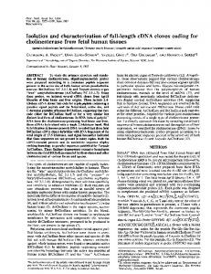
Fig. 1. Southern blot of mouse genomic DNA hybridized with HBT-l. High molecular weight DNA isolated from adult mouse testes was digested with EcoRI (lanes 1 and 3) or HindIII (lanes 2 and 4). Fragments were separated on a 1% agarose gel (10 Ag DNA per lane), transferred to nitrocellulose, and hybridized with 32P-labeled HBT-1 insert. Major fragments of -0.9 and 1.1 kb in length were detected for EcoRI and HindIII, respectively. Several weakly hybridizing bands were also visible, probably representing other mouse genes containing homeo box sequences with homology to the HBT-1 homeo box. Exposure was 4 days.
ACC GAG CAG T** *TI;
180 GAC CAC
AAA CTT CCC AAC **G ACG AAM GG * GA* GAM *G* **C AT* *** **G GGC AAA GG*
HBT-1
HBT-1
AAC CGG AGA ATG AAM TGQ_hAWAA *** *** *** **G **T *** C*C *** *** *** *** **T *** C*C *** *** **A **G **T C*C *** *AC *** *** *** C*C
CAM
EcoRl
GI TC
Fig. 2. Nucleotide sequence of HBT- 1, compared to homeo box regions of the Antp gene of Drosophila and the murine genes m6 (Colberg-Poley et al., 1985a), mS (Colberg-Poley et al., 1985b), and MolO (McGinnis et al., 1984b). The EcoRl site of HBT-1 at nucleotides 54-60 and the MboII site at nucleotides 168-173 are underlined (MboII cuts downstream of its recognition site). Asterisks indicate identical nucleotides to those found in the corresponding positions of HBT-1 (after nucleotide 54); before position 54, the asterisk indicates homology to the Antp sequence.
respect to other mouse homeo box-containing genes, Southern blot analysis was performed with restriction enzymes which had been used in studies on other homeo box-containing genes containing EcoRI sites within the homeo box region (Colberg-Poley et al., 1985a, b). The results of digesting high molecular weight DNA with EcoRI and HindlIl and probing with the HBT-1 insert are shown in Figure 1. Strongly hybridizing bands of 0.9
Mouse testis-specific transcript with homeo box homology
1
2
3
4
6
5
7
8
9
___
___
thr
tyr
thr
arg
* *
* * *
* * *
* * *
HBT-1 Antp m6
arg
lys
arg
gly
arg
gln
*
*
m5 Mol 0
gly ser
arg
* * *
* * *
* * *
* *
thr
ala
14
15
___
11 HBT-1 Antp m6
m5 Mol 0
pro
m5 Mol 0
12
* * * *
27 thr
28 arg
29 arg
arg
*
*
*
*
* *
*
thr
leu
glu
leu
glu
lys
* * *
* *
* *
leu
val
* * *
* * *
* * *
22 phe
23 asn
24 arg
25 tyr
26 leu
*
* * * *
*
*
*
* *
* *
* *
34 ile
35 ala
36 his
* *
* * *
his * * * *
* *
32 ile
*
*
leu
* *
*
HBT-1
* * *
gln
thr
mS Mol 0
20 phe
33
met
30 * * *
pro
38
39
leu
cys
40 leu
* * *
* *
asp
37 thr ala ala ala
asn
leu
46 lys
47 ile
trp
49 phe
50 gln
* * * *
45 val ile ile ile ile
* * * *
* * * *
* * * *
* * * *
* * * *
53 arg
54 met
55
56 trp
*
* * * *
iys8
lys
glu
val
* * * *
met
41 ser thr thr thr thr
42 glu
43 arg
44 gln
* * * *
* * * *
51
52 arg
asn
___
___
19 * * * *
* *
Antp m6 m5 Mol 0
HBT-1 Antp m6
18
glu
17
tyr
31 arg
m6 m5 MolO 0
16
___
HBT-1
HBT-1 Antp
13
___
21 HBT-1 Antp m6
*
10
* *
* * *
57
* * *
48
58
iys *
asn
59
60
asp glu glu
his
glu *
asn
* *
tyr
* * * *
asn
thr
lys
met
arg
ser
ser
ala
ser
ala
pro
ala
gly
pro
pro
ala
gln
thr
his
ser
pro
his
90 his
* * *
* *
lys
leu
pro
asn
thr
gly
lys
his
pro
* *
*
* * *
asn
* gln 70
80
Fig. 3. Conceptual translation of HBT-1 (homeo box region and 3' flanking region), compared with the predicted amino acid sequences of the homeo boxes of MolO (McGinnis et al., 1984c), m6 (Colberg-Poley et al., 1985a), m5 (Colberg-Poley et al., 1985b), and Antp (McGinnis et al., 1984b). Asterisks indicate amino acid homology to the corresponding position in HBT-1 (after amino acid 18); before amino acid 18, the asterisk reflects homology with the Antp amino acids.
and 1.1 kb were observed with EcoRI and HindIf digests, respectively (Figure 1). The patterns were the same with DNAs isolated from different strains and from several mouse tissues, including testis, ovary, liver, and brain (data not shown). The pattern
observed with HBT-l is distinct from the patterns characteristic for other mouse homeo box-containing genes reported to date. Weakly hybridizing fragments were also detected (Figure 1), suggesting that within the mouse genome there are additional genes containing sequences with homeo box homology - an observation consistent with data obtained by others (McGinnis et al., 1984b, c; Colberg-Poley et al., 1985a). Isolation of genomic clones for the HBT-1 gene is in progress; to date, -30 kb of genomic DNA flanking the homeo box region have been isolated in overlapping lambda clones (data not shown).
Sequence of the homeo box-containing testis cDNA clone (pHBT-1) and homology with other homeo box sequences To determine the degree of homology of the HBT-1 homeo box with other homeo box regions, the DNA sequence of the 229-bp EcoRI fragment was obtained by subcloning into M13 vectors
(Messing et al., 1981) and sequencing in both directions by the dideoxy-nucleotide chain termination method of Sanger et -al. (1977), and by sequencing in a pBR322 subclone using the chemical modification method of Maxam and Gilbert (1977). The sequence is given in Figure 2. Comparisons of homology were made to the Antp homeo box, since Antp was used to select HBT-1. In addition, the DNA sequence of HBT-1 was compared with the homeo box regions of the mouse genomic clones of MolO (McGinnis et al., 1984c) and m6 and m5 (Colberg-Poley et al., 1985a, b). M6 and m5 were included because they represent other mouse homeo box-containing genes that have an EcoRI site within the homeo box region and because both have been reported to be transcribed in the testis, among other tissues (Colberg-Poley et al., 1985b). Sequence analysis of the 229-bp insert of HBT-1 revealed that it contains only 126 bp of the 180-bp homeo box sequence and 100 bp of 3' flanking sequence. The EcoRI sites of HBT-1, m6 and m5 are in identical positions but the genes differ in 21 of the 126 (HBT-1 versus m6) and 20 of the 126 (HBT-1 versus m5) nucleotides that make up the homeo box region (83% and 84% homology, respective1231 -
D.J.Wolgemuth et al. 1
2
3
5
4
6
7
8
1
1
3 4
2
2
3
4
5
6
7
8
9
10
9,
. --1.4 kb-
i.>
9
^ A4.4 kb-
I
_
VW.s
I
b
a
Fig. 4. HBT-1 transcripts in mouse testicular RNA. (Panel a) RNA was isolated from mature mouse testes and analyzed by Northern blot analysis using the pHBT-1 cDNA probe. Lanes 1-4 are the ethidium bromide staining patterns of 1, 4 and 8 itg of poly(A)+ RNA and 25 Ag of flowthrough RNA, respectively. Lanes 5-8 represent the autoradiographic patterns of lanes 1-4 after hybridization with 32P-labeled HBT-1 insert. Exposure was 3 days. (Panel B) Poly(A)+ (4-5 Ag), lanes 1 and 3, and total RNA (>25 ytg), lanes 2 and 4, were hybridized with either the nicktranslated homeo box fragment (lanes 1 and 2) or the 3' flanking region fragment (lanes 3 and 4). Exposure was 4 days.
ly). There is only slightly less homology with Antp (80%). The sequence 3' of the homeo box region in HBT-1 is highly diverged from the 3' regions of other mouse homeo box-containing genes described to date (e.g. Figure 2). A conceptual translation of HBT-1 is presented in Figure 3 and is compared to the amino acid sequences of the homeo box regions of Antp, m6, m5 and MoIO. At the amino acid level, the degree of homology of the homeo box region of HBT- 1 to that of the Antp protein is 88%. The homology of the HBT-l homeo box with the homeo box sequences of three other mouse genes, m6 (Colberg-Poley et al., 1985a), m5 (Colberg-Poley et al., 1985b) and MolO (McGinnis et al., 1984c), is 86%, 86% and 74%, respectively.
Transcripts recognized by HBT-J and tissue specificity of expression Total, poly(A) + and poly(A) - (flow-through) RNAs were isolated from mouse testes and analyzed by Northern blot hybridization with HBT-1 at high stringency. HBT-1 recognized testicular transcripts of 1.4 kb in length (Figure 4, panel a). No hybridization was noted in the poly(A)- sample. To confirm that the transcripts being recognised were the result of hybridization to the HBT- 1 gene sequences, and not by sequences of highly homologous homeo box-containing genes (e.g. m6 or m5, Colberg-Poley et al., 1985b), the 229-bp HBT-1 fragment -
1232
ISt
Fig. 5. HBT-1 transcripts in mouse stem cell-containing tissues. Poly(A)+ and flow-through RNAs (A)- were analyzed by Northern blot hybridization for the presence of HBT-1 transcripts. Lanes 1 and 2, 30 Ag (A)- and 4 iLg (A)+ RNAs from mature mouse testes; lanes 3 and 4, 30 ug (A) - and 4 jAg (A)+ RNAs from liver; lanes 5 and 6, 30 yg (A)- and 5 ,ug (A)+ RNAs from mature thymus; lanes 7 and 8, 30 jug (A) - and 4.5 ug (A)+ RNAs from immature thymus (day 11 mice); lanes 9 and 10, 30 yg (A)- and 5 ALg (A)+ RNAs from bone marrow of adult mice. Exposure was 3 days; no additional transcripts were visualized after exposure for 12 days (not shown).
was cut by MboII into homeo box-containing and 3'-flanking segments (Figure 2). These fragments were purified, nick translated, and used for Northern blot analysis. The 1.4-kb testicular transcripts were recognized by both fragments (Figure -
4, panel b). To examine the tissue specificity of distribution of the transcripts recognized by HBT-1, RNAs from several tissues were examined. No hybridization to HBT-1 was seen with poly(A)+ or flow-through RNAs from liver, brain, uterus, or kidney when probed at high stringency (Figure 5 and data not shown), although homeo box-containing sequences have been detected in some of these tissues by other mouse probes (Colberg-Poley et al., 1985b). Since the testis contains the full range of spermatogenic cells, including stem cells, we then considered that expression of HBT-1 might be characteristic of stem cell-containing tissue in general. To this end, poly(A)+ and flow-through RNAs from bone marrow, immature thymus, and mature thymus were examined. Although the question of whether thymus contains true stem cells is unresolved, the thymus clearly contains cells which
Mouse testis-specific transcript with homeo box homology 1
2
3 4 5 6 7 8
1
2
3
4
5
a
6
1 2 3 4 5 6
7
8
-8.0 k b -6.2 kb -4.7 kb
- 1.4 Kb
b kb
L.4 kb-
b
S
I 0S 123456.
HBT- 1
a
78
c-abl
b
Fig. 6. HBT-1 transcripts in RNAs from germ cell-containing tissues. RNAs were isolated from mature testes (panel a, lanes 1 and 2; panel b, lanes 2, 3, 5 and 6), mature ovaries (panel a, lanes 3 and 4), and juvenile ovaries (day 11-18) (panel b, lanes 1 and 4) and analyzed by Northern blot hybridization with pHBT-l (panels a and b) or pabl sub9 as a control (panel b, right portion). The 1.4-kb transcripts are readily apparent in the testis poly(A)+ RNAs (panel a, lanes 1 and 2; 25 ug (A)- and 4 Itg (A)+) but not in the RNA of mature ovaries [panel a, lanes 3 and 4, 25 jig (A)and 4 Ag (A)+, respectively]. Two somatic tissues, liver and brain (panel a, lanes 5-8) also failed to reveal HBT-1 transcripts (lanes 5 and 7 each contain 25 Ag (A)- RNA and lanes 6 and 8 contain 4 ,g of (A)+ RNA. Similarly, RNAs from juvenile ovaries also lacked detectable amounts of HBT-1 transcripts (panel b, lane 1, 5 jig (A)+ RNA). As a control for integrity of RNAs analyzed in these studies, all blots were hybridized with other probes, an example of which is shown in panel b, lanes 4-6, where pabl sub9 easily detected the somatic c-abl transcripts of -8.0 and -6.2 kb in all three RNA samples, as well as the testis-specific c-abl 4.7-kb variant in testicular RNA. Lanes 1 and 4, 5 jig of juvenile ovary (A)+ RNA; lanes 2, 5, 3 and 6, 5 jig and 10 jig (A)+ mature testis RNA. Panel a, exposure 9 days; panel b, 6 days. are very early in the T-cell lineage (Rothenberg and Lugo, 1985). Within the limits of sensitivity of detection of Northern blot analysis, no HBT-1 transcripts were detected (Figure 5), although control hybridizations with EC 1 gene probes (Stacey and Evans, 1983) readily revealed ECI transcripts (G.L.Mutter, A.J.Stacey and D.J.Wolgemuth, unpublished observations). Of additional interest to us was an examination of the other germ cell-containing tissue, namely the ovary. No hybridization was observed to RNAs obtained from ovaries of mature or juvenile animals (Figure 6, panel a, lanes 3 and 4; panel b, lanes 1 and 4). In all Northern blot analyses, the quality of the RNAs examined was verified by the ability to detect discrete transcripts homologous to other genes known by concurrent studies in our laboratory to be expressed in these tissues [e.g. the protooncogene c-abl (Ponzetto and Wolgemuth, 1985; Wolgemuth and Ponzetto, 1985), (Figure 6, panel b); the gene homologous to pEC (Figure 5, data not shown); or f3-actin (Figure 7, panel b)]. HBT-J expression during testicular development To determine whether the expression of HBT- I is correlated with
- 2.3 Kb - 1.65 Kb
i~ ~ qNiSqI .~ ~ ~ ~ ~ . Fig. 7. Expression of HBT-1 during development of the testis. Poly(A)+ and flow-through RNAs from testes of different developmental stages were analyzed by Northern blot hybridization for the presence of HBT-1 transcripts. No transcripts were detected in embryonic or neonatal testes, but transcripts were readily apparent in RNAs from mature testes (panel a). Control hybridizations with ,B-actin are shown in panel b. The RNAs were clearly intact at all developmental stages as demonstrated by the typical pattern of (-actin transcripts for these stages (C.P.Ponzetto and D.J.Wolgemuth, unpublished observations). Lanes 2, 4 and 6 each contain 3.8 Aig of (A)' RNA and lanes 1, 3 and 5 contain 20 jig of flow-through RNAs of mature testis (lanes 1 and 2), day 7-8 neonatal testis (lanes 3 and 4), and day 17-20 embryonic testis (lanes 5 and 6). Lanes 7 and 8 each contain 5 jig of (A)+ RNA from mature testis and day 7-8 neonatal testis, respectively. Probe was the 3' flanking region of the HBT-I homeo box region. Exposures were 8 days (panel a) and 3 days (panel b).
discrete developmental stages of testicular differentiation, or rather, is a property of the male germ cell-containing tissue throughout development of the testis, RNAs were isolated from testes of day 17-20 embryos, day 7-8 neonatal animals, and adult animals. With respect to the types of germ cells present in these tissues, the embryonic testis contains only germinal stem cells (gonocytes), whereas testes from day 7-8 neonates contain primitive Type A spermatogonia as well as the more advanced Type A and Type B spermatogonia (Nebel et al., 1961; Bellve et al., 1977). Before day 9 of neonatal development, none of the germ cells have entered meiosis. From about day 9-10 onward, cells enter pre-meiotic DNA synthesis and meiotic prophase ensues. Testes at all stages examined in the present study contain the typical somatic cellular components, including Leydig and Sertoli cells. As depicted in Figure 7a, no HBT-1 transcripts were detected in embryonic or neonatal testes, whereas transcripts were readily apparent in RNAs isolated from mature testes. The control hybridization for this Northern blot was fl-actin, the results of which are shown in Figure 7b. 1233
D.J.Wolgemuth et al.
Discussion Sequence analysis of HBT- 1 revealed that the homeo box contained in this clone is similar to, but not identical to, the homeo box of the m6 and m5 genes described by Colberg-Poley et al. (1985a, b). Both m6 and m5 as well as HBT-l homeo boxes are quite homologous to the Antp homeo box at both the DNA and protein level (Figures 2 and 3). At the protein level, HBT-1 is actually more homologous to the Antp homeo box than to the m6 or m5 homeo boxes. An analogous observation has been noted for two Xenopus genes containing homeo boxes: at the amino acid level, the Xenopus gene MM3 is more homologous to the Drosophila Antp than to the Xenopus AC 1 gene (Muller et al., 1984). In contrast to the high level of conservation of the homeo boxes, the 3' flanking sequences of HBT- 1 and m6 and m5 are completely divergent (Figure 2). This is of particular interest since it has been reported that m6 and m5 also recognize testicular transcripts (Colberg-Poley et al., 1985b). However, both m5 and m6 recognize multiple testicular transcripts and also detect transcripts in several other adult tissues, including ovary and kidney (Colberg-Poley et al., 1985a, b). In contrast, HBT-1 recognized transcripts only in the 1.4-kb size range and only in testis of all the adult tissues examined. To our knowledge, none of the other homeo box-containing genes isolated to date have the tissue-specificity of expression, as assayed by high-stringency Northern blot analysis, exhibited by HBT-1 in the testis. The testis contains a wide variety of cell types, from hormoneproducing and hormone-responsive supporting cells of somatic origin to virtually a complete germ cell lineage, from spermatogenic stem cells through to mature spermatozoa (reviewed in Bellve, 1979). It was the presence of the stem cell population in the testis that prompted our investigation as to whether other tissues containing stem cells - those of the hemopoietic lineage - would also contain HBT-1 transcripts. The fact that none were observed lends support to our postulate that the HBT-l gene product is involved primarily in male germ cell differentiation. However, the question of HBT- 1 expression in the female germ cell-containing tissue was also addressed. RNAs were isolated from adult ovaries, which contain oocytes in stages of development from primordial follicles through to Graafian follicles (Peters, 1969). Since homeo box-containing transcripts have been observed in growing X. laevis oocytes (Muller et al., 1984), RNA was also isolated from ovaries of juvenile mice. Juvenile ovaries contain a higher proportion of growing oocytes, relative to somatic tissues, than is found in adult ovaries (Peters, 1969). No transcripts were detected in any samples. We conclude that HBT-l transcripts are not abundant in the somatic components of the ovary, because the characteristic somatic c-abl transcripts were readily detected in these samples (Figure 6, panel b). Germ cells represent >90% of the cells in the adult testis (Meinstrich et al., 1973) but only at most several thousand cells in the adult ovary (Peters, 1969). Considering that HBT-1 transcripts are detected in, for example, 5 Ag of poly(A)+ testicular RNA with an exposure time of 6 h, but cannot be detected in comparable amounts of ovary RNAs after one week or longer exposure, if there are HBT-1 transcripts in the female germ cells, they are probably not very abundant. However, certain stages of oocyte development are very rare or missing; therefore if HBT-l were expressed at only one stage of oogenesis, transcripts might have been missed in this analysis. Definitive resolution of the presence -
or absence
of HBT- 1 transcripts in the female germ line will await
the results of in situ hybridization of sections of ovarian tissues from fetal through adult stages of mouse development and Nor1234
them blot analysis of RNA prepared from isolated oocytes (in progress). The tissue-specificity of expression of HBT-1 in the testis raised the intriguing possibility that HBT-l expression might be critical in particular stages of germ cell differentiation, or alternatively, that expression of HBT-l might actually be a property of the testis as a tissue, i.e., that it could function as a 'testis-determining' gene and might therefore be expressed early in testicular development. Our results to date strongly support the former hypothesis. HBT-l transcripts have been detected only in RNAs isolated from mature testes and none in embryonic or day 7-8 neonatal testes. As noted previously, embryonic and neonatal testes contain stem cells in various stages of spermatogonial differentiation as well as the various non-germinal cells of the mature testis, including Sertoli cells and Leydig cells. No meiotic cells are present, however. The presence of HBT-l transcripts thus appears to be correlated to the entry of the male germ cells into the committed meiotic state. Additional support for a role of HBT-l during meiotic differentiation may be inferred from observations indicating first, that HBT-1 transcripts can be detected in sterility mutants in which meiosis progresses as far as early spermatids but not in germ cell deficient mutant strains; and second, that enriched populations of pachytene spermatocytes indeed contain HBT-l transcripts (D.J.Wolgemuth, C.Viviano, E.Gizang-Ginsberg and E.Engelmyer, unpublished observations). It will be important to determine the precise temporal transcriptional and translational expression of HBT-1 throughout meiosis. In addition, it will be of interest to compare the expression of HBT-l with the pattern of expression of the m6 and m5 genes, which contain homeo boxes highly homologous to HBT- 1 and which are also expressed in the testis.
Materials and methods Sources of tissues Swiss Webster mice (Camm, Inc.) were used as the source of tissues in all aspects of this study. Adult males refers to mice aged 60 days or older. Adult females refers to animals aged 30 days or older; juvenile females were 11 - 18 days of age. Embryonic testes were dissected from fetuses of pregnant females at day 17 to day 20 of gestation (the day of vaginal plug was designated day 0 of gestation). Neonatal testes were recovered from animals sacrificed at day 7 or day *8 of post-natal development. Testes, ovaries, and other tissues were removed from animals sacrificed by cervical dislocation. Tissues were either used immediately for RNA isolation or were frozen in liquid nitrogen for subsequent use as sources of RNA or DNA. Sources of probes The following cloned probes were used in these studies. Antp - p903G, a 600-bp subclone derived from a 3' cDNA of the Drosophila Antennapedia gene (McGinnis et al., 1984b); Jiishi tarazu - pFS2, a subclone of the 3' region of the Drosophila fushi tarazu gene (Kuriowa et al., 1984); engrailed - pFS7036, a 920-bp subclone from the Drosophila engrailed gene (Fjose et al., 1985); pMolO, a 1.4-kb mouse genomic homeo box-containing clone (McGinnis et al., 1984b). All the homeo box-containing clones were generously provided by W.Gehring. Control probes for ascertaining efficiency of hybridization and integrity of RNAs were (i) psub 9, a permuted v-abl clone which hybridizes to c-abl transcripts (Goff et al., 1980, note Ponzetto and Wolgemuth, 1985; and Wolgemuth and Ponzetto, 1985); (ii) pEC 1, a cDNA clone isolated from a mouse teratocarcinoma cDNA library (Stacey and Evans, 1985; the gift of A.Stacey); and (iii) pAl, a chicken ,B-actin cDNA clone (Cleveland et al., 1983; the gift of D.Cleveland). RNA isolation and Northern blot analysis RNA was isolated using the LiCI precipitation method described by Cathala et al. (1983). Poly(A)+ RNA was selected by oligo-dT cellulose chromatography according to Aviv and Leder (1972). RNA samples were electrophoresed on denaturing 1 % agarose/2.2 M formaldehyde gels, blotted onto nitrocellulose for 8-12 h according to Maniatis et al. (1982), and baked under vacuum for 3 h at 80°C. Prehybridization used sonicated salmon sperm DNA (100 Ag/ml); hybridizations in the presence of dextran sulfate were performed essentially as
Mouse testis-specific transcript with homeo box homology described by Wahl et al. (1979). Probes for hybridization were labeled with [32p]dXTPs by nick translation (Weinstock et al., 1978). Following hybridization, filters were sequentially washed for high-stringency hybridization: 20 min each in 2 x SSC, 0.1% SDS at 25°C and at 65°C; 2 x in 1 x SSC, 0.1% SDS at 65°C; and 2 x 0.1 x SSC, 0.1 % SDS at 65°C. Filters were air dried and exposed to autoradiographic film at -70°C for 6 h to 21 days. cDNA library preparation and screening RNA was isolated from total testis using the LiCI precipitation method, poly(A)selected as described above, and used to prepare a cDNA library in lambda gtlO according to procedures of Huynh et al. (1984). Oligo-dT was used to prime the first strand synthesis using reverse transcriptase. Second strand synthesis was obtained with the Klenow fragment of DNA PolI, using natural hairpin primers. The double stranded cDNAs were trimmed with SI nuclease, methylated with DNA methylase, and EcoRI linkers were added. cDNAs of greater than - 300 bp in length were cloned into the EcoRI site of the vector. Titration on E. coli Hfl strain revealed a library of -4 x 10⁵ independent clones, which was then amplified. Recombinant phages were plated, grown to near confluency, and plaques were lifted as described by Maniatis et al. (1982). Duplicate filters were prepared and hybridized to nick translated inserts of the Antp and MolO homeo box genes. Southern blot analysis High molecular weight DNA was isolated from testis, ovary, liver and brain of mature mice. The methods used were essentially those described by Wigler et al. (1979) with minor modifications described by Ponzetto-Zimmerman and Wolgemuth (1984). DNA digestions were performed with various restriction enzymes purchased from commercial sources and performed according to the manufacturers' recommended conditions. Digested DNAs were run on 1 % agarose gels and transferred to nitrocellulose filters according to Maniatis et al. (1982). High-stringency Southern blot analysis was performed as described by McGinnis et al. (1984b), using [32P]HBT-1 insert, nick translated according to Weinstock et al. (1978). DNA sequencing The 229-bp HBT-1 insert was subcloned in both orientations into the EcoRI site of M13 derivatives (M 13mpl 1, Messing et al., 1981) and used as template for DNA sequencing by the enzymatic dideoxynucleotide chain termination sequencing method of Sanger et al. (1977). The majority of the sequence was determined by reading both strands. In addition, regions of ambiguous sequence were confirmed by sequencing both strands using the chemical modification method of Maxam and Gilbert (1977).
Acknowledgements We wish to thank Dr W.J.Gehring for generously providing plasmids containing the Drosophila homeo box probes and the mouse probe MolO, and for helpful discussions throughout the course of this study, Dr Eric Schon for help in setting up dideoxynucleotide DNA sequencing protocols, Dr M.-C.Nguyen-Huu and M.Rubin for help in preparing the mouse testis cDNA library, George Grills for photographic assistance, and Drs J.L.Roberts and R.D.Palmiter for critical review of this manuscript. This work was supported in part by grants NIH S07RR 0535-24, NIH RO1 18122, and a Research Scientist Career Development Award from the I.T.Hirschl Trust to D.J.W.
Huynh,T.V., Young,R.A. and Davis,R.W. (1984) In Glover,D. (ed.), DNA Cloning: A Practical Approach, IRL Press, Oxford, UK. Kuroiwa,A., Hafen,E. and Gehring,W.J. (1984) Cell, 37, 825-831. Laughon,A. and Scott,M.P. (1984) Nature, 310, 25-31. Levine,M., Rubin,G.M. and Tjian,R. (1984) Cell, 38, 667-673. Lewis,E.B. (1978) Nature, 276, 565-570. Maniatis,T., Fritsch,E.F. and Sambrook,J. (1982) Molecular Cloning, A Laboratory Manual. Cold Spring Laboratory, Cold Spring Harbor, NY. Maxam,A.M. and Gilbert,W. (1977) Proc. Natl. Acad. Sci. USA, 74, 560-654. McGinnis,W., Levine,M., Hafen,E., Kuroiwa,A. and Gehring,W.J. (1984a) Nature, 308, 428-433. McGinnis,W., Garber,R.L., Wirz,J., Kuroiwa,A. and Gehring,W.J. (1984b) Cell, 37, 403-408. McGinnis,W., Hart,C.P., Gehring,W.J. and Ruddle,F.J. (1984c) Cell, 38, 675-680. Meistrich,M.L., Bruce,W.R. and Clermont,Y. (1973) Exp. Cell Res., 79, 213-227. Messing,J., Crea,R. and Seeburg,P.H. (1981) Nucleic Acids Res., 9, 309-321. Mlodzik,M., Fjose,A. and Gehring,W.J. (1985) EMBO J., 4, 2961-2969. Muller,M.M., Carrasco,A.E. and DeRobertis,E.M. (1984) Cell, 39, 157-162. Nebel,B., Amarose,A. and Hackett,E. (1961) Science, 13, 832-833. Peters,H. (1969) Acta Endocr., 62, 98-116. Poole,S.J., Kauver,L.M., Drees,B. and Kornberg,T. (1985) Cell, 40, 37-43. Ponzetto,C.P. and Wolgemuth,D.J. (1985) Mol. Cell. Biol., 5, 1791-1794. Ponzetto-Zimmerman,C. and Wolgemuth,D.J. (1984) Nucleic Acids Res., 12, 2807-2822. Rothenberg,E. and Lugo,J.P. (1985) Dev. Biol., 1121, 1-17. Sanger,F., Nicklen,S. and Coulson,A.R. (1977) Proc. Natl. Acad. Sci. USA, 74, 5463-5467. Scott,M.P. and Weiner,A.J. (1984) Proc. Natl. Acad. Sci. USA, 81, 4115-4119. Shepherd,J.C.W., McGinnis,W., Carrasco,A.E., DeRobertis,E.M. and Gehring.,J. (1984) Nature, 310, 70-71. Southern,E.M. (1975) J. Mol. Biol., 98, 503-517. Stacey,A.J. and Evans,M.J. (1984) EMBO J., 3, 2279-2285. Wahl,G.M., Stern,M. and Stark,G.R. (1979) Proc. Natl. Acad. Sci. USA, 76, 3683-3687. Weinstock,R., Sweet,R., Weiss,M., Cedar,H. and Axel,R. (1978) Proc. Natl. Acad. Sci. USA, 75, 1299-1303. Wigler,M., Sweet,R., Sim,G.K., Wold,B., Pellicer,A., Lacy,E., Maniatis,T., Silverstein,S. and Axel,R. (1979) Cell, 16, 777-785. Wolgemuth,D.J. and Ponzetto,C.P. (1985) In Slavkin,H. (ed.), New Technologies and New Discoveries. Alan R.Liss, Inc., New York, NY, in press. Received on 25 February 1986; accepted on 20 March 1986.
References Aviv,H. and Leder,P. (1972) Proc. Natl. Acad. Sci. USA, 69, 1408-1412. Bellve,A.R. (1979) In Finn,C.A. (ed.), Oxford Reviews of Reproductive Biology, Oxford University Press, London, Vol. 1, pp. 159-261. Bellve,A.R., Cavicchia,J.-C., Millette,C.F., O'Brien,D.A., Bhatnagar,Y.M. and Dym,M. (1977) J. Cell. Biol., 74, 68-85. Carrasco,A.E., McGinnis,W., Gehring,W.J. and DeRobertis,E.M. (1984) Cell, 37, 409-414. Cathala,G., Savouret,J.-F., Mendez,B., West,B.L., Karin,M., Martial,J.A. and Baxter,J.D. (1983) DNA, 2, 329-335. Cleveland,D.W., Lopata,M.A., MacDonald,R.J., Rutter,W.J. and Kirschner,M.W. (1983) Cell, 20, 95-105. Colberg-Poley,A.M., Voss,S.D., Chowdhury,K. and Gruss,P. (1985a) Nature, 314, 713-718. Colberg-Poley,A.M., Voss,S.D., Chowdhury,K., Stewart,C.L., Wagner,E.F. and Gruss,P. (1985b) Cell, 43,39-45. Fjose,A., McGinnis,W. and Gehring,W.J. (1985) Nature, 313, 284-289. Gehring,W. (1985) Cell, 40, 3-5. Gizang-Ginsberg,E. and Wolgemuth,D.J. (1985) Dev. Biol., 111, 293-305. Goff,S.P., Gilboo,E., Witte,O.N. and Baltimore,D. (1980) Cell, 22, 777-785. Hafen,E., Kuroiwa,A. and Gehring, W. (1984) Cell, 37, 833-841.
1235