Journal of Information Technology & Software Engineering is an international, peer-reviewed journal publishing an overview of IT research and algorithmic processes that create, describe and transform information to formulate suitable abstractions to model complex systems. Journal of Information Technology & Software Engineering (JITSE), a broad-based journal was founded on two key tenets: To publish the most exciting researches with respect to the subjects of Information Technology & Software Engineering. Secondly, to provide a rapid turnaround time possible for reviewing and publishing, and to disseminate the articles freely for research, teaching and reference purposes.
ISSN: 2165-7866
Journal of Information Technology & Software Engineering
http://omicsgroup.org/journals/jitsehome.php
K R. Laviers
M Bassiouni
Air Force Institute of Technology, USA
University of Central Florida, USA
R Mulaveesala
Ashutosh Gupta
PDPM-Indian Institute of Information, India
M. J. P. Rohilkhand University, India
Marco Vieira
University of Coimbra Portugal
T Hau Lee
Multimedia University Malaysia
K I Bin Ghauth
University Cyberjaya Malaysia
F Fang Chua
Multimedia University Cyberjaya Malaysia
S Senthilkumar University Sains Malaysia, Malaysia
Tongning Wu
Telecommunication Metrology Centre Chaina
Sima Ajami
Isfahan University of Medical Sciences, Iran
Journal of Information Technology & Software Engineering Open Access is using online manuscript submission. Submit your manuscript at http://www.omicsonline.org/submission/
OMICS Publishing Group
5716 Corsa Ave., Suite 110, Westlake, Los Angeles, CA 91362-7354, USA, Phone: +1- 650-268-9744, Fax: +1-650-618-1414, Toll free: +1-800-216-6499
Information Technology & Software Engineering
Farronato et al. J Inform Tech Soft Engg 2012, 2:1 http://dx.doi.org/10.4172/2165-7866.1000107
Research Article
Open Access
Direct 3D Cephalometric Analysis Performed on CBCT Farronato G*, Perillo L, Bellincioni F, Briguglio F, Farronato D and Dominici AD Università degli Studi di Milano – Dipartimento di Scienze Chirurgiche, Ricostruttive e Diagnostiche – IRCCS Ospedale Maggiore Policlinico, Mangiagalli e Regina Elena, Italy
Abstract Cone Beam Computed Tomography produces distortion-free and accurate images of the craniofacial anatomy, and can be considered as a useful instrument to perform a correct diagnosis and treatment using a cephalometric analysis. Now a days the 3D cephalometric analysis are made by adapting on 3D what was born on 2D, so it can be necessary to find a new technique that was born directly on 3D. Sixty-five Ricketts’ first skeletal classes were chosen in an archive of 700 CBCT acquisitions. 3 easily repeatable reference planes were found on each case and then 15 points were selected on the skull, for a total of 18 cephalometric points. Each point of these 65 patients creates a cloud of points that could represent the range of normality in the 3D space. The coordinates of each analyzed point presented a standard deviation between 0, 00 and 5, 64. This range of values may be considered representative for the first skeletal class. Authors agree that further studies are needed but the Direct 3D cephalometric analysis may be considered a new easy, precise and rapid way to reach a correct diagnosis and treatment with a cephalometric analysis born directly on 3D.
Keywords: 3D cephalometric analysis; 3D cephalometry; Cone
quality of CBCT compares favorably to that of multi-slice CT, as it has minimum image noise and a maximum signal-to-noise ratio [10-20].
Introduction
Sixty-five Rickett’s first skeletal classes patients were selected for the study. The cephalometric analysis was performed by three operators repeating the measurements twice (15 days apart) after a calibration meeting [21,22].
beam CT; First class values
A correct diagnosis is the primary need for an orthodontic treatment. Cephalometrics appears to be a useful diagnostic tool for identifying skeletal disharmony, malocclusion, and normal occlusion for orthodontics and oro-maxillo-facial surgery. To image craniofacial anatomy, low-dose cone beam computed tomography (CBCT) uses a conical beam of X-rays instead of the conventional fan beam of multislice computed tomography (CT). Thus, CBCT results in reduced adsorption of radiation compared with traditional CT and provides a greater quantity of information than two-dimensional (2D) exams [13]. The effective radiation dose to which a patient is exposed is far lower with a CBCT examination than with a multi-slice CT examination. Specifically, the effective dose from an I-Cat cone beam CT machine is 20 times lower than that from a Toshiba 64-slice machine [4-6]. In addition, the dose distribution to the various organs examined differs between CBCT and multi-slice CT; the dose absorbed by organs such as the thyroid and salivary glands is 20-40 times lower with CBCT [7,8]. Three-dimensional (3D) diagnostic imaging using CBCT produces distortion-free and accurate images of the craniofacial anatomy, without the magnification and superimposition problems of 2D imaging. This allows unobstructed views of otherwise hidden structures [9]. The present study evaluated the application of a new, simplified cephalometric protocol based on a completely new concept. It uses a personal computer for the analysis of the enormous amount of information available from low-dose CBCT. An important advantage of this technique is that it allows real measurements. Moreover, it is possible to analyze volumes in order to evaluate disproportions in a more representative way than with segments. This new cephalometric method has the potential to reduce time, costs, and human error. It is the result of a selection of 65 patients from an archive of 700.
Materials and Methods A low-dose CBCT machine was used to acquire the radiographic images in this study of the new 3D cephalometry method. The image J Inform Tech Soft Engg ISSN: 2165-7866 JITSE, an open access journal
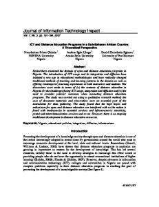
The position of the maxilla and mandible in 3D space was determined using low-dose CBCT by assigning three reference planes to obtain the (x, y, z) position of each point of the skull relative to point S with coordinates (0, 0, 0), which was automatically determined by the computer as the intersection of the reference planes. 18 easily identified and repeatable cephalometric points have been assigned on the skull [23,24]. To obtain a reference system that is repeatable and is not influenced by changes in the position of cranial points due to growth, the best solution in terms of simplicity and precision is to use three reference planes, defined using three points: the Sella (S), Nasion (N), and Basion (Ba). The three planes are the mid-sagittal plane passing through S-NBa, the mid-axial plane passing through S-N and perpendicular to the mid-sagittal plane, and the coronal plane passing through S and perpendicular to the other two planes. The intersection of these three planes defines the reference point S (0, 0, 0) (Figure 1). These planes are repeatable for each patient and they can be matched on the same planes of other patients. After determining the reference planes, each case has been scaled to perfectly match the points S and N of the other cases. This scaling has been performed by
*Corresponding author: Giampietro Farronato, dipartimento di scienze chirurgiche, ricostruttive e diagnostiche Clinica odontoiatrica - via Commenda 10 20122-Milan, Italy, E-mail:
[email protected] Received December 07, 2011; Accepted January 19, 2012; Published January 21, 2012 Citation: Farronato G, Perillo L, Bellincioni F, Briguglio F, Farronato D, et al. (2012) Direct 3D Cephalometric Analysis Performed on CBCT. J Inform Tech Soft Engg 2:107. doi:10.4172/2165-7866.1000107 Copyright: © 2012 Farronato G, et al. This is an open-access article distributed under the terms of the Creative Commons Attribution License, which permits unrestricted use, distribution, and reproduction in any medium, provided the original author and source are credited.
Volume 2 • Issue 1 • 1000107
Citation: Farronato G, Perillo L, Bellincioni F, Briguglio F, Farronato D, et al. (2012) Direct 3D Cephalometric Analysis Performed on CBCT. J Inform Tech Soft Engg 2:107. doi:10.4172/2165-7866.1000107 Page 2 of 3
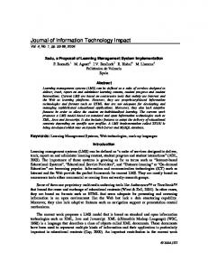
evaluating the median value of SN segment that was 62 mm with a standard deviation of 5 mm. The SN length to use as referral value has been found by subtracting the half standard deviation to the median value: 62 mm- 5 mm/2 = 59,5 mm. The remaining 15 cephalometric points identified in the program for the 3D analysis are the Right gonion (Go r), Left gonion (Go l), Menton (Me), Left supraorbital (Sor l), Right supraorbital (Sor r), Right condylar point (Cd r), and Left condylar point (Cd l), Upper incisor (Ui), Lower incisor (Li), point (A), point (B), the Right jugal point (Mx r), the Left jugal point (Mx l), the Anterior nasal spine (ANS) and the Posterior nasal spine (PNS) (Figure 2-4). Each point has 3 coordinates in space (x,y,z). By comparing the same coordinates of the same points in all the 65 first skeletal class cases, the computer calculated a cloud of points (Figure 2-4) that could represents the range of normality. Comparing images with the subsequent acquisitions, the computer automatically produces a vector that indicates the direction and growth module of the jaws in three dimensions. This allows a clear graphical
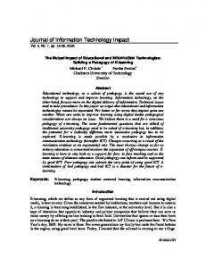
Figure 3: Another representation of the cloud of points on the skull. It can be noticed the perfect superimposition of the cloud of points of point S and point N.
Figure 4: ¾ view of the skull. Figure 1: The system of reference points obtained from the points S, N and Ba and based on three perpendicular planes.
representation that provides simple, intuitive information about the forces that a clinician should consider to correct pathological growth. The Direct 3D cephalometric analysis allows the immediate analysis of the characteristics of clinical cases, visually and simply.
Results This study shows cephalometric measurements directly performed on 3D, in 65 clinical cases. All the coordinates (x,y,z) of each point were compared and was performed the standard deviation, the minimum value, the maximum value, the average, the first and the third quartile and the percentile 0,975 and 0,025 (Table 1 as supplementary file). The coordinates of each analyzed point presented a standard deviation between 0,00 and 5,64.
Discussion
Figure 2: The 18 points with theit cloud of points. They represent the range of values in the first skeletal class.
J Inform Tech Soft Engg ISSN: 2165-7866 JITSE, an open access journal
This three-dimensional cephalometric method allows the analysis of anomalies in three spatial planes (sagittal, frontal, and axial), directly and visually, without the need to interpolate different measurements obtained in each of the three spatial planes. The analysis is mainly based on the cloud of points created by the coordinates of each point. It uses a 3D reconstruction of the patient’s skull, starting from the DICOM-
Volume 2 • Issue 1 • 1000107
Citation: Farronato G, Perillo L, Bellincioni F, Briguglio F, Farronato D, et al. (2012) Direct 3D Cephalometric Analysis Performed on CBCT. J Inform Tech Soft Engg 2:107. doi:10.4172/2165-7866.1000107 Page 3 of 3
3-compatible files derived from low-dosage volumetric CBCT of the patient. Using specific software (Mimics®, Materialise and Catia®, Dassault) dedicated to medical research and supported by high-speed computers, the patient’s skull is reconstructed in three dimensions, and the orientation of the skull is determined using three well-defined perpendicular planes and a point (0, 0, 0), which allows the (x, y, z) coordinates of the patient’s skull to be mapped. For the data collected from the 65 patients analyzed here, the standard deviations of the clouds of points are included in a compact range of values (Table 1). The additional information provided by a direct 3D cephalometry helps to solve postural and growth problems more easily and faster than with 2D cephalometry. The information is presented in a highly intuitive graphical way that facilitates orthodontic diagnosis. Consequently, treatment is quick and efficient. The small number of points to be selected and the automatic measurements made by the computer drastically reduce human error, making the diagnosis much more reliable and repeatable. In addition, the inter- and intra-individual variation is decreased. The data presented here show that the Direct 3D cephalometry method is reliable and repeatable and provides clinicians with more information than 2D methods, in a simple and intuitive graphical representation.
Conclusions Three-dimensional imaging provides information and images of craniofacial structures free from perspective distortion, with no magnification or superimposition associated with 2D images. The Direct 3D cephalometric analysis is easier to interpret than 2D cephalometric analysis (interpolation of cephalometric values on different projections) because it allows movement from a purely mathematical interpretation (evaluation of angles and linear measurements) to a graphical interpretation, with verification of the results using mathematical values (volumetric). Another aid to the clinician is the repeatability and reproducibility of this method, which reduces human error in cephalometric analysis. Although further studies are needed, the use of the cloud of points has given encouraging results. We believe that this method saves time and increases precision, offering a valuable aid to orthodontic diagnosis. This study expands the landscape of diagnostic methods, allowing for more extensive studies to confirm the clinical effectiveness and validation of the Direct 3D cephalometric analysis. Acknowledgments The authors give a special thanks to Dr. Sandro De Nardi for the technical support. Also thanks to Mr. Angelo Magni and Ing. Fabio Sonzogni for statistical calculations and software support. The authors certify that there is no conflict of interest with any financial organization regarding the material discussed in this article.
References 1. Holberg C, Steinhäuser S, Geis P, Rudzki-Janson I (2005) Cone-beam computed tomography in orthodontics: benefits and limitations. J Orofac Orthop 66: 434-444. 2. Kau CH, Richmond S, Palomo JM, Hans MG (2005) Three-dimensional cone beam computerized tomography in orthodontics. J Orthod 32: 282-293. 3. Ludlow JB, Laster WS, See M, Bailey LJ, Hershey HG (2007) Accuracy of measurements of mandibular anatomy in cone-beam computed tomography images. Oral Surg Oral Med Oral Pathol Oral Radiol Endod 103: 534-542.
J Inform Tech Soft Engg ISSN: 2165-7866 JITSE, an open access journal
4. Frederiksen NL (2004) Specialized radiographic techniques. In: White SC, Pharoah MJ. Oral Radiology: Principles and Interpretation. (5thedn), St. Louis, Mosby 245-264. 5. Compagnone G, Angelini P, Boni S (2003) Valutazione della dose efficace collettiva alla popolazione emiliano-romagnola per esposizioni a scopo medico. Associazione Italiana di Fisica Medica, Atti del III Congresso Nazionale AIFM. 6. Compagnone G, Angelini P, Pagan L (2006) Monitoring of the medical radiological exposures of the population of the Emilia-Romagna Region. Radiol Med 111: 469-480. 7. Strocchi S, Colli V, Novario R (2007) Dedicated dental volumetric and total body multislice computed tomography: a comparison of image quality and radiation dose. Medical Imaging 2007: Physics of Medical Imaging. Edited by Hsieh, Jiang, Flynn, Michael J, Proceedings of the SPIE 6510: 65102I. 8. (2006) International Commission on radiological protection. 9. Jacobson A, Jacobson RL (2006) Radiographic cephalometry: From basic to 3-D Imaging. Quintessence Publishing. 10. Stratemann SA, Huang JC, Maki K, Miller AJ, Hatcher DC (2008) Comparison of cone-beam computed tomography imaging with physical measures. Dentomaxillofac Radiol 37: 80-93. 11. Suomalainen A, Vehmas T, Kortesniemi M, Robinson S, Peltola J (2008) Accuracy of linear measurements using dental cone beam and conventional multislice computed tomography. Dentomaxillofac Radiol 37: 10-17. 12. Periago DR, Scarfe WC, Moshiri M, Scheetz, JP, Silveira AM, et al. (2008) Linear accuracy and reliability of cone beam CT derived 3-dimensional images constructed using an orthodontic volumetric rendering program. Angle Orthod 78: 387-395. 13. Pinsky HM, Dyda S, Pinsky RW, Misch KA, Sarment DP (2006) Accuracy of three-dimensional measurements using cone-beam CT. Dentomaxillofac Radiol 35: 410-416. 14. Moshiri M, Scarfe WC, Hilgers ML. Scheetz JP, Silveira AM, et al. (2007) Accuracy of linear measurements from imaging plate and lateral cephalometric images derived from cone-beam computed tomography. Am J Orthod Dentofacial Orthop 132: 550-560. 15. Cevidanes LH, Styner MA, Proffit WR (2006) Image analysis and superimposition of 3-dimensional cone-beam computed tomography models. Am J Orthod Dentofacial Orthop 129: 611-618. 16. Nakajima A, Sameshima GT, Arai Y, Homme Y, Shimizu N, et al. (2005) Twoand three-dimensional orthodontic imaging using limited cone beam-computed tomography. Angle Orthod 75: 895-903. 17. Kamiishi H, Miyasato Y, Kosaka M (2007) Development of the 3D-cephalogram: a technical note. J Craniomaxillofac Surg 35: 258-260. 18. Olszewski R, Cosnard G, Macq B, Mahy P, Reychler H (2006) 3D CT-based cephalometric analysis: 3D cephalometric theoretical concept and software. Neuroradiology 48: 853-862. 19. Maeda M, Katsumata A, Ariji Y, Muramatsu A, Yoshida K, et al. (2006) 3D-CT evaluation of facial asymmetry, in patients with maxillofacial deformities. Oral Surg Oral Med Oral Pathol Oral Radiol Endod 102: 382-390. 20. Hayashi I (2003) Morphological relationship between the cranial base and dentofacial complex obtained by reconstructive computer tomography images. Eur J Orthod 25: 385-391. 21. Kumar V, Ludlow JB, Mol A, Cevidanes L (2007) Comparison of conventional and cone beam CT synthesized cephalograms. Dentomaxillofac Radiol 36: 263-269. 22. Santoni F, Bruno E, Farronato GP (1987) Verifica inter- ed intra-operatores nella pratica cefalometrica. Odontoiatria Oggi 4: 99-103. 23. Farronato G, Garagiola U, Dominici A, Periti G, de Nardi S, et al. (2010) “Tenpoint” 3D cephalometric analysis using low-dosage cone beam computed tomography. Prog Orthod 11: 2-12. 24. Farronato G, Farronato D, Toma L, Bellincioni F (2010) A synthetic threedimensional craniofacial analysis. J Clin Orthod 44: 673-678.
Volume 2 • Issue 1 • 1000107