© 2000 Nature America Inc. • http://structbio.nature.com
letters Crystal structure of a naturally occurring parallel right-handed coiled coil tetramer
a
Jörg Stetefeld, Margrit Jenny, Therese Schulthess, Ruth Landwehr, Jürgen Engel and Richard A. Kammerer
© 2000 Nature America Inc. • http://structbio.nature.com
Department of Biophysical Chemistry, Biozentrum, University of Basel, Klingelbergstrasse 70, CH-4056 Basel, Switzerland.
The crystal structure of a polypeptide chain fragment from the surface layer protein tetrabrachion from Staphylothermus marinus has been determined at 1.8 Å resolution. As proposed on the basis of the presence of 11-residue repeats, the polypeptide chain fragment forms a parallel right-handed coiled coil structure. Complementary hydrophobic interactions and complex networks of surface salt bridges result in an extremely thermostable tetrameric structure with remarkable properties. In marked contrast to left-handed coiled coil tetramers, the right-handed coiled coil reveals large hydrophobic cavities that are filled with water molecules. As a consequence, the packing of the hydrophobic core differs markedly from that of a right-handed parallel coiled coil tetramer that was designed on the basis of left-handed coiled coil structures. The α-helical coiled coil is the most frequently encountered subunit oligomerization motif in proteins1–4. Typically, coiled coils consist of two to five right-handed amphipathic α-helices that are intertwined into a left-handed superhelix. The sequences of coiled coils are characterized by a heptad repeat of seven residues denoted a–g with a 3,4-hydrophobic repeat of mostly apolar amino acids at the a and d positions and polar residues generally at the other positions5,6. As a consequence, the reduced periodicity of 3.5 residues per turn compared to that of ∼3.6 residues in an undistorted α-helix results in a left-handed twist of α-helices7,8. The next periodicity >3.6 that allows residues to assume quasi-equivalent positions after a small number of turns is 3.67 (in this case, three turns or 11 residues). It has been demonstrated that application of this principle to the de novo designed RH4 peptide comprising 11-residue repeats results in winding of the supercoil in a right-handed fashion9. Recently, an assembly of two right-handed antiparallel coiled coils has been found in a naturally occurring protein, the Mnt repressor tetramerization domain of the Salmonella bacteriophage P22 (ref. 10). However, right-handed coiled coils containing 11-residue repeats have not as yet been observed in nature, although this motif has been identified in several proteins4,11,12. Based on the presence of 11-residue repeats, a right-handed coiled coil structure has been proposed for the C-terminus of the surface layer glycoprotein tetrabrachion from the hyperthermophilic archaebacterium Staphylothermus marinus11,13. The 1.7 MDa tetrabrachion protein complex consists of four identical subunits that form an α-helical coiled coil stalk 70 nm long that is anchored to the cell membrane at its C-terminal end and branches into four arms each 24 nm long at its N-terminus. The arms form a canopy-like meshwork by end-to-end contacts that encloses a ‘quasi-periplasmic space’. Two molecules of the 150 kDa subtilisin-like protease STABLE are noncovalently bound near the center of the stalk. The protease is probably 772
b
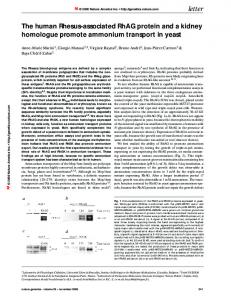
Fig. 1 Sequence and crystal structure of the right-handed parallel RHCC tetramer. Helix backbones are shown in different colors and side chains are colored according to atom type. a, 11-residue repeat positions, indicated by lowercase letters, are assigned according to the model proposed by Lupas4 and the results of the current work. Hydrophobic core positions a and h are highlighted in red. The continuity of 11-residue repeats is interrupted by a four-residue insertion (stutter) between Ile 11 and Thr 16. The first two N-terminal residues, Gly 1 and Ser 2, are not part of the tetrabrachion coding sequence. Numbering of amino acids is indicated on the left of the sequence. Amino acid residue Ile 3 corresponds to position 1238 of the tetrabrachion sequence11. b, Side view (left) with the N-terminus at the bottom and axial view (right) from the N-terminus of the RHCC tetramer. As a consequence of the stutter discontinuity between Ile 11 and Thr 16, helices are substantially more supercoiled in the N-terminal third. All figures have been prepared using the program DINO (http://www.biozentrum.unibas.ch/∼xray/dino/).
required for peptide fermentation by the sulfur-dependent organism with an optimum growth temperature of 92 °C. The interaction between stalk and protease is highly resistant towards extremely harsh conditions at neutral pH (ref. 14). Overall structure Here we present the crystal structure of a recombinant 52-residue polypeptide chain fragment (designated RHCC for right-handed coiled coil; Fig. 1) comprising the protease binding region of the tetrabrachion complex at 1.8 Å resolution. The structure was solved by multiple isomorphous replacement using the excellent phasing quality of three heavy atom derivatives with unusual binding properties (Table 1). The RHCC polypeptide chain fragment forms a parallel right-handed coiled coil tetramer with an average length of 72 Å and an average diameter of 25 Å (Fig. 1b). In the N-terminal third of the RHCC structure, the helices are substantially more supercoiled than in the C-terminal two thirds. This stronger supercoil of helices is the consequence of a four-residue insertion, called a stutter (stutter discontinuity15; Fig. 1) between Ile 11 and Thr 16 that results in local overwinding nature structural biology • volume 7 number 9 • september 2000
© 2000 Nature America Inc. • http://structbio.nature.com
letters
© 2000 Nature America Inc. • http://structbio.nature.com
a
c
of the coiled coil. The slight right-handed twist at the C-terminus of the RHCC tetramer is similar to that observed for the designed tetramer RH4 peptide9 (Table 1). Overall, the helical and superhelical parameters of the two structures reveal that parallel righthanded coiled coil tetramers are significantly less supercoiled than their left-handed counterparts (Table 1). Hydrophobic core The tetramer structure is stabilized by a complex pattern of complementary hydrophobic interactions between neighboring helices (Fig. 2a,b). The residues at positions a, e, and h of the 11-residue repeats are strictly hydrophobic and the residues at position k are predominantly hydrophobic, whereas those at positions b, d, and g are strictly hydrophilic (Fig. 1a). Two types of knobs-into-holes interactions7,8 between helices are observed with knobs formed by the side chains at positions a and h packing into nature structural biology • volume 7 number 9 • september 2000
b
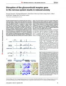
Fig. 2 Packing of the RHCC tetramer in comparison with the designed tetrameric RH4 peptide. a, Schematic projection of amino acid residues Ile 19–Leu 29 (capital letters) of the RHCC tetramer as seen from the N-terminus. 11-residue repeat positions are indicated as subscripts and are labeled as in Fig. 1a. Hydrophobic core a and h positions are highlighted in red. b, Helix cross sectional layers centered at the a, d, e, and h positions are depicted. Helices are colored as in Fig. 1b. Only residues at the a and h positions point into the center of the helix and are packed in a perpendicular and parallel manner, respectively. Residues at the d and e positions form a ring of interacting residues. c, Schematic projection of amino acid residues Leu 3–Ile 13 (capital letters) of the RH4 peptide. Hydrophobic core a, d, and h positions are highlighted in red. d, Helix cross sectional layers of RH4 centered at the a, d and h positions are depicted. In contrast to the RHCC hydrophobic core, residues at the a, d and h positions point into the center of the helix and are packed in a perpendicular, parallel and perpendicular manner, respectively.
holes formed between the side chains at positions k′–a′ and h′–i′, respectively (primes indicate residues from an adjacent chain). The side chain packing of the core residues at the a and h positions in the 11-residue repeat point into the center of the tetramer and are packed in a perpendicular and parallel manner, respectively (Fig. 2b). These packing geometries are defined by the position of the Cα-Cβ vector of the knob residue relative to the CαCα vector of the hole residues. This packing of core residues is consistent with a 7,4-hydrophobic repeat (repeat of the hydrophobic residues which point into the hydrophobic core (a and h positions)). Interestingly, the packing of side chains differs from that of left-handed coiled coils, in which β-branched Ile residues were found to have a strong bias against perpendicular packing16. As proposed in the model of Lupas and colleagues11, the d and e positions of the RHCC sequence are less hydrophobic than those at the a and h positions. Equivalent to the so-called ad-layer17 (the plane defined by all residues at a given position) the aliphatic part of the side chains at the d positions point away from the center of the tetramer and form a ring of interacting residues with the side chains at the e′ position, (Fig. 2b). This unique packing of hydrophobic residues explains why the large bulky side chain of Tyr 23 can be accommodated at an e position. The extensive hydrophobic interface of the RHCC tetramer is completed by a ring of side chains at the k positions forming cores that are not close-packed with side chains at the a′–b′ positions (Fig. 2a). A large buried surface of ∼9,500 Å2, or ∼50% of the total solvent accessible area of the four helices, further supports the notion that hydrophobic interactions are most likely the main cause for the extraordinary thermal stability of the tetrabrachion stalk domain,
d
773
© 2000 Nature America Inc. • http://structbio.nature.com
letters
© 2000 Nature America Inc. • http://structbio.nature.com
Fig. 3 Stereo view of a portion of the 1.8 Å resolution 2Fo Fc electron density map (1 σ contour level) superimposed on the current model. The network of complex surface salt bridges and polar interactions in the DDRYES region is seen (for details refer to text). The multiple conformations of Asp 21 can be seen. Helices are colored as in Fig. 1. The cluster of nine water molecules trapped in the cavity formed between Ile 19 and Tyr 23 is indicated by red spheres.
which resists heating at 130 °C in the presence of strong denaturants such as 1% (w/v) dodecyl sulfate or 6 M guanidine11,13,14. High thermal stabilities, with a midpoint of thermal transition >90 °C, were also observed for the recombinant RHCC polypeptide chain fragment (data not shown) and the designed RH4 tetramer9. However, the RHCC structure reveals marked differences to that of the designed tetrameric RH4 peptide9 (Fig. 2c,d). In the RH4 structure the residues at the a, d, and h positions all point into the interior of the tetramer and are packed in a perpendicular, parallel and perpendicular manner, respectively (Fig. 2a,d). To achieve this packing of core residues, the a and h positions in the RH4 sequence were occupied by Leu and Ile residues, respectively, whereas non-natural alloisoleucine (aIle) residues were introduced at the d positions. This packing of core residues is consistent with a 3,4,4-hydrophobic repeat (repeat of the hydrophobic residues which point into the hydrophobic core (a, d, and h positions)). Ionic interactions A characteristic feature of the RHCC sequence is the high content (∼27%) of charged residues (Fig. 1a). On the basis of distance criteria, all charged residues with the exception of Glu 6 and Asp 43 appear to form intrahelical and/or interhelical salt bridges. The three intrahelical and four interhelical salt bridges are occupied in all chains of the tetramer structure. This frequency of favorable electrostatic interactions has not been observed previously in any other coiled coil structure and may be explained in terms of the hyperthermophilic nature of the protein. Consistent with this notion, it has been proposed that a main discernible difference between thermophilic and mesophilic proteins lies in the greater number of salt bridges on the surface of the thermophilic protein18.
a
b
774
In addition to the classic i, i + 3 (Glu 24–Lys 27; f–i) and i, i + 4 (Asp 10–Arg 14; g–g intrahelical salt bridges, an i, i + 1 favorable electrostatic interaction between Asp 21 and Arg 22 (c–d) is observed in the tetramer. Besides two i, i′ + 2 type interhelical ion pairs (Arg 33–Asp 35′, d–f ′; and Arg 36–Glu 38′, g–i′), which are also found in the crystal structures of the four-stranded p-LI mutant of the GCN4 leucine zipper and the five-stranded coiled coil domain of cartilage oligomeric matrix protein16,19, the RHCC tetramer exhibits two types of favorable interhelical electrostatic interactions not present in any coiled coil structure. In contrast to the canonical interchain salt bridges, which span one and two hydrophobic core layers, there is no core layer positioned between Asp 20 and Arg 22′ (i, i′ + 2; b–d′) and only one positioned between Asp 9 and Arg 14′ (i, i′ + 5; f–g ′). Based on the crystal structures, the combinations of interactions of surface salt bridges referred to as complex salt bridges have been proposed as a mechanism for the stability of thermophilic proteins20,21. This is supported by the observation that introduction of complex surface salt bridges into the GCN4 leucine zipper results in an increase in thermal stability of the mutant protein relative to the wild type by 22 °C (ref. 22). In the RHCC tetramer the salt bridges are organized in three networks of complex salt bridges involving residues Arg 14, Arg 22 and Lys 27, and Arg 33 and Arg 36, which are flanked by polar interactions. This is illustrated by the DDRYES motif comprising residues 20–25 of the RHCC sequence (Fig. 3). Asp 20 (Oδ2) forms an interhelical salt bridge with Arg 22 (Nη1 and Nε), which interacts with Asp 21 of the same chain (Asp 21 Oδ1 with Arg 22 Nη2). Interestingly, in each of the three chains, Asp 21 has two conformations; in the second conformation Asp 21 is connected via a water molecule to Asp 20 of the same chain. Tyr 23 (Oη) forms an interhelical hydrogen bond to Ser 25 (Oγ), and only in one helix to Lys 27 (intrahelical). Glu 24 (Oε1) forms a classic intrahelical salt bridge with Lys 27 (Nγ). A prominent polar interhelical hydrogen bond is
Fig. 4 The RHCC tetramer contains a hydrophobic channel that is filled with water molecules. a, Cross section of the channel; the N-terminus of the tetramer is to the left. The protein part of three helices is shown by van der Waals spheres and ribbon representation is used for the main chain of the fourth helix (in green). The molecular surface is shown in white and positively and negatively charged side chains are colored red and blue, respectively. Four large cavities (in yellow), with volumes ranging from 145 to 300 Å3 can be seen between the a and e layers. The cavities are surrounded by networks of complex salt bridges and polar interactions. Ile residues at the a positions in the 11-residue repeat are depicted as stick models on the fourth helix in red. b, Cross section of the second cavity viewed along the tetramer axis from the N-terminus reveals a symmetric cluster of nine water molecules. The molecular surface is gray and water molecules are red spheres. Hydrogen bond distances between water molecules and between the carbonyl oxygen of Ile 19 and the amide nitrogen of Tyr 23 are indicated as dotted lines. The symmetry of the water cluster does not coincide with the noncrystallographical symmetry of the tetramer structure.
nature structural biology • volume 7 number 9 • september 2000
© 2000 Nature America Inc. • http://structbio.nature.com
letters
© 2000 Nature America Inc. • http://structbio.nature.com
Table 1 Data collection, phasing and refinement statistics and supercoil and helical parameters for four-stranded coiled coils Native 1 Data collection statistics Resolution (Å) 1.8 Observed reflections 854,694 Unique reflections 46,116 Completeness (%) 100 Rsym4 0.052 Rderiv5 Phasing statistics (30–2.9 Å) Heavy atom sites Rcullis6 (centric / acentric) Phasing power7 (centric / acentric) Refinement statistics R-factor8 (%) 19.77 Rfree (%) 21.65 Mean B-factor (Å2) 29.72 Bonds (Å)9 0.004 Angles (°)9 0.72 Dihedrals (°)9 13.65 Impropers (°)9 0.63 Superhelical parameters10 RHCC (1–18) Supercoil radius (R0; Å) 7.44 Residues per turn11 (2π / ω0) 219 Supercoil pitch (Å) 322 α-Helical parameters Residues per turn 3.65 Rise per residue (d; Å) 1.5 α-Helix radius (RI; Å) 2.24 Helix-crossing angle (Ω; °) -21.7 Interhelix distance (D, Å) 9.7
Native 2
PTCN1
MEPBAC2
AUCN3
2.9 79,623 11,500 97.3 0.074
3.2 63,410 8,854 99.7 0.075 0.302
3.4 43,099 6,977 94.6 0.109 0.143
4.5 15,872 2,470 76.9 0.115 0.279
1 0.65 / 0.58 2.1 / 3.1
4 0.62 / 0.56 2.5 / 3.2
4 0.56 / 0.56 3.6 / 4.1
tease binding region approximately at Leu 15. In the model, a pattern of highly conserved charged residues in the serine protease forms ionic interactions with Asp 9, Arg 14, Asp 20, Asp 21, Glu 24 and Asn 28 of the RHCC tetramer. This finding may explain the observation that, below pH 4.5 at room temperature, the native protease dissociates from the stalk in the presence of 0.1% SDS13.
Hydrophobic cavities The four large cavities of the tetramer are the most extraordinary feature of the molecule (Fig. 4a) and have not been seen in the left-handed tetrameric GCN4p-LI tetramer. They are connected to a continuous central channel that is lined exclusively with aliphatic side chains. The RHCC GCN4-pLI RH4 diameter of the channel, as (19–52) (1–31) (1–33) defined by the van der Waals radii 7.44 7.6 7.49 of all atoms in the hydrophobic 852 140 560 channel, varies between 2.0 and 12 1280 204 N.d. 8.4 Å and, therefore, even exceeds the maximal opening of 6 Å of the 3.65 3.59 3.67 pore in the five-stranded coiled 1.5 1.52 1.5 coil domain from cartilage 2.24 2.26 2.24 oligomeric matrix protein20. The -5.4 18.3 -4.2 cavities lie in between the middle 9.7 10.6 10.4 of the a and e layers. In the native 1PTCN, bi-potassium hexacyano platinate. structure, the cavities are occu2MEPBAC, trimethyllead acetate. pied by water molecules (Fig. 4b). 3AUCN, potassiumdicyano aureate. Because of the lack of buried 4R = Σ|Ι | / ΣΙ. sym 5R polar groups and the resulting deriv = Σ||FPH| - |FP|| / Σ|FP|. 6R cullis = Σ||FPH| - |FP + FH|| / Σ||FPH| - |FP||. weak water–protein interactions, 7Phasing power = [Σ|F |2 / Σ(|F | - |F |)2]1/2. H PH P these water molecules are clus8R-factor = Σ||F | |F || / Σ|F |. obs calc obs 9Root mean square error. tered into groups. Clusters of nine 10The paramaters16,31 were obtained by fitting the Cα coordinates of the ideal model calculated according to and five water molecules are Crick to the experimental structure. found in cavities two and three, 11Number of residues per helix turn was derived with the formula 2π[ ω - ω × cos(κ / 2)], where κ is the super1 0 respectively. Cavity one at the helix crossing angle and ω1 and ω0 are fitting parameters7. 12Not determined. N-terminus and cavity four at the C-terminus are occupied by two and one water molecule, respecalso seen between Asn 42 and Asn 44, which flank the complex tively. No electron density is seen for other regions of the channel. surface salt bridge involving Arg 33 and Arg 36. Analysis of the heavy atom derivatives, however, reveals that the major heavy atom binding sites are located within the cavities. Protease interaction They are primarily seen in the C-terminal cavity but also in the Besides the binding of the STABLE protease to the tetrabrachion other cavities. The distribution of the heavy atoms most likely stalk domain, only a few interactions between coiled coils and accounts for the excellent quality of the derivative parameters other protein structures have been described in the literature. (Table 1). The interaction is one of the most stable noncovalent protein–protein contacts known; at neutral pH it requires over Conclusion 100 °C and 1% sodium dodecyl sulfate (SDS) for dissocia- The tetramer structure of RHCC, which contains an axial channel tion11,13,14. Modeling experiments with the related serine protease with large hydrophobic cavities, may serve as a soluble model for subtilisin-BPN′ indicate that the molecule docks to the region of four-stranded transmembrane ion channels. Similar sequences the kink in the RHCC tetramer (data not shown). This result is that contain Ser residues at the hydrophobic core positions have consistent with scanning transmission electron microscopy been shown to conduct small cations23. Substituting Ser for Leu (STEM) mass measurements11 that locate the center of the pro- and Ile residues in the crystallographic coordinates of the RHCC nature structural biology • volume 7 number 9 • september 2000
775
© 2000 Nature America Inc. • http://structbio.nature.com
© 2000 Nature America Inc. • http://structbio.nature.com
letters tetramer generates a model with an enlarged interior channel lined by the hydroxyl groups of the Ser side chains. Likewise, the crystal structure of the coiled coil domain of cartilage oligomeric matrix protein has been proposed as a structural prototype for pentameric channels formed from parallel α-helices19. The RHCC structure should also be of particular interest for protein de novo design. It will be interesting to see whether twostranded and three-stranded right-handed coiled coils can be designed based on the RHCC polypeptide chain fragment. It has been argued that right-handed coiled coils are unlikely to be twostranded, except in the special case of a core consisting almost exclusively of Gly and Ala residues4. Furthermore, the RHCC tetramer may serve as a model system for understanding the stability of hyperthermophilic proteins and thus may help to improve the de novo design of proteins. Mutational analysis and pKa measurements should reveal to what extent the salt bridges contribute to the stability of the RHCC structure. Finally, because of its unique heavy atom binding properties, RHCC also has a potential practical application in X-ray crystallography. The polypeptide chain fragment may be used as a phasing tool to determine the X-ray structures of attached proteins. Methods Protein expression and purification. The recombinant RHCC polypeptide chain fragment was expressed in Escherichia coli strain JM109(DE3) (Novagen). A synthetic gene encoding residues Ile 3– Ile 52 was prepared with optimal codon usage for E. coli 24 and ligated into the BamHI/EcoRI site of pHis, a derivative of pET-15b (Novagen). Production and purification of the His6-tagged fusion protein by affinity chromatography on Ni2+-Sepharose (Novagen) was performed under denaturing conditions as described in the manufacturer’s instructions. Separation of the RHCC polypeptide chain fragment from the His6 tag was carried out as described25. The recombinant polypeptide chain fragment contains two additional N-terminal Gly and Ser residues that originate from the expression plasmid and are not part of the tetrabrachion coding sequence. Crystallization. Crystallization experiments were performed at 4 °C employing the vapor diffusion technique. Sitting droplets were made by mixing 4 µl protein solution (12.8 mg ml-1) with 2 M ammonium sulfate and 200 mM Tris-HCl (pH 7.9). The crystals belong to the space group P3121, with dimensions of a = 110.02 Å, b = 110.02 Å and c = 70.96 Å. Data collection and processing. A medium resolution data set (Native 2) and all derivative data sets were collected using monochromatized CuKα radiation (λ = 1.5418 Å) from an Elliott GX-20 rotating anode on a MAR imaging plate detector. Diffraction images were processed using the program suite MOSFLM26 and the structure factors were scaled and reduced using SCALA from the CCP4 package27. Statistics of the merged data are given. The derivative data were scaled to Native 2 data using SCALEIT27. The high resolution data set (Native 1) has been collected at the DESY synchrotron (beamline BW7B).
776
Structure determination and refinement. The structure was determined with the multiple isomorphous replacement method. The derivative data are highly isomorphous. Initial heavy atom positions were obtained from difference Patterson maps and analyzed in a common origin with crossphased difference Fourier maps. Heavy atom parameters were refined with SHARP28.The MIR phases (30–2.9 Å; mean figure of merit 0.5443) were greatly improved and extended to 1.8 Å by use of DM27 with solvent flattening, histogram matching and phase extension. Electron density interpretation and model building were done with MAIN29. The structure was refined with CNS30 against the high resolution native data set (Native 1) without using noncrystallographic symmetry restraints. The current native model includes 205 amino acid residues and 277 water molecules. Coordinates. The coordinates for the structure have been deposited in the Protein Data Bank (PDB accession number 1FE6).
Acknowledgments We thank A. Philippsen for providing the DINO program and G.Orriss for critical reading of the manuscript. This work was supported by the Swiss National Science Foundation.
Correspondence should be addressed to R.K. email: richard
[email protected] or J.S. email:
[email protected] Received 13 March, 2000; accepted 14 July, 2000. 1. 2. 3. 4. 5. 6. 7. 8. 9. 10. 11. 12. 13. 14. 15. 16. 17. 18. 19. 20. 21. 22. 23. 24. 25. 26. 27. 28. 29. 30. 31.
Cohen, C. & Parry, D.A.D. Proteins 7, 1–15 (1990). Kammerer, R.A. Matrix Biol. 15, 555–565 (1997). Kohn, W.D., Mant, C.T. & Hodges, R.S. J. Biol. Chem. 272, 2583–2586 (1997). Lupas, A. Trends Biochem. Sci. 21, 375–382 (1996). McLachlan, A.D. & Stewart, M. J. Mol. Biol. 98, 293–304 (1975). Sodek, J., Hodges, R.S., Smillie, L.B. & Jurasek, L. Proc. Natl. Acad. Sci. USA 69, 3800–3804 (1972). Crick, F.C.H. Acta Crystallogr. 6, 689–697 (1953). O’Shea, E.K., Klemm, J.D., Kim, P.S. & Alber, T. Science 254, 539–544 (1991). Harbury, P.B., Plecs, J.J., Tidor, B., Alber, T. & Kim, P.S. Science 282, 1462–1467 (1998). Nooren, I.M.A., Kaptein, R., Sauer, R.T. & Boelens, R. Nature Struct. Biol. 6, 755–759 (1999). Peters, J., Baumeister, W. & Lupas, A. J. Mol. Biol. 257, 1031–1041 (1996). Dure 3rd, L. Plant J. 3, 363–369 (1993). Peters, J. et al. J. Mol. Biol. 245, 385–401 (1995). Mayr, J. et al. Curr. Biol. 6, 739–749 (1996). Brown, J.H., Cohen, C. & Parry, D.A.D. Proteins 26, 134–145 (1996). Harbury, P.B., Zhang, T., Kim, P.S. & Alber, T. Science 262, 1401–1407 (1993). Lupas, A. et al. J. Mol. Biol. 248, 180–189 (1995). Perutz, M.F. Science 201, 1187–1191 (1978). Malashkevich, V.N., Kammerer, R.A., Efimov, V.P., Schulthess, T. & Engel, J. Science 274, 761–765 (1996). Yip, K.S. et al. Structure 3, 1147–1158 (1995). Pappenberger, G., Schurig, H. & Jaenicke, R. J. Mol. Biol. 274, 676–683 (1997). Spek, E.J., Bui, A.H., Lu, M. & Kallenbach, N.R. Protein Sci. 7, 2431–2437 (1998). DeGrado, W.F. & Lear, J.D. Biopolymers 29, 205–213 (1990). Dong, H., Nilsson, L. & Kurland, C.G. J. Mol. Biol. 260, 649–663 (1996). Kammerer, R.A. et al. J. Biol. Chem. 273, 10602–10608 (1998). Leslie, A.G.W. MOSFLM users guide (MRC-LMB, Cambridge; 1994). Collaborative Computational Project Number 4. Acta Crystallogr. D 50, 760–763 (1994). De La Fortelle, E. & Bricogne, G. Methods Enzymol. 276, 472–493 1997. Turk, D.C. Ph.D. thesis (Technische Universität München, Munich, Germany; 1992). Brunger, A.T. et al. Acta Crystallogr. D 54, 905–921 (1998). Harbury, P.B., Kim, P.S. & Alber, T. Nature 371, 80–83 (1994).
nature structural biology • volume 7 number 9 • september 2000