© 2000 Nature America Inc. • http://structbio.nature.com
letters Flt3 ligand structure and unexpected commonalities of helical bundles and cystine knots Savvas N. Savvides1–3, Tom Boone4 and P. Andrew Karplus1,2 1
Program in Biophysics, Cornell University, Ithaca, New York 14853, USA. Department of Biochemistry and Biophysics, Oregon State University, Corvallis, Oregon 97331, USA. 3Present address: Department of Biochemistry and Molecular Biophysics, Washington University School of Medicine, St. Louis, Missouri 63110, USA. 4AMGEN, Thousand Oaks, California 91320-1789, USA. © 2000 Nature America Inc. • http://structbio.nature.com
2
Human Flt3 ligand (Flt3L) stimulates early hematopoiesis by activating a type III tyrosine kinase receptor on primitive bone marrow stem cells. The crystal structure of soluble Flt3L reveals that it is a homodimer of two short chain α-helical bundles. Comparisons of structure-function relationships of Flt3L with the homologous hematopoietic cytokines macrophage colony stimulating factor (MCSF) and stem cell factor (SCF) suggest that they have a common receptor binding mode that is distinct from the paradigm derived from the complex of growth hormone with its receptor. Furthermore, we identify recognition features common to all helical and cystine-knot protein ligands that activate type III tyrosine kinase receptors, and the closely related type V tyrosine kinase receptors. Hematopoiesis, the making of blood cells, originates from a small, self-renewing population of hematopoietic stem cells1, and is mediated by the activities of soluble and cell-bound growth factors that oligomerize their respective cell surface receptors to trigger appropriate signaling pathways2. While
many hematopoietic growth factors are known, most are involved in late hematopoiesis, with stem cell factor (SCF) and the fms-like tyrosine kinase receptor 3 ligand (Flt3L) the only ones known to act directly on stem cells3. Flt3L and SCF have both overlapping and synergistic activities, but clear distinctions exist. Flt3L is a more potent stimulator of stem cell proliferation and mobilization, and has the unique ability to potently stimulate the generation of dendritic cells which are crucial for antigen capture and presentation3. As a result, Flt3L has biomedical potential in the areas of stem cell regeneration and mobilization, and in cancer immunotherapy3. Flt3L is a noncovalently linked homodimer, and is biologically active both as a transmembrane form and as a soluble form that is generated by proteolytic cleavage of the extracellular domains of the transmembrane protein 4,5. Even though the relative importance of the two forms is still unclear, the extracellular domain (residues 1–134) alone has been shown to be sufficient for bioactivity6. Despite a lack of appreciable sequence similarity with other proteins, Flt3L is predicted to be a helical cytokine (Fig. 1) homologous to SCF and macrophage colony stimulating factor (MCSF)4,5,7. Intriguingly, these three dimeric hematopoietic cytokines are unique among helical cytokines in that they bind to type III tyrosine kinase receptors (Flt3L with Flt3R, MCSF with c-fms, and SCF with c-kit) from the platelet-derived growth factor receptor (PDGFR) subfamily of tyrosine kinase receptors; this receptor family is otherwise associated with the binding of cystine-knot growth factors such as PDGF and vascular endothelial growth factor (VEGF)8 (Fig. 1). PDGFR-like receptors thus constitute a rare example of a receptor family that interacts with protein ligands having fundamentally different folds. The crystal structure of VEGF in complex with Ig-like domain 2 of Flt1 (ref. 9) has recently provided a structural paradigm for the association of cystine-knot protein ligands with their respective PDGFR-like receptors (Fig. 1). The striking structural differences between cystineknot and helical proteins, however, make it difficult to apply this Fig. 1 Classification of cytokines according to their structure and receptor. Cytokines for which a structure is known are in bold, those for which a structure of their complex with receptor exists are marked with an asterisk, and the dimeric cytokines are marked with a ‘(d)’. The helical cytokines can be divided into the long chain, short chain, and interferon-like11,23. The first panel shows growth hormone (GH) with its receptor binding epitopes marked in red (site I) and yellow (site II)19. Site I includes residues from helix D, helix A, and the loop connecting helices A and B, and site II includes residues from helices A and C. The second panel features the structure of MCSF10 as a representative of the three cytokines that bind to type III tyrosine kinase receptors. The third panel shows VEGF with the receptor binding sites that are seen in its complex with domain 2 of Flt1 (ref. 9) marked in red. The ‘?’ highlights the question whether the receptor binding mode will be driven more by the receptor family or the cytokine family. Ribbon diagrams are drawn to scale and were created using MOLSCRIPT40 and RASTER3D41. Abbreviations used are: GH, growth hormone; EPO, erythropoietin; GCSF, granulocyte colony stimulating factor; GMCSF, granulocyte macrophage colony stimulating factor; LIF, leukemia inhibitory factor; OSM, oncostatin; PRL, prolactin; TPO, thromobopoietin; IL, interleukin; IFN, interferon; MCSF, macrophage colony stimulating factor; Flt3L, fms-like tyrosine kinase 3 ligand; SCF, stem cell factor; PDGF, platelet derived growth factor; VEGF, vascular endothelial growth factor; PLGF, placental growth factor.
486
nature structural biology • volume 7 number 6 • june 2000
© 2000 Nature America Inc. • http://structbio.nature.com
letters
© 2000 Nature America Inc. • http://structbio.nature.com
information to gain insights into the binding modes of Flt3L, MCSF, and SCF. Instead, the expectation7,10–18 has been that these helical cytokines follow the paradigm derived from the complex of growth hormone (GH) with its type I cytokine receptor (GHR)19. This GH paradigm has guided structurefunction studies on all helical cytokines, and has been validated by receptor complexes of interferon-γ20, erythropoietin21, and interleukin-4 (IL-4)22, and structure-function studies for many other helical cytokines11,12,14,18. Here, we present the crystal structure of the extracellular domain (residues 1–134) of dimeric human Flt3L. We then evaluate available structure-function data for Flt3L, MCSF, and SCF and conclude that the GH paradigm does not apply to these cytokines. Finally, we document some receptor recognition features that are common to all protein ligands that interact with receptors from the PDGFR family. Structure of the Flt3 ligand The structure of the extracellular domain of human Flt3L was determined by multiple isomorphous replacement (MIR) at 3.2 Å resolution and was refined at 2.2 Å resolution (Table 1). As predicted, Flt3L is a homodimer of subunits that adopt the short chain helical cytokine fold23, and that assemble in a man-
a
c
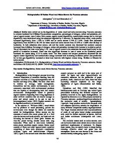
ner analogous to dimeric MCSF10 (Fig. 2). Notable features of the Flt3L fold are a 310-helix that is essentially continuous with αC, and a triplet of proline residues (Pro 89, Pro 90, and Pro 91) in the middle of the loop connecting the 3 10-helix to β2. Of the three intramolecular disulfide bridges the two (Cys 4–Cys 85 and Cys 44–Cys 127) required for bioactivity6 are buried, while the one that is dispensable (Cys 93–Cys 132) is solvent accessible, suggesting that these disulfide-bridges are not involved in receptor recognition but instead have a structural role. In forming the Flt3L dimer, each subunit buries ∼1,000 Å2 of largely hydrophobic surface with residues 63–68 and residues 25–30 from one monomer interdigitating with the equivalent segments of the other (Fig. 2a,b). The ∼20° shift of the relative orientation of the subunits in the dimer between Flt3L and MCSF (Fig. 2c) cautions that the dimer geometry of SCF may not be predictable based on the structures of MCSF or Flt3L. Interestingly, structural overlays of the Flt3L monomer with known short chain hematopoietic cytokines show that the best match for the Flt3L monomer is IL-4 and not the more functionally similar MCSF: with MCSF the root mean square (r.m.s.) deviation is 2.8 Å for 75 Cα atoms; with IL-2 it is 2.7 Å for 75 atoms; with IL-4 it is 2.4 Å for 82 atoms; and with GMCF it is 2.2 Å for 59 atoms. This supports the hypothesis that these
b
d
Fig. 2 Structure of Flt3L. a, Flt3L (dimensions 75 Å × 35 Å × 25 Å) is shown in its transmembrane form oriented with respect to the cell surface based on the position of the C-termini. Figure created with MOLSCRIPT40 and RASTER3D41. b, Stereo view of the electron density for one monomer surrounding Leu 27 at the heart of the dimer interface. The final σA-weighted 2Fo - Fc; αc electron density at 2.2 Å resolution is contoured at 1.1 ρrms. Figure created with CHAIN42. c, Superposition of Flt3L (red) and MCSF (cyan) based on just the lower monomer. The orientation of the upper monomers differ by a rotation of ∼20°. Figure created with MOLSCRIPT40. d, Molecular mapping of solvent accessible residues of Flt3L that are important for bioactivity24. Two orientations of Flt3L are shown. Highlighted are sites that when mutated reduced (red) or increased (blue) the bioactivity: H8R 0 %, S9G 14 %, S13F 0 %, R55L 30 %, K116E 2 %, H8Y 200 %, K84E 300 %, K84T 160 %, W118R 200 %, Q122R 200 %. Figure created with GRASP43.
nature structural biology • volume 7 number 6 • june 2000
487
© 2000 Nature America Inc. • http://structbio.nature.com
© 2000 Nature America Inc. • http://structbio.nature.com
letters cytokines have diverged sufficiently that the levels of structural loop prior to αA, the C-terminus of αD and the loop region similarity no longer correlate with evolutionary distances immediately after it), and possibly the face defined by αC and αB, (Fig. 2a). While many details of receptor recognition for implied by the levels of functional similarity23. each of the three cytokines still need to be characterized, one Flt3L receptor binding surface clear conclusion is that the GH paradigm does not apply to this A correlation of published mutational studies24 with the struc- group of helical cytokines (Fig. 3b). Since expectations are a ture allows us to identify nine surface residues whose mutation powerful driving force in experimental design and interpretaaffects bioactivity (Fig. 2d). These residues cluster around the tion, this realization should open the door to more rapid poles of the Flt3L dimer ∼45 Å apart and on a surface hypothe- progress in defining how these helical cytokines are recognized. sized to face away from the cell membrane (Fig. 2a,d), consistent Once the receptor binding epitopes of Flt3L, MCSF, and SCF with the consensus that Flt3L can act as a membrane-associated are mapped in detail, it will be interesting to see whether miniligand within the tight microenvironment of the bone marrow. mal amino acid substitutions can transfer specificity of one Each cluster includes three segments of the Flt3L scaffold: the cytokine for one receptor to another. flexible N-terminal loop prior to αA (His 8, Ser 9 and Ser 13); the C-terminus of αC (Lys 84); and the C-terminus of αD (Lys Common features of PDGFR-like receptor ligands 116, Trp 118 and Gln 122). Arg 55 is in αB, ∼20 Å from the cen- In addition to the helical cytokines Flt3L, MCSF, and SCF, the ter of a cluster; both Lys 84 and Arg 55 are in the middle of cystine-knot growth factors PDGF28 and VEGF29 also associate with PDGFR-like receptors8 (Fig. 1), and available structurehydrophobic patches. When Flt3L is viewed down one end of the dimer, the sites of function studies suggest that PDGF and VEGF employ strucmutation segregate so that a change to a positive charge is associat- turally equivalent parts of their structures in receptor ed with enhanced activity on the side of the molecule with Lys 116, binding9,28,29–33. An interesting question is whether the binding Trp 118 and Gln 122, but with decreased activity on the other (Fig. of these structurally distinct molecules to their respective recep2d). Interestingly, two additional positively charged residues, Arg tors involves common features. Whereas it is difficult to com121 and Arg 126, are located on the same lobe of the cluster as Lys pare structure-function relationships across the two families of 116, Trp 118 and Gln 122. The fact that not all activity-reducing ligands, we nevertheless note three striking similarities. First, Flt3L mutants were characterized24 suggests that some important Flt3L, MCSF, PDGF, and VEGF have almost identical overall mutations may have been overlooked. Nevertheless, we suggest that the identified surfaces on each Flt3L a monomer approximate the receptor binding epitope because each already accounts for ∼1,200 Å2 of surface area, which is near the upper end of the range of 700–1,200 Å2 seen for a variety of structurally characterized cytokine–receptor interaces9,19–22,25,26. Consensus for receptor recognition Available mutagenesis studies for Flt3L24, MCSF13 and SCF15,27 implicate structurally equivalent parts of all three growth factors in receptor recognition (Fig. 3a). This is consistent with a common receptor binding mode involving the distal ends of the helical bundles (N-terminal
Fig. 3 Comparisons of Flt3L, MCSF, SCF sequences and mutational data with each other and with the GH paradigm. a, Structure-based sequence alignment of human Flt3L with human MCSF10, sequence alignment with human SCF5, and the structural alignment of helices A, C, and D of human growth hormone (GH)23. Structurally equivalent regions in Flt3L and MCSF (3 Å r.m.s. deviation cutoff) are indicated by gray bars; other residues should not be considered structurally equivalent. Secondary structure elements 43 are shown as black (α-helices), brown (310-helices), and violet (β-strands) bars. Residue accesibilities for Flt3L and MCSF are shown as ‘*’ for ≥40 %, ‘-’ for 20-40 %, and ‘ ‘ for ≤20 % (calculated as accessible surface areas 44 divided by a reference value from a Gly-X-Gly tripeptide45). The black arrow indicates the point of divergence among various isoforms of each protein5,7. Highlighted are residues where mutation led to reduced (red), increased (blue), or near wild type bioactivity 13,15,24,27,46. His 8 in Flt3L (red on yellow) enhanced or inhibited bioactivity depending on the substitution. A quadruple mutant of SCF is shown as a green block with the substituted amino acids in red. GH residues directly involved in receptor binding are yellow19. b, Consensus of receptor interaction sites for Flt3L, MCSF, and SCF mapped onto the scaffold of Flt3L (red) and comparison with the GH paradigm (yellow). Sites in Flt3L, MCSF, and SCF that were found to be unimportant for bioactivity are shown in green. Figure created with MOLSCRIPT40 and RASTER3D41.
488
b
nature structural biology • volume 7 number 6 • june 2000
© 2000 Nature America Inc. • http://structbio.nature.com
letters
© 2000 Nature America Inc. • http://structbio.nature.com
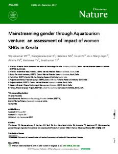
Fig. 4 Physical comparison of ligands that associate with PDGFR-like receptors and mapping of known mutagenesis data to their surfaces to allow a direct comparison of known and putative receptor interaction sites9,10,13,15,24,27,28,30,31,33. SCF is marked with an asterisk because the model presented is based on the structure of Flt3L. The two-fold axis for each molecule is indicated in green. The mapped surface for VEGF corresponds to residues that participate at the ligand–receptor interface in the complex of VEGF with domain 2 of Flt1 (ref. 9). Three red squares on the surface of PDGF correspond to the expected location of residues 27, 30, and 34 that are important for receptor binding30,31, but were not modeled in the crystal structure of PDGF-BB due to high temperature factors28. Interestingly, both Flt3L and PDGF require two or three positive charges at the poles of the dimer for optimal binding to their receptors24,28,33, while VEGF requires positive charge to bind to kinase domain receptor (KDR) and negative charge to bind to Flt132. Also shown is a general bivalent mode of ligand binding to type III (five Ig-like domains, n = domain 5) and type V (seven Ig-like domains, n = domains 5, 6, and 7) tyrosine kinase receptors through Ig-like domains 1, 2, and 3 of the receptor. The figure was created with GRASP43.
structurally diverse ligands, and vice versa, such as the diverse protein ligands binding to G protein coupled receptors34, binding of diverse ligands to the Fc fragment of human IgG35 and nerve growth factor (NGF) binding to two different classes of receptors34. Methods Purification of Flt3L. Human Flt3L (residues 1–134) expressed in Escherichia coli was purified from inclusion bodies by solubilization in 9 M urea with 10 mM cysteine and then 25-fold dilution into 20 mM Tris, pH=8.5 proprietary refolding buffer to allow Flt3L to refold overnight. Refolded Flt3L was further purified by S-sepharose cation exchange chromatography followed by butyltoyopearl 650M hydrophobic interaction chromatography, and was dialyzed into phosphate buffered saline (10 mM sodium phosphate, 150 mM NaCl, pH 7) for storage at 0.7 mg ml-1.
dimensions (∼70 Å × ∼35 Å × ∼25 Å; Fig. 4). Second, they all function as dimers and have two-fold axes of symmetry perpendicular to the longest dimension of the dimeric scaffolds (Fig. 4). Third, structure-function studies for all these ligands suggest that they bind to their respective receptors bivalently with the equivalent receptor binding sites separated by ∼45 Å (Fig. 4). We suggest that despite their diverse folds, these two classes of ligands bind to and activate PDGFR-like receptors in an equivalent manner, providing symmetric sites to bind receptor domains 2 and 3, and allowing domains 4 and higher to form receptor–receptor contacts9, (Fig. 4). Thus, it seems justified to replace the question mark in Fig. 1 with the ‘VEGF paradigm’, because the common recognition features are shared between the distinct ligands of a given receptor rather than between similar ligands binding to two distinct receptors. Assuming that cystine-knot growth factor binding to PDGFR-like receptors is more ancient, we propose that the ‘crossover’ activity of Flt3L, MCSF, and SCF originated when a dimeric helical cytokine per chance had surface features that allowed it to bind to and activate a PDGFR-like receptor. This activity probably provided a selective advantage, and later gene duplications and divergence of the cytokines and receptors led to the three distinct crossover helical cytokines of today. In retrospect, it could have been predicted that the structural features that allowed these ligands to be co-opted for their new function would not match the surfaces required for their original function. This principle may help to understand other systems in which a particular receptor family is bound by nature structural biology • volume 7 number 6 • june 2000
Crystallization and data collection. Flt3L crystals grew in one week from 10 µL drops containing a 1:1 mixture of protein solution (Flt3L at 10–15 mg ml-1 in 20 mM HEPES, 1 mM EDTA, pH 7.2) and reservoir (6–9% w/v PEG 8000, 0.2 M zinc acetate, 0.1 M sodium cacodylate, pH 6.3–6.5) over 0.5 ml of reservoir at room temperature. Zinc acetate is essential for crystal growth. For data collection at -170 °C, crystals were incubated in stabilizing solution (20% w/v PEG 8000, 0.2 M zinc acetate, 0.1 M sodium cacodylate pH 6.4) containing 25% (v/v) glycerol for 10 min, and were then flash frozen in a stream of nitrogen gas. Flt3L crystals belong to space group P21212 with a = 138.6 Å, b = 163.4 Å, c = 26.9 Å at room temperature, and a = 137.1 Å, b = 159.4 Å, c = 26.3 Å at -170 °C. Native data to 2.6 Å resolution, and data from 60 potential heavy atom derivatives, were collected at room temperature using graphite-monochromated CuKα X-rays from a Rigaku RU200 generator operating at 50 kV/150 mA with a 0.5 mm × 5 mm focus. Native data to 2.2 Å resolution were collected at -170 °C at station F1 of the Cornell High Energy Synchrotron Source (CHESS). Data reduction was carried out using the programs DENZO and SCALEPACK36 (Table 1). Structure determination and refinement. MIR phases from five heavy atom derivatives were calculated using PHASES-95 (ref. 37) and had a mean figure of merit (FOM) of 0.48 at 3.2 Å resolution (Table 1). The electron density map was largely uninterpretable, but showed clear protein-solvent contrast and two Flt3L dimers in the asymmetric unit. Noncrystallographic symmetry (NCS) relationships were generated from crude models of α-helices identified at 6 Å resolution, and iterative phase improvement was carried out using solvent flattening and improper four-fold NCS averaging37. One subunit was built as a poly-Ala chain (residues 3–132) and NCS operators were used to generate the other three subunits. The electron density for one subunit of dimer 2 appeared progressively weaker in the regions away from the dimer interface. Crystallographic calculations were performed using XPLOR 3.851
489
© 2000 Nature America Inc. • http://structbio.nature.com
letters Table 1 Diffraction, phasing, and refinement statistics1
© 2000 Nature America Inc. • http://structbio.nature.com
Data collection X-ray source Temperature Maximum resolution (Å) Unique reflections Multiplicity Completeness (%) Rmeas2 (%) I / σI
Native 1
Native 2
CHESS-F1 -170 °C 2.2 29,421 4.0 (3.3) 96.9 (95.5) 7.2 (26.8) 12.0 (3.8)
CuKα Ambient 2.6 18,530 3.2 (1.4) 92.3 (77.5) 8.1 (32.7) 8.1 (2.7)
Isomorphous replacement Concentration, time Resolution limit of phasing (Å) Number of sites Riso3 (%) Rcullis4 Phasing power (isomorphous)5 Phasing power (anomalous)6 Refinement Resolution (Å) Reflections (F > 1σ) Rcryst7 / Rfree8 (%) Non-hydrogen protein atoms Water molecules Zinc atoms R.m.s. deviations Bond length (Å) Bond angle (º) Anisotropic overall B-factor shifts
KI3
K2PtCl4 (I)
K2PtCl4 (II)
K2OsCl6
K2Pt(NO2)4
CuKα Ambient 3.2 10,356 2.9 (1.6) 94.8 (84.5) 10.6 (27.4) 8.4 (3.4)
CuKα Ambient 4.0 4,813 1.7 (1.3) 84.5 (79.3) 14.0 (41.1) 4.4 (2.2)
CuKα Ambient 3.0 11,779 2.9 (1.4) 90.1 (80.6) 12.0 (28.9) 8.0 (2.6)
CuKα Ambient 3.0 11,234 2.7 (1.4) 85.5 (70.4) 6.8 (22.0) 11.7 (3.2)
CuKα Ambient 3.5 7,980 3.2 (1.7) 94.9 (82.1) 9.8 (23.8) 10.4 (3.9)
10 mM, 3 d 3.2 8 10.2 0.48 1.90 1.90
2 mM, 16 h 4.0 3 19.4 0.67 1.10
2 mM, 1 d 3.2 2 14.3 0.71 0.95
2 mM, 20 h 3.2 4 7.7 0.73 0.70
10 mM, 3.5 d 6.0 4 13.1 0.65 1.10
20–2.2 28,346 23.9 / 28.7 4,273 217 9 0.005 1.3 B11 = -10.1 Å2, B22 = -2.5 Å2, B33 = 12.5 Å2
Numbers in parentheses correspond to values in the highest resolution shell. Rmeas = Σh (nh/nh - 1)1/2 Σi | Ih – Ih,i | / Σh Σi Ih,i, multiplicity-weighted Rmerge47. 3R iso = 100 × ||FPH| - |FP|| / ||FP |, where FP and FPH are structure factor amplitudes for native and derivative data, respectively. 4R cullis = | | |FPH | ± |FP | | - |FH | | / | |FPH | ± |FP | |, for centric reflections with F / σF = 2. 5Phasing power (isomorphous) = r.m.s. (|F | / E), where E is the residual lack-of-closure error. H 6Phasing power (anomalous) = r.m.s. (|F ″| / E), where F ″ is the anomalous correction component amplitude for F . H H H 7R = |F F | / |F | cryst o c o 8R = R for 5% of reflections against which the model was not refined. free cryst 1 2
(ref. 38). Cycles of simulated annealing with tight NCS restraints against 8–2.6 Å data (Native 2, Table 1), and model building in σA weighted 2Fo - Fc electron density maps resulted in a complete model for residues 3–132 in each monomer and six Zn atoms (Rcryst = 33%, Rfree = 38%). In support of the assignment of Zn atoms, His, Glu and Asp side chains refined within ligating distances from the sites. In addition, these sites, when modeled as water molecules, refined to unusually low B-factors and still had large positive peaks in the difference maps. Crystallographic refinement was extended to 2.2 Å resolution using synchrotron data (Native 1, Table 1). First, the protein model was placed in the new unit cell by rigid body refinement (Rcryst = 44.5%, Rfree = 45.6%). Subsequent rounds of refinement were carried out without NCS restraints against 20–2.2 Å data using a bulk solvent correction, and an overall anisotropic B-factor tensor applied to Fcs (E. A. Merritt, pers. comm.). The electron density and packing interactions of residue 41 in each subunit indicated that it is an Asp and not a Glu as in the published amino acid sequence4,5. The final model (Rcryst = 23.9%, Rfree = 28.7%) consists of residues 1–134 for each monomer in dimer 1, residues 3–132 for each monomer in dimer 2, 217 water molecules and 9 zinc atoms. The ordered zinc is presumably an artifact of crystallization, with eight zinc atoms located at crystal contacts. Residues 7–14 is the only part of Flt3L that adopts significantly different conformations among the four NCS-related chains, and the refined temperature factors of
490
this region indicate an inherent flexibility. The second monomer in dimer 2 makes few crystal packing interactions and is progressively disordered in the regions away from the dimer interface with an average main chain B-factor of 72 Å2 compared to 36 Å2 for the other three subunits. All φ,ϕ angles lie in allowed regions of the Ramachandran plot with 88% in the most favorable regions39. Considerations of published mutagenesis data. Among the characterized Flt3L mutants24, 19 mutations that decreased the bioactivity are not considered to give insight into receptor binding, because we hypothesize they induced structural perturbations or destabilization of Flt3L: 10 are at buried within the Flt3L monomer (residues 11, 15, 34, 75, 81, 87, 124); 3 (residues 26, 27, and 64) are buried at the dimer interface (Fig. 1b), emphasizing that the integrity of the Flt3L dimer is essential for bioactivity; 3 introduce cysteines (mutants R20C, R55C, R95C) and may interfere with formation of the disulfide bond network of Flt3L during folding; and 3 involve the introduction or removal of a proline (mutants P10S, P90S, S13P), thus altering the main chain structural properties. One residue, Trp118, that had been identified as buried in a homology model, is seen to be completely solvent accessible and is implicated in the receptor recognition site. It was modeled incorrectly because in Flt3L αD ends six residues earlier than in MCSF (Fig. 3a). Although the quadruple mutant of SCF does identify a single key
nature structural biology • volume 7 number 6 • june 2000
© 2000 Nature America Inc. • http://structbio.nature.com
letters residue (Fig. 3a), comparisons with mutations at equivalent positions in Flt3L and MCSF may shed some light. Arg 121 and Asp 124 each align with residues shown not to be important for the bioactivity of MCSF and Flt3L, and Lys 127 aligns with the very important Lys 116 of Flt3L. Interestingly, this lysine (Lys 116 of Flt3L, Lys 125 of MCSF and Lys 127 of SCF) is the only noncysteine residue conserved in all the proteins and may play a conserved role. Coordinates. Atomic coordinates and structure factors have been deposited in the Protein Data Bank for immediate release (accession code 1ETE).
© 2000 Nature America Inc. • http://structbio.nature.com
Acknowledgments We wish to thank the staff of MacCHESS for help with data collection, and S.-H. Kim for the complete atomic coordinates of MCSF. This work was supported in part by a NSF grant to P.A.K. and a fellowship from the International Center for Diffraction Data to S.N.S.
14. 15. 16. 17. 18. 19. 20. 21. 22. 23. 24. 25. 26. 27. 28. 29. 30.
Correspondence should be addressed to P.A.K. email:
[email protected]
31. 32. 33. 34.
Received 23 February, 2000; accepted 27 April, 2000.
35.
1. 2. 3. 4. 5. 6. 7. 8. 9. 10. 11. 12. 13.
36. 37. 38.
Ogawa, M. Blood 81, 2844–2853 (1993). Heldin, C.H. Cell 80, 213–223 (1995). Fichelson, S. Eur. Cytokine Network 9, 7–22 (1998). Lyman, S.D. et al. Blood 83, 2795–2801 (1994). Hannum, C. et al. Nature 368, 643–648 (1994). Escobar, S., Brasel, K., Anderberg, R. & Lyman, S.D. Blood 86, 21a (1995). Bazan, J.F. Cell 65, 9–10 (1991). Van der Geer, P., Hunter, T., Lindberg, R.A. Annu. Rev. Cell. Biol. 10, 251–337 (1994). Wiesmann, C. et al. Cell 91, 695–704 (1997). Pandit, J. et al. Science 258, 1358–1362 (1992). Sprang, S.R. & Bazan, J.F. Curr. Opin. Struct. Biol. 3, 815–827 (1993). Kaushansky, K. & Karplus, P.A. Blood 82, 3229–3240 (1993). Taylor, E.W., Fear, A.L., Bohm, A., Kim, S.-H. & Koths, K. J. Biol. Chem. 269, 31171–31117 (1994).
nature structural biology • volume 7 number 6 • june 2000
39. 40. 41. 42. 43. 44. 45. 46. 47.
Mott, H.R. & Campbell, I.D. Curr. Opin. Struct. Biol. 5, 114–121 (1995). Matous, J.V., Langley, K. & Kaushansky, K. Blood 88, 437–444 (1996). Mendiaz, E.A. et al. Eur. J. Biochem. 239, 842–849 (1996). Menziani, M.C., Fanelli, F. & De Benedetti, P.G. Proteins 26, 42–54 (1996). Simpson, R.J., Hammacher, A., Smith, D.K., Matthews, J.M. & Ward, L.D. Protein Sci. 6, 929–955 (1997). de Vos, A.M., Ultsch, M. & Kossiakoff, A.A. Science 255, 306–312, 1992. Walter, M.R. et al. Nature 376, 230–235 (1995). Syed, R.S. et al. Nature 395, 511–516 (1998). Hage, T., Sebald, W. & Reinemer, P. Cell 97, 271–281 (1999). Rozwarski, D.A. et al. Structure 2, 159–173 (1994). Graddis, T.J. et al. J. Biol. Chem. 273, 17626–17633 (1998). Banner, D.W. et al. Cell 73, 431–445 (1993). Vigers, G.P.A., Anderson, L.J., Caffes, P. & Brandhuber, B.J. Nature 386, 190–194 (1997). Hsu, Y.R. et al. Biochemistry 37, 2251–2262 (1998). Oefner, C., D’Arcy, A., Winkler, F.K., Eggimann, B. & Hosang, M. EMBO J. 11, 3921–3926 (1992). Muller, Y.A., Christinger, H.W., Wells, Keyt, B.A. & de Vos, A.M. Structure 5, 1325–1338 (1997). Östman, A., Andersson, M., Hellman, U. & Heldin, C.H. J. Biol. Chem. 266, 10073–10077 (1991). Clements, J.M. et al. EMBO J. 10, 4113–4120 (1991). Keyt, B.A. et al. J. Biol. Chem. 271, 5638–5646 (1996). Schilling, D. et al. Biochem. J. 333, 637–644 (1998). Nicola, N.A. In Guidebook to cytokines and their receptors. (ed. Nicola, N.A.) 1–7 (Oxford University Press, New York; 1994). DeLano, W.L., Ultsch, M.H., de Vos, A.M., Wells, J.A. Science 287, 1279–1283 (2000). Otwinowski, Z & Minor, W. Methods Enzymol. 276, 307–326 (1997). Furey, W. & Swaminathan, S. Methods Enzymol. 277, 590–620 (1997). Brünger, A.T. X-PLOR version 3.1: a system for X-ray crystallography and NMR. (Yale University Press, New Haven, Connecticut; 1992). Laskowski, R.A., MacArthur, M.W., Moss, D.S. & Thornton, J.M. J. Appl. Crystallogr. 26, 283–291 (1983). Kraulis, Per J. J. Appl. Crystallogr. 24, 946–950 (1991). Merritt, E.A. & Bacon, D.J. Methods Enzymol. 277, 505–524 (1997). Sack, J. S. Methods Enzymol. 277, 158–173 (1997). Nicholls, B., Sharp, K.A. & Honig, B. Biophys. J. 64, 166–170 (1993). Kabsch, W. & Sander, C. Biopolymers 22, 2577–2637 (1983). Miller, S., Janin, J., Lesk, A.M. & Chothia, C. J. Mol. Biol. 196, 641–656 (1987). Hsu, Y.R. et al. J. Biol. Chem. 272, 6406–6415 (1997). Diederichs, K. & Karplus, P. A. Nature Struct. Biol. 4, 269–274 (1997).
491