Special Report
L1 (CD171) as a novel biomarker for ovarian and endometrial carcinomas Mina Fogel†, Monica Huszar, Peter Altevogt and Alon Ben-Arie
CONTENTS Structural & functional aspects of the L1 adhesion molecule L1 in ovarian cancer L1 in endometrial cancer L1 for the diagnosis of metastatic carcinoma with unknown origin Soluble L1 as a serum biomarker Expert opinion & five-year view Key issues References Affiliations
†
Author for correspondence Department of Pathology, Kaplan Medical Center, 7600 Rehovot, Israel Tel.: +972 08 944 1546 Fax: +972 08 941 8086
[email protected]
KEYWORDS: ADAM10, cell migration, endometrial carcinoma, L1, ovarian carcinoma, shedding
www.future-drugs.com
The L1 molecule has emerged as a promising new biomarker for the diagnosis and prognosis of human ovarian and endometrial tumors. It was initially described as an adhesion molecule for neural cells but its function on tumor cells is less well known. In this article, the role of L1 in promoting tumor cell adhesion and migration is discussed. The question of how L1 determination in tumor tissue samples, serum and ascites could potentially improve the diagnosis and monitoring of gynecologic tumor patients is also addressed. The presence of L1 in tissue and serum was found to be associated with recurrent disease and short survival, independently of the tumor’s histological type. This provides an alternative classification of gynecologic tumors according to their aggressiveness rather than their histology. L1 expression was correlated with disease progression even in patients with Stage I endometrioid-type endometrial tumors, identifing them as high-risk patients on preoperative curettage specimens. Monitoring of soluble L1 during the follow-up period was found to signal disease progression and recurrence before clinical symptoms occur. L1-based diagnosis and prognosis has the potential to contribute to an improved disease management and could represent the basic rationale for novel tailored therapy. Expert Rev. Mol. Diagn. 4(4), 455–462 (2004)
The expression of biomarkers is associated with an increased risk of disease. Ideally, such markers would specifically and sensitively reflect a disease state and could be used for diagnosis as well as for disease monitoring during and after therapy. Such biomarkers are urgently for the diagnosis and surveillance of cancer since the current arsenal is sadly deficient and, in most cases, nonspecific. This is well documented for epithelial ovarian cancer (EOC), the most lethal gynecologic malignancy in women. Clinical symptoms are rarely present in early-stage disease and symptoms of late-stage disease (abdominal pain, bloating, fatigue and weight loss) are nonspecific. To date, there is no proven efficient screening method. Recently, the L1 molecule has emerged as a novel biomarker for the diagnosis and prognosis of human ovarian and endometrial tumors [1]. EOC is an aggressive cancer and women generally present at an advanced stage with a large volume of disseminated disease. Most
prominently, tumors of the surface epithelium have the ability to implant and disseminate widely throughout the peritoneal cavity without the need for deep tissue or lymphatic vascular invasion. Despite the fact that many patients with advanced ovarian cancer can be treated to complete remission, most will relapse and eventually die due to incomplete eradication of microscopic disease. Standard treatment includes cytoreductive surgery and adjuvant platinum-based chemotherapy. This treatment has a very poor cure rate in disseminated disease compared with a high cure rate in early disease confined to the ovary [2]. Despite considerable effort directed at early detection, no cost-effective screening tests have been developed and women generally present with disseminated disease at diagnosis. The only biomarker approved by the US Food and Drug Administration (FDA) for monitoring (not screening) of ovarian cancer is CA-125. However, CA-125 has low sensitivity and specificity
© Future Drugs Ltd. All rights reserved. ISSN 1473-7159
455
Fogel, Huszar, Altevogt & Ben-Arie
for detecting early ovarian cancer and does not have a high positive predictive value. CA-125 is effective when used in combination with other diagnostic tests to differentiate benign from malignant tumors and to monitor disease state following treatment [3]. The present modalities of diagnosis and the treatment of ovarian and endometrial carcinomas are summarized in TABLE 1. Endometrial cancer is the most common gynecologic malignancy. The disease is often, though not exclusively, associated with the postmenopausal age group. The presence of abnormal vaginal bleeding permits early detection and treatment. The majority of endometrial adenocarcinoma (~90%) are of the endometrioid histological type, with the remainder being comprised of high-grade clear cell, papillary serous and undifferentiated carcinomas. The latter tumors are associated with poor prognosis compared with the more common endometrioid adenocarcinomas. The tumor stage remains the most important prognostic determinant. Other prognostic factors
include histological type, histological grade, depth of myometrial invasion and lymph node involvement [4,5]. CA-125 determination is of little value for these patients and currently there is no specific tumor marker for endometrial cancer. It is clear that the discovery of new markers and their application to diagnosis, in concert with the identification of the optimal use of the present tumor markers such as CA-125, are needed to detect disease at an early stage when prognosis is more favorable. In the present article, the authors review their results on the L1 adhesion molecule (CD171) as a novel biomarker for ovarian and endometrial carcinoma. The L1 molecule was initially discovered in the nervous system where it is involved in the adhesion, migration and pathfinding of neural cells. The function of L1 on tumors is less well investigated. Some essential biological features of the L1 molecule will be summarized and the question of how L1 determination could improve the diagnosis and monitoring of gynecologic malignancies will be addressed.
Table 1. Currently applied standard diagnosis and treatment of human ovarian and endometrial adenocarcinomas. Cancer
Procedure
Ovarian carcinoma Early disease
Advanced disease
Endometrial carcinoma
Diagnosis
Complete and pelvic physical examination Vaginal sonography Computed abdominal and pelvic tomography Chest x-ray Serum CA-125 Colonoscopy or barium enema in the presence of gastrointestinal symptoms
Treatment
Explorative staging laparoscopy or laparotomy TAH + BSO or USO§§ and omentectomy Adjuvant platinum-based chemotherapy§§
Diagnosis
Complete and pelvic physical examination Vaginal sonography Computed abdominal and pelvic tomography Chest x-ray Serum CA-125 Colonoscopy or barium enema in the presence of gastrointestinal symptoms
Treatment
Explorative staging laparoscopy or laparotomy Cytoreductive surgery including TAH+BSO and omentectomy Adjuvant platinum-based chemotherapy Neoadjuvant platinum-based chemotherapy followed by interval debulking surgery
Diagnosis
Complete and pelvic physical examination Endometrial biopsy Papanicolaou smear and/or endocervical biopsy Vaginal sonography Abdominal sonography or computed abdominal and pelvic tomography
Treatment
Explorative staging laparotomy TAH + BSO§ Pelvic washing for cytology Omental biopsy Pelvic and para-aortic lymph node biopsy Adjuvant pelvic radiotherapy§§
§
Disease confined to one or both ovaries. Dependent on disease stage and grade. BSO/USO: Bilateral/unilateral salpingoophrectomy; TAH: Total abdominal hysterectomy. §§
456
Expert Rev. Mol. Diagn. 4(4), (2004)
L1 biomarker
Structural & functional aspects of the L1 adhesion molecule
L1 (CD171) is a 200–220-kDa transmembrane molecule that belongs to the immunoglobulin (Ig) supergene family (FIGURE 1). The extracellular part of L1 consists of six Ig-like domains followed by five fibronectin type III repeats. The ectodomain is linked via a transmembrane sequence to a conserved cytoplasmic domain of 110 or 114 amino acids [6,7]. The molecule supports homophilic L1–L1 interactions between adjacent cells, which predominatly involves Ig domains 2–4. Ig domain 6 of L1 contains one (in humans) or two (mouse) RGD sites that support integrin-mediated cell binding by integrins, such as α5β1, αvβ3 or the platelet integrin αIIaβ3 [8]. Moreover, L1 can interact via Ig domain 1 with the proteoglycan neurocan [8] and the vascular endothelial growth factor-165 receptor neuropilin-1 [9]. In addition to cell surface expression, the extracellular part of L1 can be released from the cell surface in a soluble form [10]. This process, termed ectodomain shedding, is common to many other transmembrane proteins [11]. The shedding of L1 involves the proteolytic cleavage by the metalloproteinase ADAM10 in human tumor cell lines [12]. The cleavage results in the release of the soluble L1-200 ectodomain whereas L1-32 is retained in the cell membrane (FIGURE 1). Importantly, in a recent study, the authors provided evidence for a link between L1 shedding and cell migration [10]. The metalloproteinase-released ectodomain was capable of stimulating cell migration on extracellular matrix proteins through autocrine/paracrine binding to integrins [10]. In the nervous system, L1 plays a crucial role in cell–cell interaction, dynamic neurological processes, such as neural cell migration, neurite extension, Schwann cell–axon interaction, synaptogenesis, myelination, neuronal cell survival and the induction of long-term potentiation [6,7]. In humans, mutations in the L1 gene cause abnormal brain development, characterized by mental retardation and defects in CNS axon tracts, such as the corpus callosum and corticospinal tract [13]. The defects are believed to result from malfunction of mutant L1 protein [14]. L1 is predominantly expressed in neural tissues, such as postmitotic neurons of the CNS and peripheral nervous system (PNS), on pre- or nonmyelinating Schwann cells of the PNS and glial cells. It can be detected at much lower expression levels on human lymphoid and myelomonocytic cells, including CD4+ T-cells, a subset of B-cells, monocytes, monocyte-derived dendritic cells (DCs) and follicular DC in situ. Non-neuronal functions attributed to L1 include branching morphogenesis in the kidney [15] and the maintenance of lymph node architecture during an immune response [16]. L1 in ovarian cancer
Earlier work reported L1 expression in many human and rodent tumor cell lines including neuroectodermal tumor cell lines (melanoma and neuroblastoma), carcinomas (lung, renal and skin) and monocytic leukemias [101]. The analysis of solid human tumors was hampered by the lack of appropriate antibodies capable of detecting L1 in paraffin-embedded tissues.
www.future-drugs.com
NH2 Ig1
S S
Ig2
S S
Ig3
S S
Ig4
L1-200 soluble
Ig5 Ig6
S S S S S S
FN FN FN FN Cleavage ADAM10
FN
L1-85
L1-32
COOH
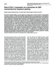
Figure 1. Structural features of the L1 adhesion molecule. L1 is a 200–220-kDa type I membrane protein composed of Ig-like domains (denoted Ig1–6) and five FN type III domains in the extracellular part. This is followed by a transmembrane and a cytoplasmic part that is highly conserved in different species. The ectodomain of L1 can be cleaved off close to the membrane by the metalloproteinase ADAM10, resulting in the release of a soluble fragment with a size of 200 kDa (L1-200). A fragment of 32 kDa (L1-32) is retained in the membrane. Additionally, L1 can be proteolytically processed by proprotein convertases or by plasmin. The position of the cleavage site and the generated fragments are indicated. FN: Fibronectin; Ig: Immunoglobulin.
The recent availability of the monoclonal antibody (mAb) L1-11A, subcloned from the original UJ127.11mAb to L1, has solved these problems and has allowed a detailed analysis of L1 in human ovarian and endometrial tumors. The classification of ovarian neoplasms relates to the nature of cell differentiation within these neoplasms, which in turn can be related to corresponding cell types of the normal ovary, the ovarian surface epithelium, the ovarian stroma and germ cells. In all cases, an understanding of the wide phenotypic potential of these cells can help explain the apparent phenotypic diversity of the corresponding neoplasms. The ovary is covered by a unique form of epithelium, the ovarian surface epithelium (OSE), which is thought to be the origin of EOC [17]. Epithelial cancers comprise 90% of ovarian tumors and are subdivided into benign, borderline and malignant categories. The incidence of ovarian cancer increases with age and is related to a variety of risk factors, such as nullparity, infertility and family history. During the last few years, a strong link was discovered between mutations at the BRCA genes and the occurence of EOC [18]. There are five major histological types of EOCs, including serous, endometrioid, mucinous, clear cell
457
Fogel, Huszar, Altevogt & Ben-Arie
A
B
C
D
E
Figure 2. L1 expression in a selected case of human metastatic ovarian carcinoma. Paraffin-embedded tissue sections were stained with monoclonal antibody L1-11A to human L1. L1-positive immunostaining was detected in primary serous ovarian carcinoma (A) and in the disseminated tumor to the omentum (B), uterine wall (C), cervix (D) and breast (E).
and transitional cell (Brenner) tumors. EOCs disseminate by the formation of peritoneal tumor implants through the abdominal cavity and invasion beneath the mesothelial layer into the underlying extracellular matrix. Cell adhesion and migration are critical steps during this process. The authors investigated expression of L1 adhesion molecule in ovarian carcinomas in a retrospective study. L1 was positive in 46 out of 58 ovarian carcinomas and was found to be a bad prognostic factor for patient survival (p < 0.0001). L1 immunohistochemical staining of a selected case of metastatic ovarian serous carcinoma is illustrated in FIGURE 2. L1 expression was found in the primary ovarian tumor (FIGURE 2A), invading into the omentum (FIGURE 2B), uterine wall (FIGURE 2C), cervix (FIGURE 2D) and metastasizing to the breast (FIGURE 2E). L1 was expressed in all the histological subtypes except for the mucinous adenocarcinoma. L1-positive tumors were usually of high disease Stage (III/IV), wheras L1-negative tumors were predominantly of Stages I/II. The early stages are associated with a better survival and therefore the knowledge of the L1 phenotype is not expected to gain a decisive role in tailoring the initial disease management. L1 in endometrial cancer
Endometrial cancer is the most frequently diagnosed gynecologic malignancy in Western countries. Approximately 38,300 new cases and 6600 deaths caused by endometrial cancer occurred in the year 2001 in the USA [19]. Several epidemiologic risk factors are known for endometrial carcinoma. These include obesity, late menopause, nuliparity and anovulation, which are all associated with prolonged exposure to unoposed estrogen. Similarly to EOC, there are five major histological subtypes that include endometrioid, serous, mucinous, clear cell and mixed endometrial carcinoma. Fortunately, as many as 75% of women with endometrial cancers present with localized disease and have an expected 5-year survival of over 75% [20].
458
Approximately 90% of the cases of endometrial adenocarcinoma are of the endometrioid histological subtype that usually harbors a very good prognosis. The remaining group is composed of high-grade clear cell, serous papillary and undifferentiated adenocarcinoma. Compared with the endometrioid subtype, these tumors are associated with worse prognosis. Stage remains the most important prognostic determinant. Other prognostic factors include histological type, histologic grade, lymph node involvement and the depth of myometrial invasion. The risk of recurrence is directly related to these prognostic factors [21]. The authors investigated expression of the L1 adhesion molecule in endometrial adenocarcinoma. L1 was positive in 20 out of 72 endometrial carcinomas and was again found to be a bad prognostic factor for patient survival (p < 0.0001). The immunohistochemical staining of a selected case of lowgrade endometrioid and serous high-grade endometrial carcinomas is illustrated in FIGURE 1. Most strikingly, L1 expression was detected in patients with early stage endometrioid type endometrial cancer usually considered to have a favorable outcome. The authors found that the prognosis for the L1-positive endometrioid tumors, including those in their early stage with an expected favorable outcome, was similarly bad as for other tumor types. Further analysis within the Stage I/II endometrioid group revealed that patients with similar clinicopathological characteristics had different clinical outcome (TABLE 2). In this cohort of patients with an expected favorable outcome, all L1-positive patients died of their disease, compared with 100% survival in patients with L1-negative tumors. The main treatment of endometrial cancer is surgery. This includes staging explorative laparotomy, total abdominal hysterectomy and bilateral salpingoophorectomy. Unsolved controversies in the treatment of endometrial cancer exist: to what extent retroperitoneal lymph node dissection is required and on the place of adjuvant radiotherapy in the subclasses of
Expert Rev. Mol. Diagn. 4(4), (2004)
L1 biomarker
Table 2. Correlation between L1 expression and clinicopathological characteristics of early stage endometrial endometrioid adenocarcinomas. Surgical stage
Histological type
Histological grade
L1staining
Outcome
Months post operation
Ib
Endometrioid
G1-G2
-
NED
72
Ib
Endometrioid
G1-G2
-
NED
45
Ib
Endometrioid
G1-G2
-
NED
68
Ib
Endometrioid
G1-G2
-
NED
55
Ib
Endometrioid
G1-G2
-
NED
49
Ib
Endometrioid
G1-G2
-
NED
28
Ib
Endometrioid
G1-G2
-
NED
70
Ib
Endometrioid
G1-G2
-
NED
24
Ib
Endometrioid
G1-G2
++
DOD
17
Ib
Endometrioid
G2-G3
-
NED
43
Ib
Endometrioid
G2-G3
-
NED
25
Ib
Endometrioid
G2
-
NED
98
Ib
Endometrioid
G2-G3
-
NED
84
Ib
Endometrioid
G2-G3
++
DOD
24
IIa
Endometrioid
G2
-
NED
80
IIa
Endometrioid
G3
-
NED
46
IIa
Endometrioid
G1
++
DOD
30
IIa
Endometrioid
G1
++
DOD
51
DOD: Died of disease; G: Grade; NED: No evidence of disease.
Stage I disease. Most textbooks and many studies advocate a tailored surgical and radiotherapy approach that is based on pre- and intraoperative prognosis factors, which include histologic type, grade of tumor and myometrial invasion. It was found that preoperative L1 immunohistochemical staining of curettage specimens is a valuable tool for the identification of aggressive endometrial tumors. L1 expression in the curettage sample and the excised tumor of one patient is compared in FIGURE 3. The figure exemplifies that the curettage sample is sufficient for tumor detection and L1 status before surgery. Curettage staining has been proven to be of high predictive value. In all 14 L1-positive endometrial tumors, the staining of curettage specimens matched the staining of the counterpart postoperative hysterectomy specimens. Likewise, in all 33 cases of L1-negative curettage specimens, the excised endometrial tumors were also negative. Therefore, L1 could be an important preoperative tool in guiding the extent of the surgical procedure and the postoperative adjuvant radiotherapy. Well-designed prospective studies are in process to validate the use of L1 expression in the classification and treatment of patients with endometrial carcinoma.
www.future-drugs.com
L1 for the diagnosis of metastatic carcinoma with unknown origin
Abdominal tumors of unknown origin are frequently encountered in clinical practice. Adenocarcinomas account for up to 60% of all metastatic neoplasms of unknown primary. The determination of the organ of origin of these tumors is often difficult and may have therapeutic consequences [22]. Some metastatic adenocarcinomas have distinctive histological features that allow for their site of determination, although the majority of metastatic adenocarcinomas have histological features that are not distinctive enough to allow diagnosis of their origin. In cases of pelvic metastatic adenocarcinomas in women, the differential diagnosis of ovarian or colonic carcinoma is very important as the standard treatment for these tumors is quite different. The authors have successfully used L1 as a marker for the analysis of unknown primary. Investigating L1 presence in 25 cases of colon cancer demonstrated that none express L1, while in nonmucinous ovarian cancer, 80% of the cases were positive. Although the number of cases of metastatic cancer of unknown primary was small, the authors can confidently conclude that whenever L1 is positive, the primary
459
Fogel, Huszar, Altevogt & Ben-Arie
The authors subsequently compared CA-125 and the soluble L1 marker in several cases of ovarian carcinoma follow-up. For example, in the case presented, the patient underwent total hysterectomy and bilateral salpingoophorectomy and omentectomy in April 2001 (FIGURE 5). Pathological evaluation indicated a bilateral endometrioid-type Stage IIIc adenocarcinoma of the ovary. The CA-125 value A B D C prior to surgery was high (544.3 µ/ml) and dropped afterwards. During chemoFigure 3. L1 expression in selected cases of human endometrial adenocarcinomas. therapy, CA-125 values were consistently L1-positive immunostaining was detected in endometrial endometrioid low-grade (A) and serous papillary high-grade (B) adenocarcinomas. Preoperative L1 staining of curettage specimen from the under cut-off values (