Jul 1, 1993 - Research Immunology/Bone Marrow Transplantation, Childrens. Hospital Los Angeles, 4650 Sunset Boulevard, Los Angeles, CA. 90027. 2567.
Proc. Natl. Acad. Sci. USA Vol. 91, pp. 2567-2571, March 1994 Medical Sciences
Lack of expression from a retroviral vector after transduction of murine hematopoietic stem cells is associated with methylation in vivo (gene therapy/gene expresion/bone marrow/long terminal repeat)
PIA-MARIA CHALLITA* AND DONALD B. KOHN*t# Departments of *Microbiology and tPediatrics, University of Southern California School of Medicine, Division of Research Immunology/Bone Marrow Transplantation, Childrens Hospital Los Angeles, Los Angeles, CA 90027
Communicated by Elizabeth F. Neufeld, November 18, 1993 (received for review July 1, 1993)
ABSTRACT We describe studies of gene transfer and expression of the human glucocerebrosidase cDNA by a Moloney murine leukemia virus (MoMuLV)-based retroviral vector in a murine gene transfer/bone marrow transplant (BMT) model. Pluripotent hematopoietic stem cells (HSCs) were assayed as the colony-forming units, spleen (CFU-S) generated after serial transplantation. Transcriptional expression from the MoMuLV long-terminal repeat (LTR) was detected at a high level in the primary (1a) CFU-S and tissues ofreconstituted BMT recipients. However, we observed transcriptional inactivity of the proviral MoMuLV-LTR in >90% of the secondary (2w) CFU-S and in 100% of the tertiary (3°) CFU-S examined. We have compared the methylation status of the provirus in the 1° CFU-S, which show strong vector expression, to that of the transcriptionally inactive provirus in the 2° and 3° CFU-S by Southern blot analysis using the methylation-sensitive restriction enzyme Sma I. The studies demonstrated a 3- to 4-fold increase in methylation of the Sma I site in the proviral LTR of 2° and 30 CFU-S compared to the transcriptionally active 1° CFU-S. These observations may have important implications for future clinical applications of retroviral-mediated gene transfer into HSCs, where persistent gene expression would be needed for an enduring therapeutic effect.
tain circumstances, with the silencing of viral control elements (6). The MoMuLV-LTR is completely inactive in embryonic stem and embryonic carcinoma cell lines, and the inactivity is accompanied by de novo methylation of the proviral sequences (7, 8). Moreover, methylation has been detected in association with the MoMuLV-LTR transcriptional inactivity in fibroblasts in vitro (9) and in vivo (2). In this study, we investigated long-term in vivo expression from the MoMuLV-LTR by transduction of murine bone marrow cells with a MoMuLV-based retroviral vector and serial bone marrow transplantation (BMT) into lethally irradiated recipient mice. We document a high rate of expression failure associated with methylation of the vector LTR in the secondary (20) and tertiary (30) colony-forming units, spleen
(CFU-S).
MATERIALS AND METHODS Retroviral Vector. The G2 retroviral vector and its corresponding high-titer amphotropic PA317 packaging cell clone have been described (5). G2 consists of the LTR from the N2 vector flanking the human GC cDNA. The packaging cell line clone used in the experiments was negative for helper virus production assayed by testing for transfer of the amphotropic env gene into 3T3 fibroblasts through PCR analysis (10). Transduction of Murine Bone Marrow Cells. Donor bone marrow cells were harvested from male C57BL/6J mice (Charles River Breeding Laboratories), prestimulated in the presence of growth factors, and cocultivated over vector producing fibroblasts according to the methods described by Weinthal et al. (5). The growth factors used for the prestimulation were 200 units of murine interleukin 3 (IL-3) per ml (Biosource, Camarillo, CA), 100 units of human IL-6 per ml (Amgen), 200 units of human IL-la per ml (Immunex) and 50 ng of mast cell growth factor per ml (or c-kit ligand; Immunex). BMT and Sample Collection. Recipient female C57BL/6J mice (8-12 weeks old) were irradiated with two split doses of 600 and 450 cGy 24 hr apart. Transduced bone marrow cells were injected into the tail vein of the irradiated mice at 1 x 10⁶ cells per mouse for isolation of CFU-S or 2-4 x 10⁶ cells for long-term reconstitution. Twelve days after BMT, two to four mice transplanted with 1 x 10⁶ bone marrow cells were sacrificed. Well-defined, individual primary (10) CFU-S were isolated and divided evenly into two portions, one for DNA
Gene therapy via bone marrow cells is a promising technique for treatment of a wide variety of human diseases, including genetic disorders, cancer, and AIDS. Effective long-term bone marrow gene therapy requires the fulfillment of two main criteria. The exogenous gene should be introduced into a high percentage of long-lived pluripotent hematopoietic stem cells (HSCs). Subsequently, the introduced gene should be persistently expressed in the mature hematopoietic progeny cells of the stem cell, thereby maintaining the effects of gene therapy for the lifetime of the individual. Although Moloney murine leukemia virus (MoMuLV)-based retroviral vectors are currently the most efficient vehicles for gene transfer into a variety of cell types including HSCs (reviewed in ref. 1), the long-term in vivo expression from the viral promoter/enhancer elements has been unsatisfactory. Lack of gene expression from the 5' MoMuLV long-terminal repeat (LTR) has been observed in several systems including primary fibroblasts (2) and hematopoietic cells (3, 4). Previous studies by our laboratory, using a retroviral vector in which a normal human glucocerebrosidase (GC) cDNA is controlled by the enhancer/promoter of the 5' MoMuLV-LTR, demonstrated a high rate of lack of expression in cells derived from HSCs (5). Methylation of cytosine residues has been shown to be associated with suppression of gene expression and, in cer-
Abbreviations: MoMuLV, Moloney murine leukemia virus; LTR, long terminal repeat; HSC, hematopoietic stem cell; GC, glucocerebrosidase; CFU-S, colony forming unit, spleen; BMT, bone marrow transplantation; 10, primary; 20, secondary; 30, tertiary. tTo whom reprint requests should be addressed at: Division of Research Immunology/Bone Marrow Transplantation, Childrens Hospital Los Angeles, 4650 Sunset Boulevard, Los Angeles, CA 90027.
The publication costs of this article were defrayed in part by page charge payment. This article must therefore be hereby marked "advertisement" in accordance with 18 U.S.C. §1734 solely to indicate this fact. 2567
2568
Proc. Natl. Acad Sci. USA 91
Medical Sciences: Challita and Kohn Table 1. Expression of G2 in the mouse model of gene transfer/BMT Exp. 2 Exp. 1 RNA RNA DNA DNA 5/5 6/6 12/12 9/9 10 CFU-S 10 tissues (1.5 months) Spleen Thymus Marrow 10 tissues (3 months) 1/1 Spleen Thymus 1/1 Marrow 10/15 0/10* 20 CFU-S (1.5 months) 1/7 20/20 20 CFU-S (3 months) 20 tissues (3 months) 1/2 2/2 Spleen 0/2 2/2 Thymus 1/2 2/2 Marrow
Total
Exp. 3 RNA DNA 8/8 8/8
DNA 29/29
RNA 19/19
2/2 2/2 2/2
2/2 2/2 2/2
2/2 2/2 2/2
2/2 2/2 2/2
2/2 0/2 2/2 3/28 37/38
2/2 0/2 2/2 0/3 3/35
2/2 0/2 3/3 57/58
2/2 0/2 2/2 0/13 4/42
2/2 2/2 2/2 7/29
1/2 0/2 1/2 0/7
13/43
0/7 7/29 30 CFU-S *This set of 2° CFU-S was analyzed for RNA expression by reverse transcription/PCR.
and one for RNA analysis. Animals transplanted with 2-4 x 10⁶ bone marrow cells were sacrificed after 1-3 months. Hematopoietic tissues were collected for nucleic acid analysis, and bone marrow cells were used directly to reconstitute a second generation of lethally irradiated female mice. Twelve days after the secondary BMT, the 2° CFU-S were isolated for DNA and RNA analysis. In one experiment, 30 BMT was performed from bone marrow of long-termreconstituted 20 recipient animals in order to generate 30 CFU-S. DNA and RNA Analysis. Genomic DNA was isolated by SDS/proteinase K and RNase digestion at 550C for 3-4 hr. The digested tissues were extracted with phenol/chloroform; the DNA was precipitated in ethanol and resuspended in TE buffer. The presence of proviral GC sequences in the CFU-S and hematopoietic tissue samples was assayed by PCR using the human GC-specific oligonucleotide primers described by Weinthal et al. (5), followed by Southern blotting and hybridization with' a 32P-end-labeled internal oligonucleotide (8). Provirus DNA was also detected by Southern blot analysis after digestion of genomic DNA with the Sst II and Xho I restriction enzymes (BRL). These digestions release the 1.65-kb GC cDNA detected by hybridization with the 1.5-kb (Sst II/BamHI) human GC cDNA probe. The probe was labeled with [32P]dCTP by the random-priming method. Individual provirus integrants in the CFU-S and long-term hematopoietic tissues were detected by Southern blot analysis of genomic DNA digested with BamHI, which cuts at one site in the provirus. Again, the Southern blot was hybridized with the 1.5-kb 32P-labeled human GC cDNA probe. RNA was isolated from the tissues by the acid guanidinium thiocyanate/phenol/chloroform method (11). RNA (15 ug) was electrophoresed on a 1.2% formaldehyde gel, denatured, neutralized, and transferred to a nylon membrane by capillary blotting. The filter was hybridized with the' human GC cDNA probe. After a satisfactory exposure was obtained, the filter was stripped and rehybridized with the mouse 3-actin DNA probe. For reverse transcription/PCR, 1 pg of RNA was reverse transcribed using the human GC-specific oligonucleotide primers, followed by PCR amplification of the cDNA as described above for the DNA samples. Methylatlon Analysis. The methylation status of the proviral 5' LTR in the CFU-S was determined by digestion of genomic DNA (15-25 pg) with BamHI to reduce the size of the DNA fragments, followed by Pvu II digestion. The DNA was then precipitated with ethanol, redissolved in TE buffer,
(1994)
and divided into two equal portions, one of which was
subjected to digestion with the methylation-sensitive enzyme Sma I. Completeness of the genomic DNA digestions was monitored by mixing a sample of the digestion mixture with either adenovirus type 2 DNA or A DNA (BRL), which were subsequently run on a 1% gel. Pvu II and Pvu If/Sma digested DNA were electrophoresed and blotted to nylon membranes. The blots were probed with a 32P-labeled fragment of the G2 vector from the Spe I site in the untranslated leader region to a Pvu II site near the 5' end of the GC gene (see Fig. 3). Densitometric analyses were performed with the United States 'Biochemical SciScan 5000, measuring the relative densities of the 1.8-kb Sma I-resistant band and the 1.5-kb Sma I-sensitive band in each lane.
RESULTS Expression of G2 in Vivo in Murine HSCs. Results of G2-mediated gene transfer and expression in, the mouse 2 CFU-S
1 CFU-S
G2
1--1
G2
PA317 X- -)G2
Human 3q93 GC
12 13
Mouse .,
S. b
3-Actin
1
2
3
4
5
do 6
7
8
to
9 10 '4
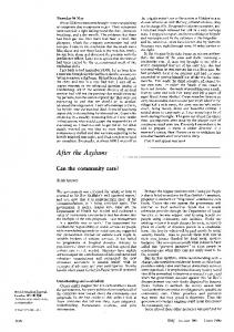
FIG. 1. Representative Northern blot analysis of the CFU-S generated in the mouse model of gene transfer/BMT (experiment 1 ofTable 1). RNA from 10 CFU-S (lanes 1-6) and 20 CFU-S generated 1.5 months after primary BMT (lanes 7-11). Bone marrow cells used for BMT were transduced with G2 (lanes 1-4 and 7-11) or with the neomycin-containing control retroviral vector (lanes 5 and 6). RNAs from the fibroblast cell line PA317 (lane 12) and PA317 transduced with G2 (lane 13) were used as negative and positive controls, respectively, forGC mRNA. (Upper) Northern blot was probed with the human GC cDNA identifying the two proviral transcripts, the full-length 4.0 kb, and the spliced 3.5 kb. (Lower) Same blot was stripped and rehybridized with the mouse P-actin probe except for the control samples (lanes 12 and 13).
Medical Sciences: Challita and Kohn rCo)
rc',
1'tissues
a:L~