Abstract. Anatomical and ultrastructural details of a translocating 10-cm leaf of sugar beet (Beta vulgaris L. var. Klein Wanzleben) were correlated with ...
Plant Physiol. (1969) 44, 45-54
Leaf Structure and Translocation in Sugar Beet' D. R. Geiger and D. A. Cataldo Department of Biology, University of Dayton, Dayton, Ohio 45409 Received July 17, 1968. Abstract. Anatomical and ultrastructural details of a translocating 10-cm leaf of sugar beet (Beta vulgaris L. var. Klein Wanzleben) were correlated with translocation rate datia. The minor veins were found to be 13 times as extensive as the major veins and measure 70 cm/cm2 leaf lamina. Measurements disclosed that a 33-,u length of minor vein services 29 mesophyll cells with the result that translocate moves an average of 73 ,u or 2.2 cell diameters during transport from mesophyll cells to a minor vein. High-resolution. freeze-dry autoradiography revealed that assimilates accumulate in organelle-rich cells of the minor vein phloem. Correlation of phloem volume and loading rate for minor veins yielded an uptake rate of 735 Amoles of sucrose per g fresh weight of phloem. The arrangement and structural features of minor veins appeared to be consistent with the concept that vein loading precedes translocation.
pounds in a translocating leaf. In addition, anatomical measurements of a translocating leaf were correlated with translocation rate data to ascertain the feasibility of vein loading of minor veins as a means of driving translocation of organic compouinds.
In the search for the mechanism of translocation of organic compounds, various workers have proposed models based on active transport of materials by phloem. The proposed mechanisms can be divided into those in which the active driving process occurs all along the translocation path and those in which this process is centered in the phloem of the source regions such as the vein endings of the leaf blade (1,8,14,15,22,25,30,34). Although inhibitor studies offer some hope of distinguishing between the alternatives, to date this approach has failed to differentiate conclusively between the 2 groups of proposed mechanisms. Difficulties include the failure of the effects of chemical inhibitors to be sufficiently restricted to the point of application (15) and the production of chilling effects beyond the inhibition of metabolism (29). Results of recent localized-chilling experiments suggest that in the phloem outside of source and sink regions, metabolism serves primarily to maintain structural organization while in source and possibly in sink regions it serves to move the translocate (10,29). Findings from experiments employing low temperature treatment thus favor mechanisms which include active vein-loading in the source leaf (1, 3, 8, 25, 30, 33). From a histochemical standpoint, previous studies have suggested a possible active loading function for border parenchvma cells and companion cells in 1 case (30) and for companion and transfer cells (Ubergangszellen) in another (1). In the present studv, several recently devised techniques (6, 7, 18, 19) were used to locate cells which accumulate water-soluble organic com-
Materials and Methods Plan1t Material. Sugar beet plants (Beta viulgaris L. var. Klein Wanzleben) were grown by solution culture in controlled environment cabinets as described earlier (11). Studies were performed on 10-cm leaves of 5 to 7-week-old plants pruned to a simplified translocation system (11, 12). Measurement of Venation. To visualize the vein pattern, leaves were allowed to take up a 0.25% (w/v) water solution of acid fuchsin until the dye had reached the minor veins. Injected leaves were fixed in 80 % ethanol, dehydrated, and then cleared in methvl salicylate. Extent of venation was measured in photographic enlargements of the cleared leaves. Histological Procedures. Details of vascular tissue were studied in 2-, thick sections of leaf tissue fixed in 3 %o (v/v) glutaraldehyde in pH 7.2 phosphate buffer or in 10 % acrolein in tap water. Tissues were dehydrated in acetone and embedded in hydroxyethvl methacrylate by the method of Ruddell (26). Tissue for ultrastructural studies was prepared by fixing 1 mm2 pieces of leaf in 3 % (v/v) glutaraldehyde in pH 7.2 phosphate buffer for 1.5 hr at 4' or in 2 % (w/v) KMnO4 for 15 min at room temperature. Glutaraldehyde fixed tissues were washed for 1.5 to 3 hr in buffer, and postfixed in 2 % (w/v) osmium tetroxide in pH 7.4 phosphate buffer for 2 hr at 4'. Sections were poststained in Reynolds' lead acetate.
1 This research was supported in part by Grant GB2470 from the National Science Foundation.
45
46
PLANT PHYSIOLOGY'
(.4:
'I~ ~
~
~~.
.-/
'F
YI
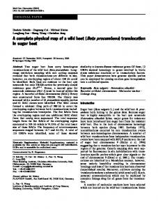
FIG. 1. (Upper left) Clearedl 10-cm sugar beet leaf, xylem-injected with acid fuchsin, showing major venation. FIG. 2. (Upper right) Detail of small branches of major venation. White rectangle designates positioni of 0.5 111n112 area sholwn in figur-es 3 and 4. Minor veins are not apparent at this magnificationi. X 2.6. FIG. 3. (Lo-wer left) Mesoplhyll cells overlying the minor veins showvn in figure 4. X 125. FIG. 4. (Lox-er right) Pattern of iminor venation in 0.5 mm2 area of 10-cmi sugar beet leaf. X 125. c) connectionis of minor veint net with major veins: cc) presumed companiion cell; ch) chloroplast; m) mesophyll cell; mv) minor vein; o) oxalic acid crystal p) procalnbial cell: pd) plasimiodesimi; pp) phloem parenchyma or companion cell; s) sieve tube: sp) sieve plate; v ) -acuole.
47
GEIGER AND CATALDO-LEAF STRUCTURE AND TRANSLOCATION
3 and 4). A drawing of a representative pattern of minor venation in a 0.8 mm2 area of lamina is shown in figure 5A. The minor vein net connects with a tertiary vein on 1 side and with quaternary branches on 2 other sides. Examples of paradermal and cross sections of a minor vein have been reconstructed from low magnification electron micrographs
Freeze-dry Autoradiography. Details of steady-
state labeling with "4CO2 were described previously ( 12). Leaves were sampled after 2 or 4 hr of labeling at which time tissue activity was 30 to 50 Squares of leaf several millimeters on /ic/dm2 leaf. quickly frozen -vith 8 % (v/v) methyla side were cyclohexane in isopentane at -170 to -180( and stored under powdered dry ice prior to dehydration by a modificationl of the mlethod of Jensen (18). Moisture content of the air streamii was mlaintained by equilibration with ice at -35o* Dehydration was carried out at -450 hile the trap was held at -78°. Following 36 to 72 hr of drying. the tissue was vacuunm infiltrated in 560 paraffin for 30 to 45 min. Sections 6 u in thickness were pressed against a thin film of Ilford K-5 emulsion, in total darkness (18). Films of froml 0.2 to 1 u thickness w-ere made by v-arying the speed of w-ithdrawal of the coverglass from liquified emlitilsion at 580 ( 19). Autoradiographic slides were sampled after periods of 3 to 15 days exposure at 40 and developed in Kodak Microdol X at 140 (18).
(figs 5-7).
To evaluate the relative accessibility of the leaf mesophyll to major and minor veins respectively, the extent of each of these classes of venation was measured. A value of 70 + 10 cm of minor vein per cm2 leaf was obtained from a sampling of 36 1-mm2 fields. By comparison, the major veins measure 5.5 cm per cm' leaf. Consequently, the minor veins have more than 10 times greater accessibilitv to the mesophvll than the major veins. Calculations based on measurements from cleared and sectioned leaf material yielded an estimate of 3 X 10⁵ mesophyll cells per cm2 leaf lamina. Combining data for cell size, vein extent and cell population (table I). it was calculated that a minor vein length, corresponding to the diameter of an average mesophvll cell, receives translocate from approximately 29 mesophvll cells. By contrast, a similar length of major vein is accessible to about 370 cells. In a leaf 6.7 cell diameters thick, the cells serviced would be within an average lateral distance of 2.2 cell diameters or 73 ,u of a minor vein. Wylie (32) found the average width of tissue bordering the minor veins in 66 species of dicotyledons to be 65 K. A sampling of cross sections of 24 minor veins disclosed that the average perimeter of contact between the phloem in a minor vein and the adjacent mesophyll is
Results and Discussion As in previous translocation rate studies (10.11, 12, 13,29), 10-cni sugar beet leaves were used as experimental mlaterial. Veiins of the mlidrib and the first 3 or 4 branches wxere visibly injected with acid fuchsin (figs 1 and 2). Beyond these major veins, the vascular system is comiposed of a network of minor veins with relatively few elemlients (figs
Table 1. .A1 (lt)I1liCll (hi(d Ph \sioloi(cal Porometcrs for Mintor Veinis in Loz7linina of 10-cmi Leaves of Beta vulgaris
Extent ot minor venation
cells serviced by a 33-, length of minor veinl
cni, 'cni-
cells/33-,u length
Surface area of bundle at interface with mesophyll cells in leaf2 cni2/cm2 leaf
29
0.49
No. of mesophyll
70 2
4
+
10
plhloem
Volume of minor vein phloem in a cm2 of leafs mm3/ccm2 leaf 0.31
Rate of export of sucrose from leaf4
,Ug/cm2± min
Average diamleter of mesophyll cell is 33 + 5 ,. Average perimeter of conitact between phloem of minor vein cross section and mesophyll is 70 Average cross sectional area of minor vein phloem is 440 + 9: ,. Data fromi Geiger and Swanson (13).
Table I[.
0.4
1.3
+
11 ,u.
)istrilbittioni of 14C Bet.keein V'ariozns Categories of Comtpounds in the Lainina of a 10-cmi Snlgar Beet Leaf Afte UVarious Pcriods of Steady-State Labelintgl 120 min
80 min ,c
13.5 80 % Ethanol insoluble compounds 7.7 Sucrose 7.1 Other soluble compounds 28.4 Total radioactivity in lamina Details o i method in Geiger and Swanson (13).
% 48
13.8
% 49
27
5.6
20
25 100
9.0 28.4
31 100
Ac
Labeling period 160 min /c
19.1 3.7 7.5 30.3
% 63 12 35 100
240 min c
%
19.3 3.9 8.0 31.2
62
13 25 100
4PLANT PIIYSIOLOGY
AO
M\1INOR XVENATION A
m...; .
C.
S
.4 ..
...:
*S.
:..
.. ..: ..eL. a;.
`!-
... pI e
@
.:.:
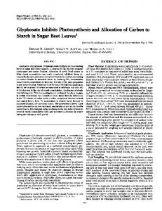
FIG. 5. (Upper) Diagram of minor venation from leaf area in figure 4. A) pattern of minor veins shoving connection to branches of major veins; B) paradermal section through minor vein phloem; C) cross section through minor vein.
FIG. 6. (Lower left) Electron micrograph of cross section through phloem of minor vein similar to figure 5C. X 7500. FIG 7. (Lower right) Electron micrograph of paradermal section similar to figure jB showing phloemii parenchyma and sieve tubes. X 5800. Insert (rectangle) shows details of sieve pore with endoplasmic reticulum traversing the pore. X 31,000. See figure 4 for key.
.4>, .:+~
49
GEIGER AND CATALDO-LEAF STRUCTURE AND TRANSLOCATION
|v
..sf
...
......
..
......
..... ......
MEMM...
..........
*. *
.
S ~~~~~~~~~~~~~~~~~S
N
l
ef
:6.: .;.
;-*
:k
-4...
i:.:::
4-:
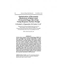
FIG. 8. Freeze-dry autoradiograph of paradermal section of minor vein. A) (U pper) Phase contrast x iewv phase dark and showing concentration of silver grains over minor vein tissue. Some silver grains slightly different bright. B) (Lower) Brightfield view of sanme field as A showing pattern ot silv grains at level of focus. X 1750. See figure 4 for keev. are
some
a
er
LIBRARY
THE
PUBLIC OF THE
TN TITUTE RI~EARCH Y-.R
HEALTH OF NEW
CITY
4
,
5, [FIR 11 A;
N'
916
.
are
50
PLAN-T PHYSIOLOGY
70 + 11 u giving a combined surface of 0.49 cm2 for minor vein phloem per cm2 leaf (table I). The greater accessibility and the higher surface to volume ratio of the minor veins is consistent with the thesis that they accumulate assimilate as part of the translocation process. Several workers (1 30) have proposed that parenchyma cells associated with the minor veins function in the transfer of assimilates into the sieve tubes (1, 8, 14, 30) though they do not agree on the specific cell type involved. To investigate the possible transfer role of cells associated with leaf veins, we undertook a histoautoradiographic study to locate cells capable of accumulating assimilates in a translocating leaf. Sugar beet leaves were supplied with '4CO" under conditionis whiclh result in a high rate of translocation of labeled sugar (12). Previous studies have shown tllat during the first 100 min of steady-state labeling the sucrose in the leaf reaches isotopic saturation (12. 13); thereafter, the spatial distribution of soluble compounds suich as sucrose, which have reaclhed tlleir mlaximiunm specific activity, is accurately reflected in the distribution of radioactivity throughout the tissue. At the same tinme of sampling, sucrose constittutes 12 to 20 % of the label (table II); this proportion decreases with time as a result of accumulttion of insoluble mlaterials in the leaf. The distribution of radioactivity in a paradermlal section of freeze-dried leaf tissue, as revealed bhigh-resolution autoradiography. is shown in figures 8 to 10. The grain density in figures 8 to 10 indicates a greater amount of labeled material present itn the phloem than in the cytoplasmn of adjacent mesophyll cells. The sieve tubes are too small to be located with certainty, but the labeling appears to be greatest over the cells wvith the dense cytoplasnm. Control autoradiographs (fig 11) show the absence of significant numbers of silver grains produced by chenmical actionl or by background radiation. Because a large portion of the label in the leaf is in glycogen (13), protein aind other- insoluble compotunds, autoradiographs of 80 % ethanol extracted tissue were prepared (fig 12). In contrast to the pattern for non-extracted tissue, there is a highlier concentration of silver grains over the chloroplasts and cytoplasm of the nmesophyll cells than over the minor veins in the extracted tisstue. Extraction caused some ioss of resoluition as evidenced by the presence of grains at the periphery of tiie vacuoles and slightly beyond the edges of the tissue sections. These comparative autoradiographs indicate that a large portion of the radioactivity in the phloenm is in ethanol-soluble compotunds, prestumably, largely in sucrose. This pattern of accumulation b- organelle-ricl cells resembles the previously reporlted distribution of energy-related compounds localized in cells postulated to function in vein loading during translocation (1, 14, 30). On the basis of phosphatase and tetrazolium reactions, Bauer implicated companion and transfer cells of the minor veins of Madia dissitifloira
and Vicia faba in the active loading of tranislocate (figs 10-13 of 1). He also found a lesser conceIntration of phosphatase and of formazan in the sieve tubes and little activity in the border parenchyma anid mesophyll cells. The recent work of Gunning. Pate and Briartv (14) demonstrates an accumulation of insoluble materials in transfer cells of the minor veins followN-ing xylem injection wvitlh 'H-leucine solution. These workers also found acid phosplhatase localized in the cell walls of the transfer cells. An examination of the tultrastructure of the minor veins revealed specializations which may relate to the proposed vein loading process. Typicallv. a minor vein is composed of 1 to 3 files of xylem vessels and 4 to 12 files of phloem cells (fig 5C). In the light of vein loading hypotheses. 2 features of the phloem appear noteworthy. First is the abundance of mitochondria and other cytoplasmic organelles in the companion cells and parenchynma cells of the phlo,em (figs 6. 7, 13), wvhich would be expected in cells involved in active transport. The second feature is the relatively large size of the organelle-riclh comiipanion cells in the minor v-eins (fig 13) as coml)ared with similar cells found in the petiole (fig 14). This size relationship. previously noted by Morretes (23. 24), may represent an additional adaptationi for vein loading. A number of calculations were made to correlate the structural features described above wvith transslocation rate data. During photosxynthesis uilder saturating light initensity, a 10-cm sugar beet leaf exports sucrose at the rate of 1.3 pAg/min cm2 or 7.8 mg/hr dm2 leaf. On the assumiption that loading of minor veins is aIn integral part of translocation. the entry of sugar inlto the minor vein phlloemI wX-ill occur at this same rate. The phloem of the miinor -eins occupies a volume of approximately 70 cm X 440 p2 (table I) or 3.1 X 10⁻⁴ cm23/cm2 leaf; the estimated fresh weight of this phloem is 310 ug/cm2 leaf. From this it follows that the sugar uptake rate Nvould be 1.3 pg sucrose/min per 310 pg fr w-t phloem tissue or 4.2 mg sucrose/min g fr wt phloen tissue. On a molar basis this is 735 pmoles sucrose/hr g fr wt phloenm in the minor veins. This rate of vein loading is equal to the rate of uptake of sugar from a 10 % sucrose solution reported by \Veatherlev for leaf disks of Atro pa belladonna (31). In the latter case, nitrogen anaerobiosis reduced the uptake rate to 25 %, suggesting an active transport mechanism is responsible for a major part of the uptake. Another indicariui- of the presence of an active transport mechanism is the high uptake rate by minor vein phloem of the sugar beet. In this regard. Bieleski (2) reported that phloem tissue excised from stemls actively accumulates 9 to 16 ,umloles sucrose/hr g fr wt of tissue from a 10-1 AI -sucrose solution, a concentration representative of the sugar content of photosynthesizing leaf tissue. The rate calculated for nminor veins of the intact sugar beet leaf is 50 to 80 times the rate found b-
Al
.*stF -
GEIGER AND CATALDO-LEAF STRUCTURE AND TRANSLOCATION
51
'..:. 1
..
.
....
:: .5
:
s:
.::
s^ m.s-... X..., w.
.. e
..
'i, ...
..
xitv~~~~~~~~~~~~~~~~~~~~~~~~~~~~~~~~~~~~~~~E5
AL
4s.
.c
-Z
.R~A
FIG. 9,10. (Upper) Phase contrast view of minor veins in a paradermal section of source leaf; identityc of individual cell types difficult to discern. Similar to figure 8A. X 950. FIG. 11. (Lower left) Freeze-dry autoradiograph of a paradermal section of unlabeled control tissue. X 950. FI(;. 12. (Lower right) Autoradiograph of 80 % (v/v) ethanol-extracted freeze-dried tissue showing labeling pattern from insoluble materials. X 950. See figure 4 for key.
*Xs}'-ot;,Z7iwe>.|'fxE@gb61C:as5n°N_S*4F-q/.X!l:>b,t;axewZd°-'.20s
52
PLANT PHYSIOLOGY
o#
;r
-
SF
..
'6?i
i
M f
p
.::
.: ,,.
-3 i
-: 0
3ffi.
;
..
,: ;. #.' ... '.
°2
e xz
e
'#
..01 W.,:
.,:!l:;l'0-1.0 I.... -. -ld ...!w
S
*,'
=:
:: . . . . . .
.
^
,
r
'.-.-=L,.
>;X.
FIG. 13. (Upper left) Electron micrograph of cross section of minor vein of leaf showing relationship between mesophlyll cell, phloein- parenchyma cell and sieve tube. X 22,000. FIG. 14. (Uppet right) Ultrastructure of phloeni in cross section of petiole. Note relative sizes ot plhloemii parenchylma cells and sieve tubes in minor vein of leaf (figs 6,7,13) and in petiole. X 5 800. FI(;. 15. ( Lower left) Plasmodesmiiata between sieve tube andl companion cell in cross sectioni of iminlor vein. X 20,000. FIG. 16. ((Lower right) Cytoplasmiiic connlectionls between mesolphyll cell, phloeni parenchyiima cell, and(l sieve tube. See figure 4 for key. X 17,400.
GEIGER AND CATALDO-LEAF STRUCTURE AND TRANSLOCATION
Bieleski for phloem excised froml the stemii. This differenice in rates is consistent with the relatively greater abundance of organelle-rich cells in the phloem of the translocating leaf than in the petiole. In the leaf lamina, the minor vein phloem consists of 80 to 90 %, by volume, of organelle-rich parenchyma cells while in the path or petiole portion of the leaf these cells occupy 20 to 30 % of the volume. Consequently, the surface to volume ratio is nmore conducive to sieve tube loading in the minor veins thaln in the petiole (figs 14, 15). Althotugh there is evidence favoring accumulation of assimilates by the phloem parenchyma cells of the minor veinis prior to translocation, many of the details *of the imiodel are tentative and remlain to be examined. From studies of chloroplasts isolated by non-aqueous methods, it appears that sucrose is first produced in the chloroplasts (4, 28) and quickly moves into the cytoplasm (28). Evidence for the intercellular miovemient of sucrose in the free space via the cell walls and intercellular spaces has been presented by Hawx-ker (16) and by Kriedemann (20). Several modes of entry of sugar into the phloeni have been suggested as a result of various experienlts. Sotmie -studies give evidence for the hydrolysis of sucrose prior to its accumulation in cells (5, 27) wvhile others indicate that phosphorylation precedes entrV- (22) ; still others indicate no hvdrolv-sis prior to uptake (17, 21). The contribution of cell surface structutres in the minor veins is not active clear. Cell wall protuberances postullate(d in vein loading ( 14), were not observed in sugar as
beet. Numierous plalsmodesmiata were observed in the w-alls of both mieFophyll and phloem cells (fig 16) but their inlportance in vein loading is conjectulral (9). The l)reseut study denmolnstrates that orgaliellerich miinlor -eini cells of the translocating leaf accumulate assimilaltes. The structural features of these cells anid tleier relationship to the mesophvll cells is consisteint with the concept that parenchyma cells of the mn,inor veins actively accumulate sugar lrior to its enltr- inLto the sieve tubes. Correlation of phvsiological anid structural mleasuremiielnts reveal nlo major inconlsi siencies wxi.hI resl)ect to vein-loading theories.
Acknowledgments
2. 3. 4.
5.
6.
7. 8. 9.
10. 11. 12.
13. 14.
15. 16.
17.
53
Wand:rung von Fluorochromen. Planta 42: 36745 1. BIELESKI, R. L. 1966. Accumulation of phosphate, sulfate, and sucrose by excised phloem tissues. Plant Physiol. 41: 447-54. BTELESKI, R. L. 1966. Sites of accumulation in excised phloem and vascular tissues. Plant Physiol. 41: 4.55--66. BIRD, I. F., H. K. PORTER, ANtD C. R. STOCKING. 1964. Intracellular localization of elnzymes associated with sucrose synthesis in leaves. Biochim. Biophys. Acta 100: 366-75. BROVCHENKO, M. I. 1964. On the movement of sugars from the mesophyll to the conducting bundles in sugar-beet leaves. Soviet Plant Physiol. 12: 230-37. CARO, L. G., R. P. VAN TUBERGEN, AND J. A. KOLB. 1962. High-resolution autoradiography. I. Methods. J. Cell Biol. 15: 178-88. CARO, L. G. 1962. High-resolutionl autoradiography. II. The problem of resolution. J. Cell Biol. 15: 189-99. ESAU, K., H. B. CURRIER, AND V. I. CHEADLE. 1957. Physiology of phloemii. Armi. Rev. Plant Physiol. 8: 349-74. ESAU, K., J. CRONSHIAW, AND L. L. HIOEFERT. 1967. Relation of beet yellows virus to the phloem and to moveement in the sieve tube. J. Cell Biol. 32: 71-87. GEIGER, D. R. 1966. Effect of sinik regioni cooling on translocation. Plant Physiol. 41: 1667-72. GEIGER, D. R. AND C. A. SWN-ANSON. Sucrose translocation in the sugar beet. Plant Phvsiol. 40: 685-90. GEIGER. D. R. AND C. A. SWANSON. 1965. Evaluation of selected paramleters in a sugar beet translocation system. Plant Physiol. 40: 942-47. GEIGER, D. R. AND J. BATEY. 1967. Translocation of '4C sucrose in sugar beet d(ur-iig darkness. Plant Physiol. 42: 1743-49. GUNNING, B. E. S., J. S. PATE, AND) 1L. G. BRIARTY. 1968. Specialized "transfer cells" in minor veins of leaves and their possible significance in pholem translocatiotn. 1. Cell Biol. 37: C7-C12. HAREL, S. AND L. REINHOLD. 1966. Effect of DNP on translocationi in the phloem. P'hysiol. Plantarum 19: 634-43. HAWKER, J. S. 1965. The sugar content of cell walls arid intercellulai- spaces in sugar-canie stems and its relationi to sugar transpo)rt. Australian J. Biol. Sci. 18: 959-69. HUMPHREYS, T. E. AND L. A. GARRARD. 1967. The storage of exogenous sucrose by corn scutellum slices. Phytochemistry 7: 701-13.
18. JENSEN, W. A. 1962. Botanical Histochemistry: grateful to Drs. Mf. Arif Hayat of the Ifniversitv of D)avton and Robert M. Giesy of the Ohlio State Universitv for their assistance in the electron microscope study alnd to the Ohio State University College of Biology for the generous use of their electron microscrope facilities. WNe alsoz thanik 'Miss Sharon NIcCloskey and fMiss Mary Anthony for their competent technical assistance. authors are
Literature Cited 1. BAUER. L. 1953. Zur Frage Stoffbewegungen in der Pflanze mit besonder-er Beriicksichtigung der
Principles and Practice. W. H. Freeman and Company, San Francisco, California. 19. KOPRIwVA, B. M. 1966. A semiautomatic instru.nent for the radioautographic coating technique. J. Histochem. Cytochem. 14: 923-28. 20. KRIEDEMANN, P. 1966. Sugar uptake and translolocation in the castor bean seedling III. An autoradiographic study of the absorption pathway. Planta 73: 175-80. 21. KRIEDE-MANN, P. AND H. BEEV-ERS. 1966. Sugar uptake and translocation in the castor bean seedling. II. Sugar transformiiation during uptake. Planlt Physiol. 42: 174-80.
54
PLANT l'HYS1OLOGY
22. KURSANOV A. L. ND M. I. BROVCHENKO. 1961. Effect of ATP on the entry of assimilates into the conducting system of sugar beets. Soviet Plant Physiol. 8: 211-17. 23. MORRETES, B. L. DE. 1962. Terminal phloem in vascular bundles of leaves of Capsicum annuumii and Phaseoliis viulgaris. Am. J. Botany 49: 56067. 24. MORRETES, B. L. DE. 1967. Flemia terminal em feixes vasculares do mesofilo de Syringa vulgaris e Boerhaav'ia coccuwaii. Botanica 22: 291-312. 25. MULLER, K. AND A. C. LEOPOLD. 1965. The mechanism of kinetin-induced transport in corn leaves. Planta 68: 186-205. 26. RUDDELL, C. L. 1967. Hydroxyethyl methacrylate combined with polyethylene glycol 400 and water: an embedding medium for routine 1-2 micron sectioning. Staini Techlnol. 42: 119-23. 27. SACHER, J. A., MI. D. HATCH, AND K. T. GLASZIOU. 1963. Sugar accumulation cycle in sugar cane. III. Physical and metabolic aspects of cycle in immature storage tissues. Plant Physiol. 38: A
348-54.
28. STOCKING, C. R., G. R. WILLIASI. AND A. ONGUN. 1963. Intracellular distribution of the early products of photosynthesis. Biochem. Biophys. Res. Commun. 10: 416-21. 29. SWANSON, C. A. AND D. R. GEIGER. 1967. Time course of low temperature inhibition of sucrose translocation in sugar beets. Plant Physiol. 42: 751-56. 30. WANNER, II. 1952. Phosphataseverteilung und Kohlenhydrattransport in der Pflanze. Planta 41: 190-94. 31. WNEATHERLEY, P. E. 1954. Onl the uptake of sucrose and water by floating leaf disks under aerobic and anaerobic conditions. -New Phy tologist 54: 13-28. 32. WYLIE, R. B. 1939. Relations between tissue organization and vein distribution in dicotyledon leaves. Am. J. Botany 26: 219-25. 33. ZIEGLER, H. 1956. Untersuchungeni uber die Leitung und Sekretion der Assimilate. Planta 47: 447-500. 34. ZIMMERMAN, M. H. 1957. Tranislocatioln of organic substances in trees. 11. On the translocation mechanism in the phloem of white ash (Fraxinns amitericania L.). Plant Phvysiol. 32: 399-404.