J Neurol Neurosurg Psychiatry 2001;71:813–816
813
SHORT REPORT
Leber’s hereditary optic neuropathy (LHON/11778) with myoclonus: report of two cases V Carelli, M L Valentino, R Liguori, S Meletti, R Vetrugno, F Provini, G L Mancardi, F Bandini, A Baruzzi, P Montagna
Abstract The previously unrecognised association of myoclonus in two patients with LHON with the 11778/ND4 pathogenic mutation is described. EEG failed to disclose epileptic figures, and a back averaging study suggested that myoclonus was cortical in origin in both patients. (J Neurol Neurosurg Psychiatry 2001;71:813–816) Keywords: LHON; myoclonus; mitochondria
Doheny Eye Institute, University of Southern California, Los Angeles, California V Carelli Istituto di Clinica Neurologica, Universita’ di Bologna, Bologna, Italy V Carelli M L Valentino R Liguori S Meletti R Vetrugno F Provini A Baruzzi P Montagna Department of Neurological and Visual Sciences, University of Genoa, Genoa, Italy G L Mancardi F Bandini Correspondence to: D Valerio Carelli, Istituto di Clinica Neurologica, Universita’ di Bologna, Via U.Foscolo 7, 40123 Bologna, Italy
[email protected] Received 30 April 2001 and in revised form 28 June 2001 Accepted 6 July 2001
Leber’s hereditary optic neuropathy (LHON), a disease due to mtDNA pathogenic mutations, is characterised by bilateral loss of central vision, most often in young males.1 A few cases of LHON may be associated with more widespread neurological involvement and they are referred to as “LHON-plus”.1–3 Various movement disorders, including dystonia, parkinsonism, and postural tremor, with evidence of basal ganglia lesions, can occur in LHON with one of the typical pathogenic mutations.1–3 A multiple sclerosis-like syndrome is now established as the most frequent association of molecularly definite LHON with extraocular neurological involvement.4 Other associations were reported with epilepsy, peripheral neuropathy, skeletal abnormalities, deafness, dementia, cerebellar atrophy, and brain stem involvement.1 We present a novel extraocular association of LHON in two patients (11778/ND4 pathogenic mutation) with myoclonic jerks accompanying the classic evolution of the optic neuropathy. In both cases there was MRI evidence of brain lesions. A reversible unilateral lesion was present in one patient, whereas in the other a multiple sclerosis-like syndrome overlapped with LHON. Electrophysiological investigation documented the cortical origin of myoclonus, without any evidence of epilepsy. Case reports PATIENT 1
A 19 year old man with negative family history had bilateral sequential visual loss characterised by photopsias followed by blurred vision. He smoked two or three cigarettes a day. Brain MRI showed a lesion involving the left optic tract and hypothalamus. Retrobulbar injections of corticosteroids were ineVective.
www.jnnp.com
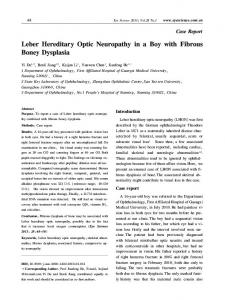
We first saw the patient 10 months after onset. His visual acuity was 1/50 on the right and counting fingers at 40 cm on the left. He had bilateral central scotomas and pale optic discs, diVuse hypotonia, postural tremor of the upper limbs, massive myoclonic jerks of the lower limbs, and partial repetitive myoclonias aVecting the hands and feet. Myoclonic jerks were not evoked by diVerent sensorial stimuli. Deep reflexes were brisk in the lower limbs. The 11778/ND4 pathogenic mutation was found slightly heteroplasmic (a small amount of normal mtDNA was coexisting with the majority of mutant genomes) in his leucocytes. Serum lactic acid at rest and after standardised cycloergometer eVort was normal. Muscle biopsy was normal at light microscopy, but subsarcolemmal aggregates of mitochondria with lipid and lipofuscin accumulation were noted on electron microscopy. Pattern visual evoked potentials (P-VEPs) were absent bilaterally; an electroretinogram (ERG) was normal bilaterally. Motor evoked potentials (MEPs) and somatosensory evoked potentials (SSEPs) of the median and the tibial nerves were normal. The patient underwent videopolygraphic recording: an EEG was normal, and in particular no paroxysmal activity was recorded. Electromyographic activity recorded from different muscles in the upper and lower limbs disclosed frequent, brief (40–100 ms), erratic myoclonia at rest (fig 1 A). The onset of EMG signal of the right extensor carpi radialis was employed as a trigger to back average the simultaneously recorded EEGs. The averaged scalp activity disclosed a small (amplitude 15 µV) biphasic, positive-negative EEG transient that was localised over the contralateral central (C3) scalp region. The positive peak of the EEG potential preceded the onset of the muscle contraction by about 16 ms (fig 1 B). Treatment with the coenzyme Q analogue idebenone was started (270 mg/day). We saw the patient at follow up 4 and 11 months later. Neurological examination was unchanged after 4 months. The myoclonic jerks were still evident 11 months later, although markedly decreased in intensity and frequency. A new brain MRI was normal, without any evidence of the previously observed lesion. His visual acuity had slightly worsened and therapy with idebenone was discontinued.
814
Carelli, Valentino, Liguori, et al
A F4 – A1 C4 – A1 P4 – A1 Cz – A1 F3 – A2 C3 – A2 P3 – A2 R SCM R Delt R ECR R APB R BF R RF L RF R TA L TA 1s
B
C4 – 10 µV + Cz
C3
10 mV
Right ECR
–300
–200
–100
0
100
200
300
Time (ms) Figure 1 EEG and back average study of patient 1. (A) Representative polygraphic sample showing frequent myoclonic twitches in diVerent muscles not associated with paroxysmal EEG discharges. EEG activity collected from scalp electrodes positioned according to the 10–20 International System (EEG band pass 0.1–60 Hz; monopolar montage with contralateral ear reference is shown); EMG activity collected from several muscles (R SCM=right sternocleiomastoideus; R Delt=right deltoideus; R ECR=right extensor carpi radialis; R APB=right abductor pollicis brevis; R BF=right biceps femoris; R RF=right rectus femoris; L RF=left rectus femoris; R TA=right tibialis anterior; L TA=left tibialis anterior). (B) Superimposition of two back averages of EEG activity (each of 50 trials). Reference to a digital marker positioned at the beginning of the myoclonic contraction in the R ECR with a time window of + and—300 ms. A positive-negative potential (amplitude 15 µV) is evident over the C3 electrode. The latency between the positive peak and the onset of EMG activity is 16 ms.
PATIENT 2
A 45 year old man, with an older sister diagnosed with multiple sclerosis, experienced blurred vision on the right and complained of fatigue of the lower limbs in the past few months. He did not smoke and drank wine moderately. His visual acuity was reduced to 2/10 on the right. Visual field examination showed a profound central defect in the right eye. Therapy with systemic corticosteroids and antiaggregants was begun, suspecting either a multiple sclerosis or a vascular aetiology. Three months later the visual field defect had almost cleared and visual acuity recovered to 7/10 in the right eye. Shortly afterwards, he had a new loss of vision in the right eye. Cerebrospinal
www.jnnp.com
fluid showed oligoclonal bands. Corticosteroids failed to improve his vision this time. During the next year the patient complained of paraesthesia and loss of tactile sensation on the right hand and fatigue of the lower limbs and right arm. Brain and cervical MRI showed a few small white matter lesions, hyperintense in T2 weighted scans, compatible with a demyelinating process; enhancement of the retrobulbar portion of the right optic nerve was also noted. A year and a half after the right eye onset the patient had a progressive loss of visual acuity also in the left eye. Fundus examination showed an oedematous and hyperaemic optic disc in the left eye and a pale atrophic disc in the right eye. The visual loss in the left eye
815
Leber’s hereditary optic neuropathy and myoclonus
A F4 – A1 C4 – A1 O2 – A1 Cz – A1 F3 – A2 C3 – A2 O1 – A2 L ADM L APB L ECR L Delt R ECR R TA L TA R VM L VM 1s
B
C4
– 10 µV
Cz
+
C3
10 mV
Left ECR
–300
–200
–100
0
100
200
300
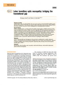
Time (ms) Figure 2 EEG and back average study of patient 2 (A) Polygraphic recording showing myoclonic erratic activities without a recognisable related EEG activity. EEG activity collected from scalp electrodes positioned according to the 10–20 International System (EEG band pass 0.1–60 Hz; monopolar montage with contralateral ear reference is shown); EMG activity collected from several muscles (L ADM=left abductor digiti minimi; L APB=left abductor pollicis brevis; L ECR=left extensor carpi radialis; L Delt= left deltoideus; R ECR=right extensor carpi radialis; RTA=right tibialis anterior; L TA=left tibialis anterior; R VM=right vastus medialis; L VM=left vastus medialis). (B) Superimposition of two back averages of EEG activity (each of 50 trials). Reference to a digital marker positioned at the beginning of the myoclonic contraction in the L ECR with a time window of + and—300 ms. A positive-negative potential (amplitude 10 µV) is evident over the C4 electrode. The latency between the positive peak and the onset of EMG activity is 23 ms.
progressed slowly despite corticosteroid therapy and therapy with â-interferon 1a was started. We saw the patient 4 months after the loss of vision in the left eye. Visual acuity was hand motion in the right eye and 3/10 in the left eye, with pale disc and sluggish pupillary reflex in the right eye and slight microangiopathy in the left eye. Neurological examination also showed sporadic fasciculations in both hands, occasional segmentary myoclonic jerks of the lower limbs, and rare spontaneous or postural partial myoclonic jerks aVecting the distal limbs. Dysaesthesia in the right hand, impaired vibration sense at lower limbs and slight impairment of proprioceptive sensitivity in the left foot were also found.
www.jnnp.com
Leucocyte mtDNA investigation showed a homoplasmic 11778/ND4 pathogenic mutation. He had slight anaemia and the screening of autoimmunity markers for vasculitic and collagen diseases was negative. Serum lactic acid at rest was normal, but raised after standardised cycloergometer eVort (60 mg/dl, normal range 9–18). Muscle biopsy showed a few fibres with enhancement of subsarcolemmal SDH staining. Aggregates of mitochondria with increased lipid and lipofuscin accumulation were evident on electron microscopy. P-VEPs were absent bilaterally and an ERG was normal bilaterally. The SSEPs of the median and of tibial nerves showed cortical responses with increased latencies and increased central conduction time bilaterally.
816
Carelli, Valentino, Liguori, et al
Brain stem auditory evoked potentials and MEPs were normal. Videopolygraphic recording showed normal EEG with no paroxysmal activity. The EMG disclosed myoclonic erratic activities without recognisable related EEG activity (fig 2 A). The onset of EMG signal of the left extensor carpi radialis was employed as a trigger to back average the simultaneously recorded EEGs. The averaged scalp activity disclosed a small (amplitude 10 µV) biphasic, positive-negative contralateral central (C4) EEG transient, that preceded the myoclonic EMG activity with a latency, from the positive peak of the EEG potential, of about 23 ms (fig 2 B). High dose idebenone was started (450 mg/day). The next month the patient complained of a further progression of visual impairment. Three months later, he had 1/50 visual acuity in the right eye and 1/20 in the left eye and he subjectively reported improved vision in the right eye. The idebenone dosage was then increased to 675 mg/day. Serum lactic acid after standardised eVort was still raised. At a further follow up, 3 months later, the patient’s vision in the right eye was still subjectively improving and he did not complain any more of fatigue and paraesthesias. At neurological examination he was still having sporadic partial myoclonic jerks of the hands and feet. His visual acuity was improved to 1/20 in the right eye and unchanged in the left eye (1/20). Discussion Myoclonus is common in mitochondrial disorders such as MERRF, MELAS, KSS, and Leigh’s disease.5 However, except for a few cases of oculopalatal myoclonus associated with a reversible brain stem lesion,6–8 limb myoclonus has never been reported in LHON.1–3 We also reviewed the collection of papers reporting LHON/multiple sclerosis-like syndrome recently listed by Bhatti and Newman,4 failing to find any such case. In our patients with LHON, polygraphic recordings showed small erratic myoclonias of all four limbs (figs 1 A and 2 A). Jerk locked averaging analysis disclosed a contralateral EEG transient preceding the EMG activity (figs 1 B and 2 B). The latency between the positive peak of the EEG transient and the onset of contraction was 16–23 ms, consistent with the time interval between cortical magnetic stimulation and the activation of the muscles (20–21 ms).9–10 Therefore, myoclonus in our patients can be attributed to activation of the fast conducting corticospinal system. These findings, together with the absence of EEG spike and wave discharges and the lack of EEG photosensitivity and of action myoclonus, diVerentiate the myoclonus in these patients with LHON from myoclonic epilepsy in MERRF syndrome. The pathophysiological mechanisms underlying the myoclonic activity in our patients remain unknown. The second patient showed the LHON/multiple sclerosis-like syndrome. Myoclonus in multiple sclerosis is rare or has been considered coincidental.11–12 Likewise, the
www.jnnp.com
relation between LHON and multiple sclerosis is poorly understood at present.1 Our first patient did not fulfill the criteria for the LHON/MS-like syndrome. However, the first brain MRI showed a single lesion unilaterally involving the optic tract and hypothalamus. Unfortunately this patient refused CSF examination and subsequent brain MRI was normal. A few patients with LHON with reversible lesions in the brain stem have been reported.6–8 The myoclonus in both patients was similar, aVecting all four limbs in the form of massive or partial multifocal jerks. Back averaging studies suggested cortical origin in both patients. However, cortical involvement was not immediately evident on neuroimaging. Multifocal and generalised myoclonic jerks may indeed occur in conditions such as posthypoxic encephalopathy and some drug intoxications or in syndromes of progressive myoclonic ataxia and progressive myoclonic epilepsy,13 where extensive cortical involvement occurs. Alternatively, the subcortical lesions present in both patients could be responsible for a dysfunctional control of cortical excitability. Cortical myoclonus associated with white matter lesions was documented in coeliac disease encephalopathy,14 and a few reports documented an immune process in some forms of myoclonus.15–16 Although its origin remains elusive, myoclonus in these two patients represents a novel neurological association that widens the clinical presentation of LHON. We acknowledge Dr Piero Barboni, Dr Sabina Cevoli, and Giuseppe Plazzi for their help in studying these patients. We also acknowledge Dr Alfredo A Sadun for helpful comments. We are indebted to the patients for participating in this study. 1 Carelli V. Leber’s hereditary optic neuropathy. In: Schapira AHV, DiMauro S, eds. Mitochondrial Disorders in Neurology. 2nd edition, Blue Book series in Neurology. ButterworthHeinemann, 2001, in press. 2 Newman NJ. Leber’s hereditary optic neuropathy: new genetic considerations. Arch Neurol 1993;50:540–8. 3 Nikoskelainen EK, Marttila RJ, Huoponen K, et al. Leber’s “plus”: neurological abnormalities in patients with Leber’s hereditary optic neuropathy. J Neurol Neurosurg Psychiatry 1995;59:160–4. 4 Bhatti MT, Newman NJ. A multiple sclerosis-like illness in a man harboring the mtDNA 14484 mutation. J Neuroophthalmol 1999;19:28–33. 5 DiMauro S, Bonilla E, Davidson M, et al. Mitochondria in neuromuscular disorders. Biochim Biophys Acta 1998;1366: 199–210. 6 Diehl GE, Wilmes E. Etiology and clinical aspects of palatal myoclonus. Laryngorhinootologie 1990;69:369–72. 7 Paulus W, Straube A, Bauer W, et al. Central nervous system involvement in Leber’s optic neuropathy. J Neurol 1993; 240:251–3. 8 Funalot B, Ranoux D, Mas JL, et al. Brainstem involvement in Leber’s hereditary optic neuropathy: association with the 14484 mitochondrial DNA mutation. J Neurol Neurosurg Psychiatry 1996;61:533–4. 9 Benecke R, Meyer BU, Gohmann M, et al. Analysis of muscle responses elicited by transcranial stimulation of the cortico-spinal system in man. Electroenceph Clin Neurophysiol 1988;69:412–22. 10 Rossini PM, Barker AT, Berardelli A, et al. Non-invasive electrical and magnetic stimulation of the brain, spinal cord and roots: basic principles and procedures for routine clinical application. Report of an IFCN Committee. Electroenceph Clin Neurophysiol 1994;91:79–92. 11 Aigner ER, Mulder DW. Myoclonus: clinical significance and approach to classification. Arch Neurol 1960;2:600–15. 12 Smith CR, Scheinberg L. Coincidence of myoclonus and multiple sclerosis: dramatic response to clonazepam. Neurology 1990;40:1633–4. 13 Brown P. Myoclonus. Curr Opin Neurol 1996;9:314–6. 14 Tison F, Arne P, Henry P. Myoclonus and adult coeliac disease. J Neurol 1989;236:307–8. 15 Salmaggi A, Carella F, Ciano C, et al. Intratechal immune activation in three patients with progressive myoclonic ataxia. Mov Disord 1995;10:207–10. 16 Caviness JN, Forsyth PA, Layton DD, et al. The movement disorder of adult opsoclonus. Mov Disord 1995;10:22–7.