Coll. Antropol. 30 (2006) 1: 171–174 Original scientific paper
Leber’s Hereditary Optic Neuroretinopathy (LHON) Associated with Mitochondrial DNA Point Mutation G11778A in Two Croatian Families Irena Martin-Kleiner1, Jelka Gabrilovac1, Mario Bradvica2, Tomislav Vidovi}3,5, Branimir Cerovski3,5, Ksenija Fumi}4,5 and Milivoj Borani}1 1 2 3 4 5
Division of Molecular Medicine, Institute »Ruder Bo{kovi}«, Zagreb, Croatia Department of Ophthalmology, University Hospital Osijek, Osijek, Croatia Department of Ophthalmology, School of Medicine, University of Zagreb, Zagreb, Croatia Clinical Institute of Laboratory Diagnosis, Faculty of Pharmacy and Biochemistry, Zagreb, Croatia University Hospital »Rebro«, Zagreb, Croatia
ABSTRACT Leber's hereditary optic neuroretinopathy (LHON) is manifested as a bilateral acute or subacute loss of central vision due to optic atrophy. It is linked to point mutations of mitochondrial DNA, which is inherited maternally. The most common mitochondrial DNA point mutations associated with LHON are G3460A, G11778A and T14484C. These mutations are linked with the defects of subunits of the complex I (NADH-dehydrogenase-ubiquinone reductase) in mitochondria. The G11778A mitochondrial DNA point mutation is manifested by a severe visual impairment. In this paper two Croatian families with the LHON G11778A mutation are presented. Three LHON patients from two families were younger males which had the visual acuity of 0.1 or below, the ophthalmoscopy revealed telangiectatic microangiopathy and papilloedema, while Goldmann kinetic perimetry showed a central scotoma. The mothers and female relatives were LHON mutants without symptoms, whereas their sons suffered from a severe visual impairment. Molecular diagnosis helps to explain the cause of LHON disease. Key words: LHON, visual impairment, mitochondrial DNA, G11778A point mutation
Introduction Leber's hereditary optic neuroretinopathy (LHON) is manifested as a bilateral acute or subacute loss of central vision due to optic atrophy. LHON at first affects one eye, and central vision is lost in that eye over a period of a few weeks. One or two months later, the second eye is affected. The time when the affected person is loosing the eyesight is the acute stage of LHON. Typical funduscopic finding in the acute stage of LHON is a peripapillary microangiopathy, damage to the optic nerve and the retina. A visual defect is usually the only clinical manifestation. However, it can be associated with cardiac conduction abnormalities (pre-excitation syndrome), peripheral neuropathy and/or ataxia1,2. Mitochondria are organelles responsible for energy production by oxidative degradation of carbon substrates into water and oxygen through the respiratory chain.
These reactions are catalyzed by respiratory chain complexes I, II, III, and IV embedded in the inner membrane of the mitochondria. The mitochondrial DNA is 16569 base pairs long. It contains a double-stranded DNA molecule, no introns and replicates independently of the nuclear genome. Mitochondrial DNA is maternally inherited1,3. Defects of the mitochondrial DNA are large scale rearrangements with single deletions or duplications. Over 118 point mutations of mitochondrial DNA are known today4, associated with varying clinical phenotypes1,3,5. Among the diseases associated with defects of mitochondrial DNA, LHON was first linked to point mutations of the mitochondrial DNA1,6. Today, LHON is linked to more than 30 point mutations of the mitochondrial DNA (www.mitomap.org). The most common LHON
Received for publication August 28, 2005
171
Martin-Kleiner et al.: LHON Disease in Two Croatian Families, Coll. Antropol. 30 (2006) 1: 171–174
point mutations are G3460A, G11778A and T14484C1,2. The point mutation G3460A is associated with a functional defect in ND1 subunit1,2, G11778A with a functional defect in ND4 subunit1,6 and T14484C with a functional defect in ND6 subunit1,7 of the respiratory chain complex I. The disease usually begins in the second or third decade of life, with male predominance1,2,6,7. The average onset of LHON is at 24.3 years of age among males, at 31.8 years among females8. However, there is also a case report of a 6-year-old girl with LHON9. The point mutation G11778A has a poor prognosis1,2,6 while T14484C is linked to a mild course of the disease1,2,7. Spontaneous recovery from LHON was described in 5 out of 136 patients7. This work is the first study of Croatian LHON families using molecular diagnosis for the most common LHON mitochondrial DNA point mutations.
Materials and Methods Patients 24 persons from five families were included in this study. Bilateral loss of vision was the criterion for diagnostic analysis including the tests for the mitochondrial DNA mutations. The maternal line of inheritance was followed while testing relatives. Consent of the patients and of their relatives was obtained for the DNA analysis.
Ophthalmologic examinations A complete ophthalmologic examination was conducted on patients with visual impairment. The examination included best-corrected visual acuity using Snellen charts, color vision with Ishihara plates, ophthalmoscopy, Goldmann kinetic and/or automated perimetry. Detailed family anamnesis was recorded.
Other examinations Computed axial tomography (CAT) scan and magnetic resonance imaging (MRI) with and without contrast were done for LHON patients. Routine hematological and biochemical analyses of blood and urine analysis were performed.
DNA testing from peripheral blood lymphocytes using the PCR-RFLP method DNA was isolated from peripheral blood lymphocytes10. A polymerase chain reaction (PCR) was performed for each point mutation separately using the specific set of primers. The positions of primers in the sequence of the mitochondrial genome (Cambridge sequence, www.mitomap.org) were fw 5’ 3150 and rv 3’ 3600 for the G3460A mutation, fw 5’ 11680 and rv 3’ 12000 for the G11778A mutation and fw 5’ 14450 and rv 3’ 14608 for the T14484C mutation. The PCR reaction was performed using the PCR Core Kit (Roche, Germany). The final concentrations were: dNTP 0.2 mM, MgCl2 1.5 mM, Taq DNA polymerase 0.03 U/mL and primers 0.4 mM. 172
PCR conditions were: initial denaturation 96 °C 2 min, denaturation 94 °C 1 min, annealing 56 °C 1 min, extension 72 °C 1 min and final extension 72 °C 5 min, total 35 cycles. A GeneAmp PCR system 2400, Perkin Elmer, Applied Biosystems was used11. PCR products were verified on 1.5% agarose gels. The PCR reaction was followed by the restriction length fragment polymorphism method (RFLP). PCR products were digested overnight at 37 °C with restrictive enzymes, final concentration 25 U/mL. For each point mutation a specific restrictive enzyme was used: BsaH1 for G3460A12, SfaN1 for G11778A6 and Mbo for T14484C (Bio Labs, New England). Restrictive fragments were tested on 1.5% agarose gel using ethidium-bromide for UV visualization of DNA. Both mutated DNA as a positive control11 and wild-type DNA as a negative control were included in the analysis. In detection of LHON mitochondrial DNA mutation G11778A, wild-type DNA was cut into two fragments in the PCR-RFLP reaction, while mutated DNA was detected as one band, no restriction. Restrictive fragments were analyzed by Image Master VDS software 1.0 (Pharmacia) in order to quantify the ratio of mutated and wild-type DNA for heteroplasmy.
Results The most common three LHON mitochondrial DNA point mutations G3460A, G11778A and T14484C were tested in DNA samples of twenty four persons. Two family cases with ten LHON G11778A mutants are presented in this paper.
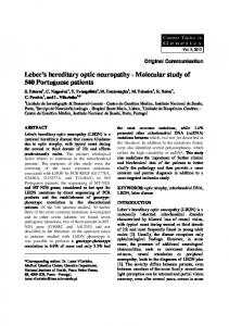
Family No. 1 Four persons positive for the LHON G11778A mitochondrial DNA mutation were two patients 27 and 25 years of age, males with severe visual impairment (visual acuity 0.01) and their mothers 50 and 53 of age with normal vision. The mothers were cousins (Figure 1). These mutants were homoplasmic for the LHON G11778A mutation. Patient #1 (26 years old) was hospitalized at the Neurology Department for a month in 2003 due to unilateral loss of the right eye vision and papilloedema. Laboratory analysis of blood and urine showed no pathological values. CAT scan and MRI with and without contrast revealed no intracranial process. Visual evoked potential revealed a suspicious neuronal lesion of the right visual pathway and conduction disturbances in crossing fibers. Brainstem evoked potential detected neuronal lesion of the right auditory pathway. Somatosensory evoked potentials of the median and tibial nerves were normal. Vision in his other eye was also lost within six months. Ophthalmoscopy revealed telangiectatic microangiopathy in the nerve fiber layer and papilloedema. Goldmann kinetic perimetry revealed a central scotoma. As the patient had a second-degree male cousin (the mothers were cousins) who also suffered from sudden visual loss in both eyes eight years ago at the age of 17 (patient #2), LHON disease was suspected. DNA testing revealed
Martin-Kleiner et al.: LHON Disease in Two Croatian Families, Coll. Antropol. 30 (2006) 1: 171–174
LHON G11778A mitochondrial point mutations in both patients #1 and #2 and in their mothers (Figure 1). The grandmother of patient #2 was heteroplasmic and expressed both wild type (80%) and mutated (20%) DNA in peripheral blood lymphocytes (Figure 1).
I II III IV
Fig. 1. Presentation of family No. 1 with LHON G11778A mitochondrial DNA mutation. LHON – Leber's hereditary optic neuroretinopathy, DNA-deoxyribonucleic acid. – females, not tested, – males, not tested, – carriers of the mutation, mothers without clinical symptoms, – patients with clinical symptoms, carriers of the mutation, – carrier of the mutation, grandmother, without symptoms, heteroplasmic.
Family No. 2 Six members of that family were carriers of the LHON G11778A point mutation: patient #3, his mother and sister, aunt, her daughter and the granddaughter (Figures 2 and 3). Patient #3 was a 26 year old male admitted for clinical care in 1994, due to unilateral visual loss in the right eye and papilloedema. Two months later his left eye vision was lost as well. Best corrected visual acuity in both eyes was 0.08 according to the Snellen chart. Goldmann perimetry showed central scotomas but no inner isopters. Ophthalmoscopy revealed bilateral papilloedema. CAT scan showed no intracranial process. Pulse steroid therapy was instituted but the recovery of visual function was minimal (visual acuity improved from 0.08 to 0.1). Standard hematological, biochemical and immunological tests were within the normal range. There were no cardial problems. Since the recovery of visual function with corticosteroid treatment was poor, LHON disease was suspected. DNA testing turned to be positive for the G11778A mitochondrial DNA point mu-
I II
III IV
Fig. 2. Presentation of family No. 2 with LHON G11778A mitochondrial DNA mutation.. – females, not tested, – males, not tested, – carriers of the mutation, female relatives without clinical symptoms, – patient with clinical symptoms, carrier of the mutation.
Fig. 3. Analysis of DNA from patient #3 with LHON mitochondrial point mutation G11778A and his female relatives. Lane 1 – DNA standard, Lane 2 – patient, Lane 3 – patient's mother, Lane 4 – patient's aunt (mother's sister), Lane 5 – positive control, Lane 6 – negative control, wild type (two restriction fragments of 222 and 98 bp), Lanes 2 – 5 reveal LHON G11778A mutation seen as one band, no restriction.
tation. The patient's sister, mother, aunt, aunt's daughter and granddaughter were also LHON G11778A mutants, but without visual impairment. All LHON G11778A individuals were homoplasmic, expressed only mutated DNA. Except for the mother who had arterial hypertension, other family members had no signs of systemic diseases, nor laboratory evidence of metabolic abnormalities.
Discussion This is the first Croatian pilot study of LHON linked to a mitochondrial DNA point mutation. The mitochondrial DNA point mutation LHON G11778A was detected in two families. Three young men (patients #1, #2, #3) had a clinically manifested LHON disease. The mothers and female relatives of these LHON patients were carriers of the G11778A mitochondrial DNA mutation, having neither visual impairment nor any other clinically manifested disease. All LHON G11778A individuals presented in this paper were homoplasmic, except for one grandmother in family No.1 who was heteroplasmic. Heteroplasmy has also been described in LHON1,2 in 1 out of 7 individuals13. LHON cases in these two families are in line with literature showing the preponderance of LHON in young males having clinically manifested disease1,2, 6–8. These three most common LHON mitochondrial DNA mutations investigated in our study were not detected in other thirteen patients from three families having optical neuropathy. Nevertheless, LHON may not be completely excluded. Over 30 mitochondrial DNA point mutations, linked to LHON and named secondary, have been described (www.mitomap.org). This possibility should be investigated further. In this study no haplotype analysis of mitochondrial DNA was made. LHON was linked most often to the J haplotype of mitochondrial DNA1,14. In the Croatian population predominantly H15,16, U, J and T haplotypes of mitochondrial DNA were detected15. 173
Martin-Kleiner et al.: LHON Disease in Two Croatian Families, Coll. Antropol. 30 (2006) 1: 171–174
Identification of LHON cannot be based solely upon clinical diagnostic tests, a genetic analysis revealing the maternally inherited mitochondrial DNA point mutation should also be made. Joint efforts of ophthalmologists, neurologists, other clinicians, biochemists and molecular biologists are required for the diagnosis of LHON. Once the diagnosis is established, genetic counseling of the family becomes important1,10. A LHON prognosis is better for female carriers of the mutated mitochondrial DNA, who will not develop the clinically manifested disease. No effective therapy is so far available for LHON, improved nutrition is recommended. Gene therapy as an experimental approach is not yet clinically relevant1.
Acknowledgements Professor Massimo Zeviani and Mr. Franco Carrara from the Unit of Molecular Neurogenetics, Carlo Besta National Neurologic Institute, Milan, Italy kindly supplied the primers and the mutant DNA controls. Dr. Rajko Ku{ec, Merkur Clinical Hospital, Zagreb, kindly donated restrictive enzymes MbO and BsaH1. Dr. Ivanka [tenc-Bradvica was responsible for clinical care of patient #1 at the Department of Neurology, Clinical Hospital Osijek. Dr. Davorka Breljak’s help with gel analysis is gratefully acknowledged.. This research was supported by the Croatian Ministry of Science, Sports and Education, Project No. 00980 94.
REFERENCES 1. ZEVIANI, M., S. DI DONATO, Brain, 127 (2004) 2153. — 2.MAN, P. Y. W., P. G. GRIFFITHS, D. T. BROWN, N. HOWELL, D. M. TURNBULL, P. F. CHINNERY, Am. J. Hum. Genet., 72 (2003) 333. — 3. SCHEFFLER, I. E., Mitochondrion, 1 (2001) 3. — 4. WESTHUIZEN, VAN DER, F. H., L. P. VAN DEN HEUVEL, R. SMEETS, J. A. VELTMAN, R. PFUND, A. G. VAN KESSEL, B. M. URSING, J. A. M. SMEITINK, Neuropediatrics, 34 (2003) 14. — 5. NAVIAUX, R. K., Mitochondrion, 4 (2004) 351. — 6. WALLACE, D. C., G. SINGH, M. T. LOTT, J. A. HODGE, T. G. SCHURR, A. M. S. LEZZA, L. J. ELSAS, E. K. NIKOSKELAINEN, Science, 242 (1988) 1427. — 7. JOHNS, D. R., K. L. HEHER, N. R. MILLER, K. H. SMITH, Arch. Ophthalmol., 111 (1993) 495. — 8. LEO-KOTTLER, B., M. CHRIST-ADLER, Ophthalmologe, 96 (1999) 698. — 9. BALAYRE, S., J. J. GICQUEL, M. MERCIE, P. J. DIGHIERO, J. Fr. Ophtalmol., 26 (2003) 1063. — 10. NIKOSKELAINEN, E. K., K. HUOPONEN, V. JUVONEN, T. LAMMINEN., K. NUMMELIN, M. L. SAVONTAUS, Oph-
thalmology, 103 (1996) 504. — 11. MARTIN-KLEINER, I., E. PAPE-MEDVIDOVI], I. PAVLI]-RENAR, @. METELKO, R. KU[EC, J. GABRILOVAC, M. BORANI], Acta Diabetol., 41 (2004) 179. — 12. CARRARA, F., P. F. CHINNERY, P. Y. W. MAN, M. ZEVIANI, V. TIRANTI, Mitochondrion, 4 (2004) 37. — 13. CHINNERY, P. F., R. M. ANDREWS, D. M. TURNBULL, N. HOWELL, Am. J. Med. Genet., 98 (2001) 235. — 14. BROWN, M. D., E. STARIKOVSKAYA, O. DERBENEVA, S. HOSSEINI, J. C. ALLEN, I. E. MIKHAILOVSKAYA, R. I. SUKERNIK, D. C. WALLACE, Hum. Genet., 110 (2002) 130. — 15. TOLK H. V., M. PERI^I], L. BARAC, I. M. KLARI], B. JANI]IJEVI], I. RUDAN, J. PARIK, R. VILLEMS, P. RUDAN, Coll. Antropol., 24 (2000) 267. — 16. CVJETAN S., H. V. TOLK, L. B. LAUC, I. COLAK, D. \OR\EVI], L. EFREMOVSKA, B. JANI]IJEVI], A. KVESI], I. M. KLARI], E. METSPALU, M. PERI^I], J. PARIK, D. POPOVI], A. [IJA^KI, R. TERZI], R. VILLEMS, P. RUDAN, Coll. Antropol., 28 (2004) 193.
I. Martin-Kleiner Division of Molecular Medicine, Laboratory for Experimental Hematology, Immunology and Oncology, Institute »Ru|er Bo{kovi}«, PO Box 180, 10002 Zagreb, Croatia e-mail:
[email protected]
LEBEROVA NASLJEDNA OPTI^KA NEURORETINOPATIJA (LHON) VEZANA UZ TO^KASTU MUTACIJU G11778A MITOHONDRIJSKE DNK U DVIJE HRVATSKE OBITELJI
SA@ETAK Leberova nasljedna opti~ka neuroretinopatija (LHON) manifestira se bilateralnim akutnim ili subakutnim gubitkom centralne vidne o{trine uslijed atrofije vidnog `ivca. LHON je vezan uz to~kaste mutacije mitohondrijske DNK i naslje|uje se po majci. Naj~e{}e to~kaste mutacije mitohondrijske DNK vezane uz LHON jesu G3460A, G11778A i T14484C. Ove mutacije vezane su uz defekte podjedinica kompleksa I (NADH-dehidrogenaza-ubikinon reduktaza) u mitohondrijima. LHON G11778A to~kasta mutacija mitohondrijske DNK manifestira se te{kim o{te}enjem vida. U ovom radu prikazane su dvije hrvatske obitelji s LHON G11778A mutacijom mitohondrijske DNA. Tri LHON bolesnika iz dvije obitelji mlade su mu{ke osobe u kojih vidna o{trina iznosi 0.1 ili manje, oftalmoskopija je otkrila telangiektati~nu mikroangiopatiju i edem papile vidnog `ivca, dok je kvantitativna kineti~ka Goldmannova perimetrija pokazala su`enje uz centralne skotome. Majke i `enski pripadnici obitelji su LHON G11778A mutanti bez simptoma, a sinovi LHON G11778A mutanti imaju te{ka o{te}enja vida. Molekularna dijagnoza poma`e u obja{njenju uzroka LHON bolesti.
174