Article available at http://www.parasite-journal.org or http://dx.doi.org/10.1051/parasite/2003102111
Le is h m a n ia (V ia n n i a )
u t in g e n s is n
.
sp .,
A PARASITE FROM THE SANDFLY LUTZOMYIA (V lANNAMY lA) TUBERCULATA in A m a z o n ia n B r a z il BRAGA R.R.*, LAINSON R.*, ISHIKAWA E A .Y .* & SHAW J.J.**
Sum m ary:
R ésu m é :
L e is h m a n ia ( V ia n n ia ) u t in g e n s is n . s p ., p a r a sit e d u P h l é b o t o m e L u t z o m y ia ( V ia n n ia m y ia ) t u b e r c u la t a e n A m a z o n ie BRÉSILIENNE
A leishmanial parasite isolated in 1 9 7 7 from a specimen o f the sandfly Lutzomyia tuberculata from Para State, A m azonian Brazil,
Une Leishmanie isolée en 1 9 7 7 d'un spécimen d e Lutzomyia
has been characterized follow in g its com parison w ith other species o f Leishmania from the same region, using isoenzyme profiles, m onoclonal antibodies and characterization of the mini exon gene repeat, using the polymerase chain reaction technique (PCR). It is described here under the name o f Leishmania {Viannia) utingensis n. sp.
tuberculata d e l'état d e Pará , Amazonie brésilienne, est diffe renciée des autres esp èces d e Leishmania d e la même région
par les profils enzymatiques, les anti-corps monoclonaux et la caractérisation, par PCR, des mini-exons répétés dans les gènes. Elle est nommée Leishmania (Viannia) utingensis n. sp. MOTS CLÉS : Leishmania (Viannia) utingensis n. sp., Lutzomyia tuberculata, phlébotome, Brésil.
KEY WORDS : Leishmania (Viannia) utingensis n. sp., Lutzomyia tuberculata, sandflies, Brazil.
INTRODUCTION
MATERIALS AND METHODS
I
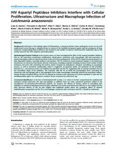
n 1977 a leishmanial parasite was isolated from a single specimen o f the phlebotomine sandfly, Lut z o m y ia (V ia n n a m y ia ) tu bercu lata (Mangabeira) (Fig. 1), taken from the trunk of a large tree in Utinga forest on the outskirts of Belém, Para State, Amazo n ian B ra z il. In itia lly th o u g h t to p o ssib ly be L. (V ia n n ia ) brazilien sis (Lainson & Shaw, 1979), it was later listed among the “unnamed parasites of the subgenus Viannia" by virtue of its peripylarian deve lopment in the sandfly host, morphology, and beha viour in hamsters and in vitro culture (Lainson & Shaw, 1987). In the present paper, we give a more complete description of the organism and show it to differ from all those species of L eish m a n ia previously recorded in the Amazon Region of Brazil, following characterization of the mini-exon gene repeat, using the polymerase chain reaction technique (PCR), isoen zyme electrophoresis profiles, and monoclonal anti bodies (McAbs).
* Departamento de Parasitologia, Instituto Evandro Chagas, Av. Almirante Barroso 492, 66090-000 Belém, Pará, Brazil. ** Departamento de Parasitologia, Instituto de Ciencias Biomédicas, Universidade de Sao Paulo, Av. Lineu Prestes 1374, 05508-900 Sao Paulo, Brazil. Correspondence: Ralph Lainson. Tel: +55 91 211 4453 - Fax: +55 91 226 1284. E-mail:
[email protected] Parasite, 2003, 10, 111-118
M
o r p h o l o g y o f a m a s t ig o t e s a n d p r o m a s t ig o t e s
F
ollowing retrieval of the parasite (Register Num ber M4964) from our cryobank, it was maintained in Diffco B45 blood-agar culture medium (Walton et al., 1977) and the skin of hamsters inoculated intradermally with promastigotes into the dorsal surface of the hind feet. For amastigotes, animals were sacrificed eight days post inoculation (d.p.i.) and impression smears made from the skin excised from the point of inoculation. Prepa rations were air-dried, fixed in aqueous Bouin’s fluid for 20 minutes, and washed repeatedly in 70 % ethyl alcohol until colourless. The smears were then stained for one hour by Giemsa’s method, differentiated in a graded series of acetone-xylol mixtures and mounted under a cover-slip in “Permount”®. Promastigotes of 7-day-old cultures were washed twice by centrifugation in pH 7.2 phosphate buffered saline (PBS) and the sediment used to prepare thin smears. These were air-dried, fixed in absolute methyl alcohol and stained by Giemsa’s method. Measurements of 50 amastigotes and 40 promastigotes were made using the oil immersion lens, x 10 oculars and an eyepiece micrometer. All measurements are in pm and given as means, followed by the standard deviation and the range in parentheses. Photomicrographs were pre pared using a Zeiss “Photomicroscope III” and Kodak TMX 100 film.
Mémoire
111
BRAGA R.R., LAINSON R„ ISHIKAWA E.A.Y. & SHAW J.J.
Fig. 1. — Lutzomyia (V ian nam yia) tuberculata. Freshly dissected female genitalia (spermathecae). Bar = 0.05 mm.
P o lym er a se
c h a in
r e a c t io n
The oligonucleotides used in the PCR were with the following sequences (Fernandes et al., 1994): S 1629:5'GGG-AAT-TCA-ATA-TAG-TAC-AGA-AAC-TG-3'and S 1630:5'-GGG-AAG-CTT-CTG-TAC-TTT-ATT-GGT-A-3'. Agarose gel (1 %) electrophoresis of mini-exon PCR amplification products and a molecular weight marker (Φ X 174 DNA/Hae III) were visualized following stai ning with ethidium bromide. A negative control, with no added DNA, was included. E nzym e
e l e c t r o p h o r e s is
We used the enzymes ASAT, ALAT, PGM, GPI, MPI, G6PD, MDH, ACON, PEP and 6PGDH (for methods and abbreviations, see Miles et al., 1980), and the pro files of isolate M4964 were compared with those of L. (V.) braziliensis (MHOM/BR/1975/M2903) from Serra dos Carajas, Para; L. (V.) braziliensis sensu lato (MHOM/ BR/1975/M2904) also from Serra dos Carajas; L. (V.) g u y an en sis(MHOM/BR/1975/M4l47) from Monte Dourado, Para; L. (V.) lain son i (MHOM/BR/1981/M6426) from Benevides, Para; L. (V .) n a i f f i (MDAS/BR/ 1979/M5533) from Monte Dourado, Para; L. (V .)sh aw i (MCEB/BR/1984/M8408) from Parauapebas, Para; L. (Leishm an ia) a m a z o n e n s is (IFLA/BR/l967/PH8) from Utinga Forest, Belem, Para; and L. in fantum ch a g a si (MCER/ BR/1981/M6445) from Salvaterra, Marajó island, Pará. M
o n o c l o n a l a n t ib o d ie s
The isolate was tested with 23 McAbs prepared against the above-listed L eishm an ia (V ian n ia) species respon sible for cutaneous leishmaniasis in the Amazon Region of Brazil and neighbouring parts of South America 112
(McMahon Pratt et al., 1986; Hanham et al., 1991). They were used together with the indirect immunofluores cence/fluorescein-labelled avidin technique (Shaw et al., 1989). E x p e r im e n t a l
in f e c t io n s
la bo ra to ry-b r e d
in
s a n d f l ie s
Using the membrane-feeding technique of Ward et al. (1978), culture forms of both M4964 and L. (V.) b ra z i liensis (M 2903) were separately fed to laboratory-bred Lutzomyia (Lutzomyia) longipalpis and Lutzomyia (Vian n am yia) fu rcata. The sandflies were dissected five days later, when all the ingested blood had been digested.
RESULTS B
e h a v io u r
in
c u l t u r e m e d iu m
G
rowth of the parasite is rapid, with the pro duction of abundant, large rosettes of dividing forms. The separate flagellates are small and extremely active - a characteristic of most of the species within the subgenus V iannia (Lainson & Shaw, 1987). B
e h a v io u r
in
th e
s k in
of
h a m ster s
Following the intradermal inoculation of enormous numbers of log-phase promastigotes, only a small number of free and intracellular amastigotes could be detected in the stained smears of skin excised from the site of inoculation eight days p.i. No visible skin lesion could be detected up to two months p.i., when para sites were encountered with great difficulty: in this res pect the parasite resembles L. (V.) n a iffi (Lainson &
Mémoire
Parasite, 2003, 10, 111-118
L eish m an ia u t in g è îs is
n . s p . in
L it z o m y ia w b e r c u l a t a
Fig. 2. - L e i s h m a n i a ( V i a n n i a ) u t i n g e n s i s n. sp. Intracellular and free amastigotes in the skin of a hamster inoculated eight days pre viously with promastigotes. a-f. Division stages: one parasite (f), shows the conspicuous fla gellar vacuoles o f the two daughter amasti gotes which are on the point of separation, gk. Smaller products of division. Bouin fixation, Giemsa staining. Bar = 10.0 pm.
Fig. 3 . - L e i s h m a n i a ( V i a n n i a ) u t i n g e n s i s n. sp. Log-phase promastigotes from an 8-day culture in Diffco B45 blood-agar medium, ab. Dividing forms, c-f. Small, non-dividing fla gellates. Methyl alcohol fixation, Giemsa stai ning. Bar = 10.0 pm.
Parasite, 2003, 10, 111-118
Mémoire
113
BRAGA R.R., LAINSON R., ISHIKAWA E.A.Y. & SHAW J.J.
Shaw, 1989; Lainson et al., 1990) and the newly des cribed L. (V.) lin d en berg i ( Silveira et a l , 2002). M o rph o lo g y o f amastigotes and prom astigotes (Figs 2 and 3) Mean measurement of amastigotes was 3.0 ± 0.7 x 2.1 ± 0.3 (2.2-4.0 x 1.5-2.2), n = 50. The parasite shows no morphological features by which to differentiate it from most of the species in the subgenus Viannia, with which it shares the characteristic of very small, com pact amastigotes in which the kinetoplast is typically large and positioned between the anterior end and the middle of the nucleus (Shaw & Lainson, 1976). The mean body measurement of the promastigotes was 8.1 ± 2.0 x 2.3 ± 0.6 (5.2-11.1 x 0.7-3-0). The mean length of the free flagellum was 9 .3 ± 2.7, n = 30, but the wide range of 4.0-21 suggests that many flagella were broken during the washing process. P olymerase chain reaction (Fig. 4) Using the oligonucleotides S1629: 5' and S1630: 5' the PCR clearly shows that isolate M4964 belongs to the
subgenus Viannia, and differentiates it from the two representatives of the subgenus L eishm an ia examined, Leishm ania infantum ch ag asi and L. (L.) am azon en sis. Among species of the subgenus V iannia, the parasite could be separated from L. (V.) lainsoni, but not from L. (V.) braziliensis, L. (V.) guyanensis, L. (V.) naiffi, and L. (V.) shawi. I so enzym e profiles (Fig. 5) Shaw et al. (1991) showed that failure of the enzyme ACON to migrate from its origin towards the negative pole was a means of separating parasites of the sub genus V ian n ia from those of the subgenus Leish m an ia, and the present result for this enzyme again supports the previous allocation of M4964 to the sub genus V iannia. All the enzymes used separated M4964 from L. in fan tum ch a g a si and L. (L.) am azon en sis, and the combi ned use of the seven enzymes ASAT, ALAT, PGM, G6PD, MPI, 6PGDH and MDH enabled separation of the parasite from all the known reference strains of species within the subgenus V iannia.
Fig. 4. - Agarose gel (1 %) electrophoresis of mini-exon PCR amplification products from neotropical L e i s h m a n i a spp., visualized by ethidium bromide staining. Lane PM: molecular weight marker (Φ X 174 DNA/Hae III). Lane 1: L e i s h m a n i a (V .) b r a z i l i e n s i s sensu lato (M2904). Lane 2: L. (V .) u t i n g e n s i s n. sp. Lane 3: L. (V .) s h a w i (M8408). Lane 4: L. (V.) g u y a n e n s i s M4147). Lane 5: L. (V .) n a i f f i (M5533). Lane 6: L. (V .) l a i n s o n i (M6426). Lane 7: L. (V .) b r a z i l i e n s i s (M2903). Lane 8: L. ( L .) a m a z o n e n s i s (PH8). Lane 9: L. i n f a n t u m c h a g a s i (M6445). Lane CN: negative control (no DNA added). Lane PM: molecular weight marker.
11 4
Mémoire
Parasite, 2003, 10, 111-118
L eishmania
utingensis n . sp. in
L utzomyia
tuberculata
Fig. 5. - Electrophoresis of the enzymes ASAT, ALAT, PGM, GPI, G6PD and ACON of Leishm ania species from Amazonian Brazil. The parasites under comparison are: 1. L. (L.) am azon en sis (PH8); 2. L. (V.) braziliensis (M2903); 3. L. (V.) guyanensis (M4147); 4. L. (V.) shaw i (M8408); 5. L. (V.) lainsoni (M6426); 6. L. (V.) n aiffi (M5533); 7. L. (V.) braziliensis sensu lato (M2904); 8. L. (V.) utingensis n. sp., (M4964); 9. L. (L.) infantum chagasi (M6445) and 10. L. (L.) am azon en sis (PH8).
Parasite, 2003, 10, 111-118
Mémoire
115
BRAGA R.R., LAINSON R., ISHIKAWA E A Y. & SH A W JJ.
Species o f Leishm ania L. L. L. L. L. L. L.
(V.) ( V.) (V.) (V.) (V.) (V.) (V.)
braziliensis guyanensis shaw i naiffi lainsoni utingensis n. sp lindenbergi
Monoclonal antibodies B18
B19
N2
N3
B2
B12
+
+
+ + +
+ + +
-
-
-
+
+
-
-
-
-
-
-
-
-
-
-
-
-
+
-
-
-
-
+ +
B5
M2
W1
WA2
LA2
+
+
-
+
-
-
-
-
-
-
+
-
-
-
-
-
-
-
-
-
-
-
-
-
-
-
-
-
-
-
-
-
-
+
-
-
-
-
-
-
-
-
+ +
-
Table I. - Comparison of the serodeme profiles of Leishm ania (V iannia) utingensis n. sp. with other species of the subgenus Viannia from the Amazon Region of Brazil.
M onoclonal a n tibo d ies (Table I)
SPECIFIC DIAGNOSIS
The subgeneric position of the parasite was once more confirmed, by the McAb B2 which is specific for the taxon V ian nia (McMahon-Pratt et al., 1982, 1985). Among the known species within the subgenus, it was distinguished from L. (V.) braziliensis and L. (V.) guyanensis by its failure to react with McAbs B18 and B19, respectively; from L. (V.) n aiffi by its non-reaction with McAb N3, which recognizes that parasite; from L. (V.) sh a w i which, unlike M4964, does not react against McAb N2; from L. (V.) lain son i by its failure to react with McAb LA2, specific for that parasite, and its posi tive reaction to McAb B2 to which L. (V.) lain son i does not react; and from L. (V.) lin den bergi (Silveira et al., 2002) which reacts with McAb B12, whereas M4964 does not. In conclusion, M4964 can be distinguished from other member of the subgenus V ian n ia by its unique sero deme, as determined by monoclonal antibodies, and a com bination of the profiles for seven different enzymes. Of particular note is the fact that mobility of the parasite’s ASAT is similar to that of L. (V.) b r a zilien sis and faster than that o f L. (V.) lindenbergi, while mobility of its G6PD is intermediate between that of L. (V.) brazilien sis and L. (V.) n a iffi and L. (V.) lin den berg. The G6PD of the latter parasite migrates less than any other named species of the subgenus V iannia. As a result of the present study we propose the name of L eish m a n ia (V ia n n ia ) utingensis n. sp for the isolate M4964, which has for so long remained incognito. D evelopment in experimentally infected sandflies In Lutzom yia (Lutzom yia.) longipalpis, both L. (V.) bra ziliensis and L. (V.) utingensis n. sp. showed typical peripylarian development in both the pylorus and ileum. In Lutzom yia (V ian n am y ia) fu r c a ta , L. (V.) utingensis n. sp. underwent a similar development, whereas L. (V.) braziliensis showed attached flagellates only in the ileum. 116
L eishmania (V iannia ) utingensis n . sp T
y p e host: the phlebotomine sandfly Lutzom yia (V ian n am y ia) tu bercu lata (Diptera: Psychodidae: Phlebotominae). Type locality: Utinga forest, on the outskirts of Belem, Para State, North Brazil. Strain designation: ITUB/BR/1977/M4964. Amastigotes: non-dividing parasites small and com pact, 3.0 ± 0.7 x 2.1 ± 0.3 (2.2-4.0 x 1.5-2.2), n = 50. Promastigotes: small. Body of non-dividing parasites 8.1 ± 2.0 x 2.3 ± 0.6 (5.2-11.1 x 0.7-3.0), n = 40. Mean length of free flagellum 9 .3 ± 2.7, ranging from 4.021.0, n = 30. Behaviour in sandfly host: prolific peripylarian deve lopment, characteristic of the subgenus Viannia, with promastigotes and paramastigotes predominantly atta ched to the wall of the pylorus. Behaviour in hamster: no visible lesion produced at the site of intradermal inoculation two months p.i. Amas tigotes rare or undetectable in the skin, but their pre sence can be confirmed by culture in blood-agar medium. Behaviour in in vitro culture: exuberant growth with abundant, large rosettes. Isoenzyme profiles: the combined use of enzymes ASAT, ALAT, PGM, G6PD, MPI, 6PGDH and MDH dis tinguishes the parasite from other members of the subgenus Viannia. Monoclonal antibodies: major epitopes in distinguishing L. (V.) utingensis n. sp. from other members of the sub genus V iannia are B12, B18, B19, LA2, N2 and N3. Type material: hapantotype slides of amastigotes and prom astigotes in RL’s collection, in the Instituto Evandro Chagas. Promastigotes and amastigotes main tained in the Institute’s cryobank. Etymology: the specific name is derived from that of the forest (Utinga) in which the infected sandfly was captured.
Mémoire
Parasite, 2003, 10, 111-118
Leishm ania
u tin gen sis n . sp. in
DISCUSSION
ACKNOWLEDGEMENTS
I
T
n addition to the above discussed characteristics separating L. (V.) utingensis from other species of the subgenus V iannia, it is of interest that in Lut zom y ia fu r c a t a the parasite developed abundant atta ched forms in the pylorus and ileum, whereas L. (V.) braziliensis produced attached forms only in the ileum. This suggests differences in the lectin receptors of the two species. Lutzom yia (V ian n am y ia) tu bercu lata was first des cribed from the forest of Aura, on the outskirts of Belém, Pará (Mangabeira, 1941), and the sandfly has subsequently been recorded in many other areas of north Brazil, French Guyana, Surinam, Venezuela, Colombia and Panamá (Young & Duncan, 1994). It is predominantly a forest species and is commonly taken from the trunks of the larger trees. It is likely, there fore, that the mammalian host of L. (V.) utingensis is an arboreal animal. To date, the parasite has not been found in man, and as Lutzom yia tu bercu lata shows no anthropophilic tendencies it is unlikely that this sandfly plays any part in the epidemiology of American leishmaniasis. It nevertheless remains possible that other sandflies are hosts of L. (V.) utingensis n. sp., and if these include an anthropophilic species it may well be that this parasite may find its way to man. In this respect, it is noteworthy that the parasite developed abundant atta ched forms in the pylorus and ileum of experimentally infected Lutzom yia (L utzom yia) longipalpis, and that we have on several occasions encountered heavy peripylarian promastigote infections in wild-caught spe cimens of another member of the same subgenus, Lut z om y ia (L utzom yia) g o m ez i, captured in the forests of Pará (Lainson & Shaw, 1979 and unpublished obser vations). Unfortunately, we have consistently failed to isolate this parasite which, like L. (V.) utingensis n. sp., did not produce a visible lesion in the skin of inocu lated hamsters. Unidentified promastigotes were recor ded in a single specimen of Lutzom yia g o m ez i taken from a tree trunk in Colombia (Young et al., 1987) and, as this sandfly feeds avidly on man, it remains a sus pected vector of L eish m an ia throughout its wide geo graphic range in Latin America. It might equally well be argued, however, that the development of abun dant attached promastigotes of L. (V.) utingensis in the pylorus of two members of the subgenus, V iannam yia (Lutzom yia tu bercu lata and Lutzom yia fu r c a ta ) indi cates a specificity of L. (V.) utingensis for sandfly spe cies of this group of sandflies. Only continued attempts to identify the parasite of Lutzom yia g o m ez i and the examination of further specimens of Lutzom yia tuber cu lata and other members if the subgenus V iannam yia will answer this question. Parasite, 2003, 10, 111-118
L utzom yia
tu bescu iata
o Constancia M. Franco, Yara L.L. Jennings, lor iando R. Barata, Raimundo N. Barbosa Pires and Antonio J. de Oliveira Monteiro for technical assis
tance. This work formed part of an MSc thesis (RRB), Centro de Ciencias Biológicas da Universidade Federal do Pará , Brasil and received the support of Grant 049426 from the Wellcome Trust, London (RL).
REFERENCES F e r n a n d e s O . , M u r t h y V . K . , K u r a t h U ., D
egrave
W ., & C am p
DA. M ini-exon g en e variation in human pathogenic L e is h m a n ia species. M o le c u la r a n d B io c h e m ic a l P a r a s i tology, 1994, 66, 261-271. bell
C.A., S h a w J.J. & L a in s o n R . M onoclonal antibodies that react with L e is h m a n ia ( V ia n n ia ) n a iffi. J o u r n a l o f
H anham
P arasito lo g y , 1991, 77, 680-687. R. & S h a w J.J. T he role o f animals in the epidem io logy o f South American leishm aniasis. In: Biology o f the Kinetoplastida Vol 2; W.H.R. Lumsden & D.A. Evans. Aca
L a in s o n
dem ic Press : London, 1979, 1-116. R. & S h a w J . J . Evolution, classification and geogra phical distribution. In. The leishm aniases in biology and m edicine Vol 1, Peters W. & Killick-Kendrick R. Academic Press: London, 1985, 1-120.
La in s o n
R. & S h a w J.J. L e is h m a n ia (V ia n n ia ) n a iffi sp . n., a parasite o f the armadillo, D asy p u s n o v em c in c tu s ( L .) in A m azonian Brazil. A n n a le s d e P a r a s ito lo g ie H u m a in e et C o m p a rée, 1989, 64, 3-9.
L a in s o n
R., S h a w J.J., S ilv eir a F.T., B r a g a R.R. & I sh ik a w a E.A.Y. Cutaneous leishmaniasis o f man due to L e is h m a n ia (V ia n n ia ) n a iffi. A n n a le s d e P a r a s ito lo g ie H u m a in e et C o m p a rée, 1990, 65, 282-284.
L a in s o n
O. Contribuição ao estudo dos F leb otom u s. V ian n a m y ia n. subg. (Diptera: Psychodidae). M em ó ria s d o I nstituto O sw a ld o Cru z , 1941, 3 6, 251-262.
M a n g a b e ir a
e n n e t E. & D a v id J.R . M onoclonal anti bodies that distinguish subspecies o f L e is h m a n ia b r a z i liensis. J o u r n a l o f Im m u n o lo g y , 1982, 1 2 9 , 926-927.
M cM a h o n - P ra tt D ., B
D., B e n n e t E., G r im a l d i G . & J a f f e C.L. Sub species and species-specific antigens o f L e is h m a n ia m ex ic a n a characterized by m onoclonal antibodies. J o u r n a l o f
M cM a h o n -P ra tt
Im m u n o lo g y , 1985, 134, 1935-1940. C.L.. B e n n e t E., D a v id J.R . & G r i Studies employing m onoclonal antibodies for ana lysis o f the genus L e is h m a n ia . Ross, 1903. In : L e is h m a n ia : T axon om ie et phylogénèse, applications éco-épid ém iologiques, J.A. Rioux. C olloque international, CNRS-INSERMOMS, IMEEE, M ontpellier, 1986, 173-178.
M c M a h o n - P r a t t D . , J a fff , m aldi
G.
M.A., P o v o a M.M., L a in s o n R . & S h a w J . J . Som e methods for the enzym ic characterization o f Latin-American L eish m a n ia , with particular reference to L e is h m a n ia m e x ic a n a a m a z o n e n s is and subspecies o f L e is h m a n ia hertigi. T ran -
M il e s
Mémoire
117
BRAGA R.R., LAINSON R„ ISHIKAWA E.A.Y. & SHAW J.J.
sactions o f the Royal Society o f Tropical Medicine and Hygiene, 1980, 74, 243-252. J.J. & L a in s o n R. Leishmaniasis in Brazil: XI. O bserva tions on the m orphology o f Leishmania o f the braziliensis and mexicana com p lexes. Journal o f Tropical Medicine an d Hygiene, 1976, 79, 9 -1 3 .
S haw
J .J ., I s h ik a w a E.A.Y. & L a in s o n R. A rapid and sensitive m ethod for the identification o f Leishmania with m on o clonal antibodies using florescein-labelled avidin. Tran
Shaw
sactions o f the Royal Society o f Tropical Medicine an d Hygiene, 1989, 83, 783-784. J.J., B r a g a R.R., L a in s o n R. & I s h ik a w a E.A.Y. Aconitase hydratase (ACON), an enzym e that distinguishes Leishmania o f the subgenus Viannia from other trypanosom atids. Transactions o f the Royal Society o f Tropical Medicine an d Hygiene, 1991, 85, 597-598.
Shaw
F.T., I s h ik a w a E.A.Y., D e S o u z a A.A.A. & L a in s o n R. An outbreak o f cutaneous leishm aniasis am ong soldiers in B e lé m , Para State, Brazil, caused by Leishmania (Viannia) lindenbergi n. sp. A new leishm anial parasite o f m an in the Amazon Region. Parasite, 2002, 9, 43-50.
S il v e ir a
B.C ., S h a w J . J . & L a in s o n R . O bservations o n the in vitro cultivation o f Leishmania braziliensis. Journal o f Parasitology, 1977, 63, 1118-1119.
W
a lto n
W
ard
Y
oung
Y
oung
R.D., L a in s o n R. & S h a w J.J. Som e m ethods for m em brane feeding o f laboratory reared, neotropical sandflies (D iptera:Psychodidae). Annals o f Tropical Medicine and Parasitology, 1978, 72, 269-276.
D .G . & D u n c a n M.A. Guide to the identification and geographic distribution o f Lutzomyia sandflies in M exico, the W est Indies, Central and South A m erica (Diptera: Psychodidae). Memoirs o f the American Entomological Insti tute, 54, A ssociated Publishers, Am erican Entom ological Institute: Gainsville, 1994, pp. 881. D . G . , M o r a l e s A ., K
redo r
A ., T
e sh
reutzer
R .B ., F er r o D
e
R.D.,
A lexa n d er
J.B .,
C a r r a s q u il l a C . & D
e
Cor
R o d r í
g u e z C . Isolations o f Leishmania braziliensis (K inetoplastida: T ypanosom atidae) from cryopreserved Colom bian sand flies (Diptera: Psychodidae). Journal o f Medical Ento mology, 1987, 24, 588-589.
Reçu le 18 février 2003 A ccepté le 4 mars 2003
118
Mémoire
Parasite, 2003, 10, 111-118