ANTIMICROBIAL AGENTS AND CHEMOTHERAPY, Sept. 2009, p. 3855–3859 0066-4804/09/$08.00⫹0 doi:10.1128/AAC.00548-09 Copyright © 2009, American Society for Microbiology. All Rights Reserved.
Vol. 53, No. 9
In Vitro Susceptibilities of Leishmania donovani Promastigote and Amastigote Stages to Antileishmanial Reference Drugs: Practical Relevance of Stage-Specific Differences䌤 Marieke Vermeersch,1 Raquel Inoceˆncio da Luz,1 Kim Tote´,1 Jean-Pierre Timmermans,2 Paul Cos,1 and Louis Maes1* Laboratory of Microbiology, Parasitology, and Hygiene (LMPH)1 and Laboratory of Cell Biology and Histology,2 Faculty of Pharmaceutical, Biomedical, and Veterinary Sciences, University of Antwerp, Groenenborgerlaan 171, 2020 Antwerp, Belgium Received 23 April 2009/Returned for modification 10 June 2009/Accepted 15 June 2009
The in vitro susceptibilities of the reference strain Leishmania donovani MHOM/ET/67/L82 to sodium stibogluconate, amphotericin B, miltefosine, and the experimental compound PX-6518 were determined for extracellular log-phase promastigotes, established axenic amastigotes, fresh spleen-derived amastigotes, and intracellular amastigotes in primary mouse peritoneal macrophages. Susceptibility to amphotericin B did not differ across the various axenic models (50% inhibitory concentrations [IC50], 0.6 to 0.7 M), and amphotericin B showed slightly higher potency against intracellular amastigotes (IC50, 0.1 to 0.4 M). A similar trend was observed for miltefosine, with comparable efficacies against the extracellular (IC50, 0.4 to 3.8 M) and intracellular (IC50, 0.9 to 4.3 M) stages. Sodium stibogluconate, used either as Pentostam or as a crystalline substance, was inactive against all axenic stages (IC50, >64 g SbV/ml) but showed good efficacy against intracellular amastigotes (IC50, 22 to 28 g SbV/ml); the crystalline substance was about two to three times more potent (IC50, 9 to 11 g SbV/ml). The activity profile of PX-6518 was comparable to that of sodium stibogluconate, but at a much higher potency (IC50, 0.1 g/ml). In conclusion, the differential susceptibility determines which in vitro models are appropriate for either drug screening or resistance monitoring of clinical field isolates. Despite the more complex and labor-intensive protocol, the current results support the intracellular amastigote model as the gold standard for in vitro Leishmania drug discovery research and for evaluation of the resistance of field strains, since it also includes host cell-mediated effects. Axenic systems can be recommended only for compounds for which no cellular mechanisms are involved, for example, amphotericin B and miltefosine. been successful, enabling promising new opportunities for drug screening and mode-of-action studies (31, 38). For field strains, infection of macrophages with metacyclic promastigotes is generally used but is subject to a high degree of variability in infectivity depending on the growth characteristics of the isolate and the level of in vitro metacyclogenesis (6). Despite the large body of literature on the subject, the central question remains which laboratory system is optimally suited for either drug-screening purposes or field strain susceptibility testing against the current firstline antileishmanial drugs. The aim of the present study was to compare the different in vitro test systems and the intrinsic susceptibilities of the various stages of a single reference strain of Leishmania donovani (MHOM/ET/67/L82) to pentavalent antimony (SbV), amphotericin B, miltefosine, and the experimental “antileishmanial lead” compound PX-6518, a saponin mixture isolated from the Vietnamese plant Maesa balansae (19) and included in the test to underline the importance of current findings for drugscreening and mode-of-action studies. To our knowledge, this is a first report on the in vitro sensitivities of all Leishmania stages (including freshly collected ex vivo amastigotes) to a broad range of standard reference drugs in a single integrated experimental setup.
Current first-line chemotherapy of leishmaniasis relies on a rather limited arsenal of drugs including sodium stibogluconate, meglumine antimoniate, amphotericin B, and miltefosine, but these entail either problems of emerging resistance, severe side effects, or high costs (5). Since vaccines are not yet on the horizon (23), maintenance and improvement of existing treatment regimens, combined with new drug discovery initiatives, appear to be the only ways to guarantee continued control of this important tropical disease (3, 11, 27). Both for drug screening and for determination of the susceptibility of field strains, different laboratory methods are being used that focus on the promastigote, the axenic amastigote, or the intracellular amastigote stage. However, it remains unclear how these models cross-validate each other. The differences in environmental conditions between promastigotes and amastigotes in vivo are reflected in their needs for in vitro cultivation. While promastigotes are easily cultured in suspension (8), amastigotes are more difficult to maintain in vitro, since they require macrophages as host cells to meet the highly acidic intracellular environment (15). Cultivation of axenic amastigotes in suspension has
* Corresponding author. Mailing address: Laboratory for Microbiology, Parasitology, and Hygiene (LMPH), Faculty of Pharmaceutical, Biomedical, and Veterinary Sciences, Antwerp University, Groenenborgerlaan 171, B-2020 Antwerp, Belgium. Phone: 32 3 265 3354. Fax: 32 3 311 6301. E-mail:
[email protected]. 䌤 Published ahead of print on 22 June 2009.
MATERIALS AND METHODS Culture media, products, reagents, and animals. Adenosine, folic acid, Dbiotin, hemin, NaHCO3, potato starch, dimethyl sulfoxide, Giemsa stain, and
3855
3856
VERMEERSCH ET AL.
ANTIMICROB. AGENTS CHEMOTHER.
TABLE 1. IC50s of antileishmanial reference compounds against extracellular stagesa and intracellular amastigotes of L. donovani MHOM/ ET/67/L82 in PMM after infection with either promastigotes, axenic amastigotes, or ex vivo amastigotes IC50 (mean ⫾ SD for at least 6 replicates) Reference compound (unit)
Amphotericin B (M) Miltefosine (M) PX-6518 (g/ml) Pentostam (g/ml) SbV (g/ml) a
Axenic evaluation model
Cellular (PMM) evaluation model
Log-phase promastigotes
Axenic amastigotes
Ex vivo amastigotes
Metacyclic promastigotes
Axenic amastigotes
Ex vivo amastigotes
0.7 ⫾ 0.2 3.8 ⫾ 2.1 ⬎32 ⬎64 ⬎64
0.7 ⫾ 0.3 0.4 ⫾ 0.2 ⬎32 ⬎64 ⬎64
0.6 ⫾ 0.3 0.9 ⫾ 0.5 ⬎32 ⬎64 ⬎64
0.3 ⫾ 0.1 2.7 ⫾ 1.6 0.7 ⫾ 0.3 22.0 ⫾ 10.1 8.8 ⫾ 1.6
0.1 ⫾ 0.1 0.9 ⫾ 0.4 0.1 ⫾ 0.0 28.4 ⫾ 14.0 9.2 ⫾ 2.3
0.4 ⫾ 0.2 4.3 ⫾ 1.1 0.1 ⫾ 0.01 28.1 ⫾ 18.3 10.5 ⫾ 2.1
Cytotoxicity (MRC-5 cells)
⬎8 ⬎32 ⬎32 ⬎64 ⬎64
Promastigotes, axenic amastigotes, and ex vivo amastigotes.
resazurin were purchased from Sigma, whereas minimal essential medium (MEM), RPMI-1640 medium, L-glutamine, fetal calf serum (FCS), and trypsinEDTA (0.25%) were supplied by Invitrogen. The powder form of sodium stibogluconate and the marketed injectable formulation Pentostam (GSK) were included along with miltefosine (pure crystalline compound obtained from WHO-TDR) and amphotericin B-deoxycholate (Fungizone; Squibb). The experimental drug PX-6518 was available from previous investigations (19). Golden hamsters and Swiss mice were supplied by Janvier (France). Animal experiments were approved by the ethical committee of the University of Antwerp. Parasite and cell cultures. Leishmania donovani MHOM/ET/67/L82 (also known as LV9 or HU3) was obtained as a cryostabilate from the Institute of Tropical Medicine in Antwerp, Belgium, and was used to infect golden hamsters serving as donor animals. The adapted axenic amastigote culture of the same strain was obtained from the Swiss Tropical Institute and further maintained in Schneider’s medium. For the specific purpose of this experiment, the axenic amastigotes were converted back to promastigotes and were maintained in MEM supplemented with 200 mM L-glutamine, 16.5 mM NaHCO3, 10% heat-inactivated FCS, 40 mg/liter adenine, 3 mg/liter folic acid, 2 mg/liter D-biotin, and 2.5 mg/liter hemin. Fresh ex vivo amastigotes were purified from the spleen of a 6-week-infected donor hamster and were immediately kept at 37°C on acidic amastigote medium. Primary mouse peritoneal macrophages (PMM) were collected from Swiss mice 2 days after peritoneal stimulation with a 2% potato starch suspension. Cells were collected and grown in RPMI-1640 medium supplemented with 200 mM L-glutamine, 16.5 mM NaHCO3 and 5% inactivated FCS. Human simian virus 40-immortalized lung fibroblasts (MRC-5 SV2) were obtained from the European Collection of Cell Cultures (United Kingdom) and maintained in MEM supplemented with 5% FCS and 1% L-glutamine. All amastigote cultures and assays and the cytotoxicity assay were conducted at 37°C under 5% CO2. The promastigote cultures and assay mixtures were kept at 25°C under normal atmospheric conditions. Compound solutions/dilutions. Stock solutions of crystalline sodium stibogluconate (NaSbV [31.3% SbV]) were prepared in preheated phosphate-buffered saline at 37°C and were immediately used in the various tests. Pentostam (100 mg SbV/ml), miltefosine, amphotericin B, and semipurified PX-6518 were dissolved/ diluted in MilliQ-water. All results with crystalline NaSbV and Pentostam are presented as SbV g/ml equivalents. The stock solutions were kept at ⫺20°C prior to use, except for miltefosin and Pentostam, which were stored at 4°C. For axenic tests, twofold serial dilutions of the reference drugs were spotted in 96-well flat-bottom microplates (Greiner) prior to addition of the biological test system. For intracellular amastigote tests, drugs were added after adhesion of macrophages and proper internalization of Leishmania stages. Therefore, dilutions were spotted in separate V-bottom plates, allowing easy transfer to the test plates after solubilization in 100 l culture medium. Axenic drug sensitivity assays. Assays were performed in sterile 96-well microtiter plates, with each well containing 10 l of the compound dilution and 200 l of parasite inoculum (1 ⫻ 105 promastigotes/well or 2 ⫻ 105 amastigotes/well). Parasite multiplication was compared to that for untreated controls (100% growth) and uninfected controls (0% growth). After 72 h of incubation, resazurin was added and cell viability was measured fluorometrically (excitation , 550 nm; emission , 590 nm) (21, 34). The results were expressed as the percentage of reduction in the parasite burden compared to that in untreated control wells, and the 50% inhibitory concentration (IC50) was calculated by Probit analysis. At least six independent replicates were performed for each observation.
Intracellular drug sensitivity assays. PMM were seeded in 96-well microtiter plates at 3 ⫻ 105 cells/well and were left for adhesion and differentiation for 48 h before infection with either promastigotes, axenic amastigotes, or ex vivo amastigotes. To assure optimal metacyclogenesis prior to infection, promastigotes were preconditioned for 24 h at 37°C in acidified promastigote medium (pH 5.4). For promastigotes, an infection ratio of 15 stages per macrophage was used. Since axenic and ex vivo amastigotes tend to multiply slightly more extensively in PMM, the infection ratio was kept at 5. Compound dilutions were added 2 h after infection, except for promastigotes, which were left undisturbed for another 24 h to allow proper internalization and transformation into amastigotes. At this stage, the medium was replaced to remove the noninternalized stages. After 5 days of incubation, total parasite burdens were microscopically assessed on Giemsa-stained wells and were compared to those for untreated infected controls. The results are expressed as the percentages of reduction in the parasite burden from that in wells with untreated infected controls, and the IC50s were calculated. At least six independent tests were performed for each observation. Electron microscopic studies. A small-scale ultrastructural study was performed with the knowledge that long-term cultivation of axenic amastigotes may promote the reappearance of promastigote-like features (16, 24). Samples for transmission electron microscopy were obtained after centrifugation of extracellular stages or trypsinized PMM with intracellular stages, fixation in a 0.1 M sodium cacodylate-buffered (pH 7.4) 2.5% glutaraldehyde solution for 2 h, rinsing (three times, for 10 min each time) in 0.1 M sodium cacodylate-buffered (pH 7.4) 7.5% saccharose, and postfixation in 1% OsO4 solution for 1 h. After dehydration in an ethanol gradient (70% ethanol [20 min], 96% ethanol [20 min], and 100% ethanol [twice, for 20 min each time]), samples were embedded in Durcupan ACM. Ultrathin sections were stained with uranyl acetate and lead citrate and were examined in a Philips CM 10 microscope at 80 kV.
RESULTS In vitro susceptibility testing. The activity of amphotericin B did not differ much across the different models and stages (Table 1). Mean IC50s against axenic log-phase promastigotes, established axenic, and ex vivo amastigotes ranged from 0.6 to 0.7 M. Slightly higher potency was observed against the intracellular amastigote stage (IC50s, 0.1 to 0.4 M), irrespective of the stages that were used for infection. A similar trend was observed for miltefosine, with almost comparable efficacies against the extracellular and intracellular stages; established axenic and ex vivo amastigotes were slightly more sensitive (IC50s, 0.4 to 0.9 M). Pentostam and NaSbV were not active against promastigotes (IC50, ⬎64 g SbV/ml), but surprisingly, no activity was observed against established axenic amastigotes or freshly collected spleen-derived ex vivo amastigotes (IC50, ⬎64 g SbV/ ml). Against intracellular amastigotes, Pentostam yielded IC50s of 22 to 28 g SbV/ml, although with a higher degree of variability among the different replicates. It is noteworthy that
VOL. 53, 2009
IN VITRO SUSCEPTIBILITIES OF LEISHMANIA STAGES
TABLE 2. Suitability of different L. donovani in vitro laboratory models for drug screening and drug susceptibility determination Suitabilitya of laboratory in vitro model for: Test system (L. donovani) Drug screening
Determination of susceptibility to the following drugb: AMB MILT
Log-phase promastigotes Established axenic amastigotes Ex vivo axenic amastigotes Intracellular amastigotes
⫺ ⫺ ⫺ ⴙ
ⴙ NA ⴙ ⴙ
ⴙ NA ⴙ ⴙ
Sb
PX-6518
⫺ NA ⫺ ⴙ
⫺ NA ⫺ ⴙ
ⴙ, suitable; ⫺, not suitable; NA, not applicable. AMB, amphotericin B; MILT, miltefosine; Sb, antimony (Pentostam or Sb ). a b
V
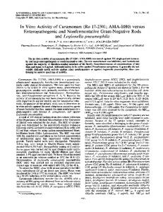
crystalline NaSbV showed about a two- to threefold increase in potency, with IC50s ranging from 8.8 to 10.5 g SbV/ml. Like Pentostam and NaSbV, PX-6518 showed no efficacy against promastigotes, established axenic amastigotes, or ex vivo amastigotes. A consistent, high activity was obtained against intracellular stages after infection with either axenic or ex vivo amastigotes (IC50, 0.1 g/ml). Infection with metacyclic promastigotes resulted in marginally lower efficacy (IC50, 0.7 g/ml), which may reflect incomplete intracellular transformation of promastigotes into amastigotes. The applicability of the different models for either drug screening or determination of the resistance of field strains is summarized in Table 2. Morphological evaluation. Specific attention was given to the established axenic amastigotes in an attempt to ascertain whether the parasite stages were either amastigote-like or promastigote-like. Light microscopy showed amastigote-like organisms, i.e., clear, immobile, rounded stages with dense cell masses, about 2 m in diameter. However, ultrastructural characteristics indicated the presence of organisms that were more promastigote-like, i.e., more-oval cell bodies, the presence of a strong protruding flagellum, and the absence of
3857
megasomes (Fig. 1a and b). In contrast, fresh ex vivo and intracellular amastigote stages (Fig. 1c) did not possess flagella beyond the margins of the cell body, were clearly rounded, and exhibited structures resembling megasomes. DISCUSSION The Leishmania life cycle comprises different developmental stages, all of which have been used for either drug-screening purposes or determination of the resistance of field isolates. However, specific recommendations are needed depending on the experimental objective. To address this, the comparative in vitro sensitivities of all possible stages of a single L. donovani reference strain (MHOM/ET/67/L82) to all current first-line drugs and one experimental drug, PX-6518, were determined (Table 1). The main observation for all reference drugs was that logphase promastigotes tended to be less sensitive than the other extracellular or intracellular stages and were fully refractory to SbV and PX-6518. These findings and the fact that promastigotes are the vector stage, differing considerably from the intracellular mammalian amastigote target, provide a convincing argument for excluding them for drug-screening purposes (Table 2). On the other hand, susceptibility to amphotericin B and miltefosine, which are known to affect membrane integrity directly or indirectly and which are able to exert antileishmanial action independently of cell-mediated parasiticidal mechanisms, has been clearly demonstrated (20, 26, 40). It is therefore not too speculative to propose that promastigotes could indeed be used for determination of the resistance of field isolates to amphotericin B and miltefosine (18, 25). This procedure will become particularly relevant for the large-scale monitoring of resistance to miltefosine in the Indian subcontinent, in view of its recent introduction as a first-line medication in the current “Kala-Azar Elimination Program” (36; WHO Regional Office for South-East Asia, meeting on guidelines and standard operating procedures for kala-azar elimination, Kolkata, India, 16 April 2007).
FIG. 1. Ultrastructural details of in vitro-established axenic and intracellular amastigotes. Shown are Leishmania donovani L82 axenic amastigotes in cell-free medium (a and b) and 72 h after infection of PMM (c). Established axenic amastigotes show oval cell bodies with a clear nucleus (n) and kinetoplast (k) and with flagella (f) protruding from the flagellar pocket. In contrast, intracellular axenic amastigotes are more rounded and possess short flagella and megasome-like (m) structures.
3858
VERMEERSCH ET AL.
Established axenic amastigotes have been positioned as an attractive alternative to the more complicated and labor-intensive cellular amastigote test in drug screening, but they obviously cannot be proposed for monitoring of the resistance of clinical field strains (Table 2). Publications on similarities between axenic and intracellular amastigotes in terms of morphology, gene regulation, enzyme profile, and sensitivity to some reference drugs may strengthen the use of established axenic amastigotes as a promising alternative drug-screening model (4, 9, 37). However, other investigators have drawn attention to marked differences between axenic amastigotes and lesion-derived ex vivo or intracellular amastigotes (17). In addition, long-term axenic cultivation may promote the reappearance of promastigote-like features, indicating the need for cautious use and continued follow-up of the amastigote-like characteristics (16, 24). Based on the drug sensitivity profile (Table 1) and the small-scale transmission electron microscopic study, our axenic amastigote model exhibited distinct promastigote-like characteristics, despite all precautions to prevent the culture from drifting back to the promastigote stage. Under light microscopy, the axenic amastigotes appeared as immobile round stages with dense cell masses (diameter, about 2 m). Ultrastructurally, flagella protruding from the flagellar pocket were frequently observed (Fig. 1a and b). On the other hand, internalized axenic amastigotes were clearly rounded, without protruding flagella, and contained megasome-like structures (Fig. 1c), which are membranebound compartments with lysosomal properties described for amastigotes of Leishmania chagasi (1) and Leishmania mexicana complex (39). Apart from these ultrastructural indications, our in vitro results with SbV and PX-6518 also confirm that the axenic amastigotes behaved as promastigotes. A general consensus is that SbV acts as a prodrug, exerting its parasiticidal effects through reduced SbIII, which is highly toxic to all stages (2, 7). Even though several reports describe selective activity of SbV on established axenic amastigotes (13, 32, 33), other researchers argue that cellular mechanisms are indeed required for the antileishmanial action of SbV (12, 22, 28). Although the mechanism of action of PX-6518 is not yet known, our results indicate that its action is mainly cell mediated as well, as shown by its lack of efficacy in the axenic models (IC50, ⬎32 g/ml) and its high potency against intracellular amastigotes (IC50, 0.1 g/ml). Besides PMM, other cells, such as U937, J774, THP-1, and bone marrow-derived and human monocyte-derived macrophages, have been used in the intracellular amastigote model. Recently, it was shown that the macrophage cell type may influence the levels of activity that are obtained with current antileishmanial drugs, necessitating harmonization if results from different laboratories are to be compared (29). No differences in the IC50s of the reference drugs were found upon infection of PMM with metacyclic promastigotes, established axenic amastigotes, or fresh spleen-derived ex vivo amastigotes (Table 1). Amphotericin B showed slightly higher potency in the cellular model than in the axenic models, a difference that is likely related to accumulation in the phagolysosome. Miltefosine showed no clear trend to support any theory of differential sensitivity. Stibogluconate was active only in the cellular model, with a higher potency for the crystalline substance than for Pentostam (IC50s, 22.0 to 28.4 g SbV/ml). A similar dif-
ANTIMICROB. AGENTS CHEMOTHER.
ferential susceptibility was observed for PX-6518, but at a much higher potency (IC50, 0.1 g/ml). The slightly lower activity after infection with metacyclic promastigotes (IC50, 0.7 g/ml) may be related to incomplete intracellular transformation into amastigotes. The latter phenomenon has been shown to occur frequently with clinical field isolates during their adaptation to in vitro culture and will affect the outcome of in vitro Sb resistance determinations (L. Maes, unpublished data). Overall, the results obtained in the present study are within the range of published IC50s for L. donovani strain L82 (10, 14, 30, 35): IC50s of 4.8 to 34.5 g SbV/ml were reported for sodium stibogluconate against intracellular amastigotes; miltefosine exhibited IC50s of 0.18 to 13.6 M on promastigotes and 2.8 to 5.8 M on intracellular amastigotes, while amphotericin B showed IC50s of 0.003 to 0.15 M and 0.026 to 0.076 g/ml against promastigotes and intracellular amastigotes, respectively. In summary, this study clearly demonstrates that the intracellular amastigote model is the only suitable approach for drug screening. The same cellular model is also required for the determination of resistance to Sb in view of its cell-mediated mode of action. For the latter, particular care must be taken to ascertain adequate intracellular transformation into amastigotes, a problem frequently encountered with poorly infective field strains. If field strains are infective to hamsters, it may sometimes be helpful to use ex vivo amastigotes as an infection inoculum for macrophages. For amphotericin B and miltefosine, the simpler axenic models could theoretically be used for resistance detection, but validation with a large set of field strains is still needed. ACKNOWLEDGMENTS This work was supported by a grant from FWO Flanders (project G.0103.06) and the UNDP/World Bank/WHO Special Programme for Research and Training in Tropical Diseases (TDR). M. Vermeersch is a BOF-NOI 20282 Ph.D. student at the University of Antwerp. P. Cos is a postdoctoral researcher awarded a grant by the Fund for Scientific Research (FWO Flanders, Belgium). REFERENCES 1. Alberio, S. O., S. S. Dias, F. P. Faria, R. A. Mortara, C. L. Barbieri, and E. F. Haapalainen. 2004. Ultrastructural and cytochemical identification of megasome in Leishmania (Leishmania) chagasi. Parasitol. Res. 92:246–254. 2. Ashutosh, S. Sundar, and N. Goyal. 2007. Molecular mechanisms of antimony resistance in Leishmania. J. Med. Microbiol. 56:143–153. 3. Berman, J. 2005. Clinical status of agents being developed for leishmaniasis. Expert Opin. Investig. Drugs 14:1337–1346. 4. Callahan, H. L., A. C. Portal, R. Devereaux, and M. Grogl. 1997. An axenic amastigote system for drug screening. Antimicrob. Agents Chemother. 41: 818–822. 5. Chappuis, F., S. Sundar, A. Hailu, H. Ghalib, S. Rijal, R. W. Peeling, J. Alvar, and M. Boelaert. 2007. Visceral leishmaniasis: what are the needs for diagnosis, treatment and control? Nat. Rev. Microbiol. 5:873–882. 6. Croft, S. L. 2001. Monitoring drug resistance in leishmaniasis. Trop. Med. Int. Health 6:899–905. 7. Croft, S. L., S. Sundar, and A. H. Fairlamb. 2006. Drug resistance in leishmaniasis. Clin. Microbiol. Rev. 19:111–126. 8. Cunningham, I. 1977. New culture medium for maintenance of tsetse tissues and growth of trypanosomatids. J. Protozool. 24:325–329. 9. Debrabant, A., M. B. Joshi, P. F. P. Pimenta, and D. M. Dwyer. 2004. Generation of Leishmania donovani axenic amastigotes: their growth and biological characteristics. Int. J. Parasitol. 34:205–217. 10. del Rayo Camacho, M., J. D. Phillipson, S. L. Croft, P. Rock, S. J. Marshall, and P. L. Schiff. 2002. In vitro activity of Triclisia patens and some bisbenzylisoquinoline alkaloids against Leishmania donovani and Trypanosoma brucei brucei. Phytother. Res. 16:432–436. 11. Drugs for Neglected Diseases Initiative. 2008. Annual report, 2007–2008. DNDi, Geneva, Switzerland.
VOL. 53, 2009 12. Dube, A., N. Singh, S. Sundar, and N. Singh. 2005. Refractoriness to the treatment of sodium stibogluconate in Indian kala-azar field isolates persists in in vitro and in vivo experimental models. Parasitol. Res. 96:216–223. 13. Ephros, M., A. Bitnun, P. Shaked, E. Waldman, and D. Zilberstein. 1999. Stage-specific activity of pentavalent antimony against Leishmania donovani axenic amastigotes. Antimicrob. Agents Chemother. 43:278–282. 14. Escobar, P., S. Matu, C. Marques, and S. L. Croft. 2002. Sensitivities of Leishmania species to hexadecylphosphocholine (miltefosine), ET-18OCH(3) (edelfosine) and amphotericin B. Acta Trop. 81:151–157. 15. Fumarola, L., R. Spinelli, and O. Brandonisio. 2004. In vitro assays for evaluation of drug activity against Leishmania spp. Res. Microbiol. 155:224– 230. 16. Gupta, N., N. Goyal, and A. K. Rastogi. 2001. In vitro cultivation and characterization of axenic amastigotes of Leishmania. Trends Parasitol. 17:150– 153. 17. Holzer, T. R., W. R. McMaster, and J. D. Forney. 2006. Expression profiling by whole-genome interspecies microarray hybridization reveals differential gene expression in procyclic promastigotes, lesion-derived amastigotes, and axenic amastigotes in Leishmania mexicana. Mol. Biochem. Parasitol. 146: 198–218. 18. Kumar, D., A. Kulshrestha, R. Singh, and P. Salotra. 2009. In vitro susceptibility of field isolates of Leishmania donovani to miltefosine and amphotericin B: correlation with sodium antimony gluconate susceptibility and implications for treatment in areas of endemicity. Antimicrob. Agents Chemother. 53:835–838. 19. Maes, L., D. Vanden Berghe, N. Germonprez, L. Quirijnen, P. Cos, N. De Kimpe, and L. Van Puyvelde. 2004. In vitro and in vivo activities of a triterpenoid saponin extract (PX-6518) from the plant Maesa balansae against visceral Leishmania species. Antimicrob. Agents Chemother. 48:130– 136. 20. Mbongo, N., P. M. Loiseau, M. A. Billion, and M. Robert-Gero. 1998. Mechanism of amphotericin B resistance in Leishmania donovani promastigotes. Antimicrob. Agents Chemother. 42:352–357. 21. Mikus, J., and D. Steverding. 2000. A simple colorimetric method to screen drug cytotoxicity against Leishmania using the dye Alamar Blue. Parasitol. Int. 48:265–269. 22. Mookerjee Basu, J., A. Mookerjee, P. Sen, S. Bhaumik, P. Sen, S. Banerjee, K. Naskar, S. K. Choudhuri, B. Saha, S. Raha, and S. Roy. 2006. Sodium antimony gluconate induces generation of reactive oxygen species and nitric oxide via phosphoinositide 3-kinase and mitogen-activated protein kinase activation in Leishmania donovani-infected macrophages. Antimicrob. Agents Chemother. 50:1788–1797. 23. Palatnik-de-Sousa, C. B. 2008. Vaccines for leishmaniasis in the fore coming 25 years. Vaccine 26:1709–1724. 24. Pan, A. A., S. M. Duboise, S. Eperon, L. Rivas, V. Hodgkinson, Y. TraubCseko, and D. McMahon-Pratt. 1993. Developmental life cycle of Leishmania—cultivation and characterization of cultured extracellular amastigotes. J. Eukaryot. Microbiol. 40:213–223. 25. Pe´rez-Victoria, F. J., M. P. Sa ´nchez-Can ˜ ete, K. Seifert, S. L. Croft, S. Sundar, S. Castanys, and F. Gamarro. 2006. Mechanisms of experimental resistance of Leishmania to miltefosine: implications for clinical use. Drug Resist. Updat. 9:26–39.
IN VITRO SUSCEPTIBILITIES OF LEISHMANIA STAGES
3859
26. Rakotomanga, M., S. Blanc, K. Gaudin, P. Chaminade, and P. M. Loiseau. 2007. Miltefosine affects lipid metabolism in Leishmania donovani promastigotes. Antimicrob. Agents Chemother. 51:1425–1430. 27. Rijal, S., F. Chappuis, R. Singh, P. A. Bovier, P. Acharya, B. M. Karki, M. L. Das, P. Desjeux, L. Loutan, and S. Koirala. 2003. Treatment of visceral leishmaniasis in south-eastern Nepal: decreasing efficacy of sodium stibogluconate and need for a policy to limit further decline. Trans. R. Soc. Trop. Med. Hyg. 97:350–354. 28. Roberts, W. L., J. D. Berman, and P. M. Rainey. 1995. In vitro antileishmanial properties of tri- and pentavalent antimonial preparations. Antimicrob. Agents Chemother. 39:1234–1239. 29. Seifert, K., P. Escobar, and S. Croft. 2009. Activity of current anti-leishmanial drugs in in vitro macrophage-amastigote model is dependent on the host macrophage, poster 398. Fourth World Cong. Leishmaniasis, Lucknow, India, 3 to 7 February 2009. 30. Seifert, K., F. J. Pe´rez-Victoria, M. Stettler, M. P. Sa ´nchez-Can ˜ ete, S. Castanys, F. Gamarro, and S. L. Croft. 2007. Inactivation of the miltefosine transporter, LdMT, causes miltefosine resistance that is conferred to the amastigote stage of Leishmania donovani and persists in vivo. Int. J. Antimicrob. Agents 30:229–235. 31. Sereno, D., and J.-L. Lemesre. 1997. Axenically cultured amastigote forms as an in vitro model for investigation of antileishmanial agents. Antimicrob. Agents Chemother. 41:972–976. 32. Sereno, D., M. Cavaleyra, K. Zemzoumi, S. Maquaire, A. Ouaissi, and J. L. Lemesre. 1998. Axenically grown amastigotes of Leishmania infantum used as an in vitro model to investigate the pentavalent antimony mode of action. Antimicrob. Agents Chemother. 42:3097–3102. 33. Shaked-Mishan, P., N. Ulrich, M. Ephros, and D. Zilberstein. 2001. Novel intracellular SbV reducing activity correlates with antimony susceptibility in Leishmania donovani. J. Biol. Chem. 276:3971–3976. 34. Shimony, O., and C. L. Jaffe. 2008. Rapid fluorescent assay for screening drugs on Leishmania amastigotes. J. Microbiol. Methods 75:196–200. 35. Smith, A. C., V. Yardley, J. Rhodes, and S. L. Croft. 2000. Activity of the novel immunomodulatory compound tucaresol against experimental visceral leishmaniasis. Antimicrob. Agents Chemother. 44:1494–1498. 36. Sundar, S., and H. W. Murray. 2005. Availability of miltefosine for the treatment of kala-azar in India. Bull. W. H. O. 83:394–395. 37. Tavares, J., A. Ouaissi, P. K. Lin, A. Toma ´s, and A. Cordeiro-da-Silva. 2005. Differential effects of polyamine derivative compounds against Leishmania infantum promastigotes and axenic amastigotes. Int. J. Parasitol. 35:637–646. 38. Teixeira, M. C., R. de Jesus Santos, R. B. Sampaio, L. Pontes-de-Carvalho, and W. L. Dos-Santos. 2002. A simple and reproducible method to obtain large numbers of axenic amastigotes of different Leishmania species. Parasitol. Res. 88:963–968. 39. Ueda-Nakamura, T., M. Attias, and W. de Souza. 2007. Comparative analysis of megasomes in members of the Leishmania mexicana complex. Res. Microbiol. 158:456–462. 40. Urbina, J. A. 2006. Mechanisms of action of lysophospholipid analogues against trypanosomatid parasites. Trans. R. Soc. Trop. Med. Hyg. 100:S9– S16.