Leishmania (Viannia) Species Identification on Clinical Samples from Cutaneous Leishmaniasis Patients in Peru: Assessment of a Molecular Stepwise Approach Nicolas Veland,a Andrea K. Boggild,b Cristian Valencia,a Braulio M. Valencia,a Alejandro Llanos-Cuentas,a,f Gert Van der Auwera,c Jean-Claude Dujardin,c,d and Jorge Arevaloa,e Instituto de Medicina Tropical Alexander von Humboldt, Universidad Peruana Cayetano Heredia, Lima, Perua; Tropical Disease Unit, Division of Infectious Diseases, Toronto General Hospital, Toronto, Canadab; Department of Parasitology, Institute of Tropical Medicine Antwerp, Antwerp, Belgiumc; Department of Biomedical Sciences, Faculty of Pharmaceutical, Biomedical and Veterinary Sciences, University of Antwerp, Antwerp, Belgiumd; Departamento de Bioquímica, Biología Molecular y Farmacología, Laboratorios de Investigación y Desarrollo, Facultad de Ciencias y Filosofía, Universidad Peruana Cayetano Heredia, Lima, Perue; and Hospital Nacional Cayetano Heredia, Lima, Peruf
We present an algorithm based on three PCR assays for Leishmania (Viannia) species identification and assessed its performance using 70 specimens from Peruvian patients. The succession of the assayed targets can be ordered according to species prevalence. Sequential progression through the algorithm reduced the number of samples here studied by approximately 30% after each step.
C
utaneous leishmaniasis (CL) is a vector-borne disease, which affects up to 1.5 million persons annually in tropical and subtropical regions worldwide (5). In several areas of endemicity like Peru, Leishmania species identification is important because different species with various clinical phenotypes coexist (11). There, higher incidence of mucocutaneous leishmaniasis (MCL) is attributed to Leishmania (Viannia) braziliensis (4), and low-pentavalent-antimonial treatment failures are associated with Leishmania (Viannia) guyanensis infections (1). Recognized associations between species and disease outcomes (1, 10, 12) have led the WHO expert committee on the control of leishmaniasis to recommend species identification for optimal treatment and control (16). PCR identification of Leishmania (Viannia) from clinical specimens (8, 14, 15) has used different targets: mannose phosphate isomerase (mpi) (17) and cysteine proteinase B (cpb) and the 70kDa heat shock protein (hsp70) (6, 7, 9). These targets have been used successfully on CL specimens collected by filter paper lesion impressions (FPLIs) (3). Here, we report the development of a three-step strategy of PCR-based assays for Leishmania (Viannia) species identification and assess its performance on CL FPLIs. Reductions of time and cost to obtain results in areas where leishmaniasis is endemic were the criteria on which the algorithm was evaluated. DNA isolation from cultured parasites and filter paper lesion impressions. International reference strains and cultured parasites obtained from 80 patients attending our reference center were harvested, and their DNA was extracted as previously described (13). DNAs from anonymized, FPLI specimens from 70 CL lesions collected from 57 Peruvian patients were obtained as reported previously (2, 3). Codes and geographical origins are presented in Table S1 in the supplemental material. PCR assays for species identification. Leishmania (Viannia) species were identified using three PCR targets. The mpi gene was amplified in two separate reactions (mpi Lp and mpi Lb for Leishmania [Viannia] peruviana and L. [V.] braziliensis, respectively) according to the work of Zhang et al. (17). cpb and hsp70 genes were amplified as previously reported (7, 9). Primer sequences and reaction conditions for each target are described in Table S2 in the supplemental material.
0095-1137/12/$12.00
Journal of Clinical Microbiology
p. 495– 498
Analysis of cultured isolates and algorithm development. Eighty cultured isolates were analyzed using the three targets, of which 41 were identified as L. (V.) braziliensis, 23 as L. (V.) peruviana, 14 as L. (V.) guyanensis, and 1 as having an L. (V.) braziliensis/L. (V.) peruviana mixed pattern (see Table S1 in the supplemental material). Only 1 out of 80 remained unidentifiable after the three targets were assayed. Each one of the targets identified the three most prevalent species: mpi for L. (V.) peruviana, cpb for L. (V.) braziliensis, and hsp70 for L. (V.) guyanensis. To differentiate genotypes in the lowest number of steps, the three targets were merged into one algorithm that decreases the number of samples to analyze in each step, reducing time and costs. According to the prevalence of species in our study site (Instituto de Medicina Tropical Alexander von Humboldt [IMTAvH], Lima, Peru), the mpi/cpb/hsp70 order provided the lowest number of steps for species identification (Table 1). PCR detection limit. The PCR detection limit was determined for each of the three targets using dilutions from 5 ng down to 0.1 pg of DNA. Amplification products were observed down to 1 pg of Leishmania DNA (approximately 10 parasites) after 30 and 45 cycles with mpi (both reactions) and hsp70, respectively. In contrast, 45 cycles of cpb PCR yielded amplification from 10 pg or more. Neither species cross-reaction nor nonspecific amplifications were observed with the targets. Inclusion criteria for species identification in clinical specimens. Kinetoplast DNA (kDNA) PCR was performed on FPLIs to diagnose leishmaniasis (2, 3). PCR-positive FPLIs (n ⫽ 70) were classified into three groups according to resulting kDNA band intensities: (i) strong positive (ⱖ10 pg of Leishmania DNA), n ⫽
Received 11 July 2011 Returned for modification 7 October 2011 Accepted 14 November 2011 Published ahead of print 23 November 2011 Address correspondence to Nicholas Veland,
[email protected]. Supplemental material for this article may be found at http://jcm.asm.org/. Copyright © 2012, American Society for Microbiology. All Rights Reserved. doi:10.1128/JCM.05061-11
jcm.asm.org
495
Veland et al.
TABLE 1 Algorithm step results according to amplification products and restriction patterns Step order
Assayc
Gel result (bp)
Species identity
Next step
1st
mpi Lp PCR mpi Lb PCR mpi Lp PCR mpi Lb PCR mpi Lp PCR mpi Lb PCR mpi Lp PCR mpi Lb PCR
312 Negative 312 312 Negative 312 Negative Negative
L. (V.) peruviana
Stop
L. (V.) braziliensis/L. (V.) peruviana Hybrid or mixed infection
Stop
L. (V.) braziliensis or L. (V.) guyanensis Not identified
2nd (cpb PCR-RFLP) 3rd (hsp70 PCR-RFLP)
2nd
cpb PCR-RFLP
543, 343, 257 543, 343, 257, 220, 123 543, 257, 220, 123 Negative
L. (V.) braziliensisa L. (V.) braziliensisb Probable L. (V.) guyanensis Not identified
Stop Stop 3rd (hsp70 PCR-RFLP) 3rd (hsp70 PCR-RFLP)
3rd
hsp70 PCR-RFLP
338, 307, 286, 265 (338 ⫹ 333), 307, 145, 120 338, 307, 286, 225 Negative
L. (V.) guyanensis L. (V.) lainsoni L. (V.) braziliensis Not identified
Stop Stop Stop Stop
a
Typical L. (V.) braziliensis cpb PCR restriction fragment length polymorphism (PCR-RFLP) pattern. Unusual L. (V.) braziliensis cpb PCR-RFLP pattern that might be confounded with L. (V.) braziliensis/L. (V.) peruviana hybrids if mpi PCR were not available. c mpi Lp and mpi Lb correspond to PCRs using reverse allele-specific primers for L. (V.) peruviana and L. (V.) braziliensis, respectively. b
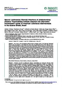
41; (ii) medium positive (between 10 and 0.1 pg), n ⫽ 20; and (iii) weak positive (⬍100 fg), n ⫽ 9. Different intensities of positive kDNA bands are clearly illustrated in Fig. 1. Only those specimens that were classified after kDNA PCR as strong positive or medium positive (Fig. 1, lanes 1 to 6) were adequate for species identification through the three-step algorithm. We established that a sample of lower intensity than the lane 6 band would not be amenable for species identification.
Algorithm performance on clinical specimens. Algorithm performance on clinical specimens was assessed using 70 FPLIs, where nine were excluded because of weak positive kDNA band after diagnostic PCR. After the three steps were performed on the remaining 61 specimens, species identification was successful in 53 (76% of all samples). From these, 17 were identified as L. (V.) peruviana, 19 as L. (V.) braziliensis, 15 as L. (V.) guyanensis, one as Leishmania (Viannia) lainsoni, and one as having an L. (V.) bra-
FIG 1 kDNA PCR band intensity categories used as references to allocate diagnosis of clinical specimens. PCR mixtures (lanes 1 to 8) contained 5 ng of human DNA plus different amounts (5 ng down to 1 fg) of Leishmania DNA. PCR product sizes correspond to 70 bp for Leishmania kDNA and 197 bp for human beta-globin gene (internal control). Abbreviations: w/o, without; M, molecular size ladder.
496
jcm.asm.org
Journal of Clinical Microbiology
Leishmania Species Identification Algorithm
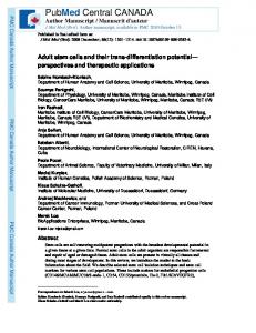
FIG 2 Reduction of numbers of clinical specimens through the algorithm steps. PCR steps are shown with black shading. Subsequent steps are indicated with arrows, while identification endpoints in the flow chart are indicated within gray-shaded boxes. The algorithm was evaluated using 70 specimens on filter paper lesion impressions, and the numbers processed in each step are indicated (n). mpi Lp and mpi Lb correspond to PCRs using reverse allele-specific primers for L. (V.) peruviana and L. (V.) braziliensis, respectively. ⴱ, two specimens that could not be typed with cpb PCR due to negative or weak cpb amplification but were identified with hsp70 PCR-restriction fragment length polymorphism (PCR-RFLP).
ziliensis/L. (V.) peruviana mixed pattern (Fig. 2). Of the eight nonidentified specimens, seven specimens did not amplify any target despite displaying a medium positive kDNA band, which may reflect low DNA levels (less than 1 pg) or highly inhibited specimens. The remaining specimen produced an incongruent pattern: an amplification product with the mpi Lb reaction allocated to either L. (V.) braziliensis or L. (V.) guyanensis and a cpb product indicative of L. (V.) guyanensis. Nevertheless, hsp70 distinguished this specimen as L. (V.) braziliensis. In our experience, this result could be attributed to nucleotide polymorphisms that modified restriction enzyme recognition sites. Progression through the algorithmic steps considerably reduces the number of specimens to be analyzed in the sequential step, thereby reducing time and resources. Thus, 30% of specimens analyzed were typed in the first 4 h (after kDNA PCR result) and 28% were identified over the next 24 h. The final 42% needed execution of the last step, which required 24 additional hours to obtain results. Consequently, one-third of patients had a definitive answer in the first 24 h after clinical specimen collection. Algorithm flexibility allows switching steps according to species prevalence in a given region of endemicity. We recommend starting with the PCR target for the most predominant species.
February 2012 Volume 50 Number 2
Our patient enrollment is principally from the Andean valleys where L. (V.) peruviana is predominant (1); therefore, mpi was the first target. Nevertheless, if this algorithm is to be applied in Cuzco, where L. (V.) braziliensis is mostly found (1, 11), the first target should be cpb. Therefore, it is expected that species identification will be obtained for most patients in the first step, leaving the minority for the subsequent hsp70 step. In areas where L. (V.) guyanensis is the most prevalent, as occurs in the central high jungle (1, 11), the order of the targets should place hsp70 in the first step followed by cpb. The proposed algorithm strategy should be useful to guide patient management as well as for epidemiological studies and clinical trials. This concept can also be applied in Old World leishmaniasis and in other pathologies, especially in public health settings where resources are limited. ACKNOWLEDGMENTS We thank Milena Alba of the Instituto de Medicina Tropical Alexander von Humboldt-Universidad Peruana Cayetano Heredia (Lima, Peru) for technical support. This study was funded by the European Community (INCO-Dev program “Control strategies for visceral leishmaniasis [VL] and mucocuta-
jcm.asm.org 497
Veland et al.
neous leishmaniasis [MCL] in South America: applications of molecular epidemiology” [contract INCO-CT2005-015407]) and the DirectorateGeneral for Development Cooperation of the Belgian Government (framework agreement 03—project 95502). A.K.B. was supported by a professional development grant (2009) and a Detweiler traveling fellowship (2010) through the Royal College of Physicians and Surgeons of Canada during the study period. A.K.B., B.M.V., and A.L.-C. contributed to sample collection. A.K.B., C.V., and G.V.D.A. contributed to data interpretation and writing of the manuscript. J.-C.D. and J.A. contributed to study design, implementation, and data interpretation. N.V. contributed to study design, data collection, analysis, and interpretation and was primarily responsible for writing the manuscript. All authors critically appraised the manuscript. All authors report no potential conflicts of interest.
REFERENCES 1. Arevalo J, et al. 2007. Influence of Leishmania (Viannia) species on the response to antimonial treatment in patients with American tegumentary leishmaniasis. J. Infect. Dis. 195:1846 –1851. 2. Boggild AK, et al. 2011. Diagnostic performance of filter paper lesion impression PCR for secondarily infected ulcers and nonulcerative lesions caused by cutaneous leishmaniasis. J. Clin. Microbiol. 49:1097–1100. 3. Boggild AK, et al. 2010. Detection and species identification of Leishmania DNA from filter paper lesion impressions for patients with American cutaneous leishmaniasis. Clin. Infect. Dis. 50:e1– e6. 4. Davies CR, et al. 2000. The epidemiology and control of leishmaniasis in Andean countries. Cad. Saude Publica 16:925–950. 5. Desjeux, P. 2004. Leishmaniasis. Nat. Rev. Microbiol. 2:692. 6. Fraga J, Montalvo AM, De Doncker S, Dujardin JC, Van der Auwera G. 2010. Phylogeny of Leishmania species based on the heat-shock protein 70 gene. Infect. Genet. Evol. 10:238 –245. 7. Garcia AL, et al. 2005. American tegumentary leishmaniasis: antigen-
498
jcm.asm.org
8.
9. 10. 11. 12. 13. 14. 15.
16.
17.
gene polymorphism, taxonomy and clinical pleomorphism. Infect. Genet. Evol. 5:109 –116. Garcia AL, Parrado R, De Doncker S, Bermudez H, Dujardin JC. 2007. American tegumentary leishmaniasis: direct species identification of Leishmania in non-invasive clinical samples. Trans. R. Soc. Trop. Med. Hyg. 101:368 –371. Garcia L, et al. 2004. Culture-independent species typing of neotropical Leishmania for clinical validation of a PCR-based assay targeting heat shock protein 70 genes. J. Clin. Microbiol. 42:2294 –2297. Llanos-Cuentas A, et al. 2008. Clinical and parasite species risk factors for pentavalent antimonial treatment failure in cutaneous leishmaniasis in Peru. Clin. Infect. Dis. 46:223–231. Lucas CM, et al. 1998. Geographic distribution and clinical description of leishmaniasis cases in Peru. Am. J. Trop. Med. Hyg. 59:312–317. Nolder D, Roncal N, Davies CR, Llanos-Cuentas A, Miles MA. 2007. Multiple hybrid genotypes of Leishmania (Viannia) in a focus of mucocutaneous Leishmaniasis. Am. J. Trop. Med. Hyg. 76:573–578. Oddone R, et al. 2009. Development of a multilocus microsatellite typing approach for discriminating strains of Leishmania (Viannia) species. J. Clin. Microbiol. 47:2818 –2825. Roelfsema JH, Nozari N, Herremans T, Kortbeek LM, Pinelli E. 2011. Evaluation and improvement of two PCR targets in molecular typing of clinical samples of Leishmania patients. Exp. Parasitol. 127:36 – 41. Rotureau B, et al. 2006. Use of PCR-restriction fragment length polymorphism analysis to identify the main new world Leishmania species and analyze their taxonomic properties and polymorphism by application of the assay to clinical samples. J. Clin. Microbiol. 44:459 – 467. World Health Organization. 2010. Control of the leishmaniasis: report of a meeting of the WHO Expert Committee on the Control of Leishmaniases, Geneva, 22 to 26 March 2010. World Health Organization technical report series no. 949. World Health Organization, Geneva, Switzerland. Zhang WW, et al. 2006. Development of a genetic assay to distinguish between Leishmania Viannia species on the basis of isoenzyme differences. Clin. Infect. Dis. 42:801– 809.
Journal of Clinical Microbiology