JMM Case Reports Retrospective PCR-based species identification of Leishmania in two patients with visceral leishmaniasis in Serbia --Manuscript Draft-Manuscript Number:
JMMCR-D-16-00044R1
Full Title:
Retrospective PCR-based species identification of Leishmania in two patients with visceral leishmaniasis in Serbia
Article Type:
Case Report
Section/Category:
Soft tissue
Order of Authors:
Zorica Dakić, Ph.D. Henrik Vedel Nielsen, Ph.D. Milorad Pavlović, Prof. Jasmina Poluga, Asst. prof. Goran Stevanović, Asst. prof. Lidija Lavadinović, MSc Branko Milošević, Asst. prof. Mijomir Pelemiš, Prof. Aleksandar Urošević, Asst. Snežana Jovanović, MSc Christen Rune Stensvold, Ph.D., Senior Scientist
Abstract:
Introduction: Retrospective molecular identification of Leishmania parasites in two patients with visceral leishmaniasis (VL) previously treated in Serbia was carried out. DNA was isolated from unstained bone marrow smears (BMS) kept for 11 and 8 years, respectively. Genus-specific real-time PCR was combined with conventional PCR and sequencing for detection and species identification. Case Presentation: In 2003, a 40-year-old Serbian male was admitted to the Clinical Centre of Serbia (CCS) with fever, sweating, fatigue, and splenomegaly developing over a period of seven weeks. He had frequently travelled around Europe. VL was confirmed by microscopy of Giemsa-stained BMS. Treatment by pentavalent antimonials was successfully completed. Two years later, the patient developed postkala-azar dermal leishmaniasis. Treatment resulted in symptom resolution. Later on, L. infantum was identified as the causative agent of VL by sequencing of the ITS region; mixed Leishmania sp. infection could not be excluded. In 2006, a 33-year-old female from Vojvodina, Serbia, with pre-existing diabetes mellitus, chronic meningoencephalitis, and with a history of frequent visits to the Montenegrin seacoast, was admitted to CCS with fever, pancytopenia, and moderate hepatosplenomegaly. A stained BMS revealed abundant Leishmania amastigotes. Indirect hemagglutination analysis was positive with a titre of 1:2048, and a rapid dipstick rK39 test was also positive. Treatment by liposomal amphotericin B was successful; however, shortly after, the patient developed neural infection and pneumonia and deceased. The causative agent was identified as L. infantum. Conclusion: Molecular diagnosis of VL and species delinieation using DNA from unstained BMS stored for several years is possible.
First Author:
Zorica Dakić, Ph.D.
Corresponding Author:
Zorica Dakić, Ph.D. Clinical Center of Serbia Belgrade, SERBIA
Downloaded from www.microbiologyresearch.org by IP: 181.215.111.6 Powered by Editorial Manager® and ProduXion Manager® from Aries Systems Corporation On: Thu, 09 Mar 2017 13:03:37
Manuscript Including References (Word document)
Click here to download Manuscript Including References (Word document) JMM CaseReport Dakic et al.
JMM CASE REPORTS Case report template TITLE OF CASE Retrospective PCR-based species identification of Leishmania in two patients with visceral leishmaniasis in Serbia Running title: Leishmania species delineation in Serbian patients Author Name: Zorica Dakić1, Henrik Vedel Nielsen2, Milorad Pavlović3,4, Jasmina
Poluga3,4,
Goran
Stevanović3,4,
Lidija
Lavadinović3,4,
Branko
Milošević3,4, Mijomir Pelemiš3,4, Aleksandar Urošević3,4, Snežana Jovanović1, Christen Rune Stensvold2 Address: 1Parasitological Laboratory, Department of Microbiology, Clinical Centre of Serbia, Bulevar oslobodjenja 16, 11000 Belgrade, Serbia 2
Laboratory of Parasitology, Department of Microbiology and Infection Control,
Division of Diagnostics and Infection Control, Statens Serum Institut, 5 Artillerivej, DK–2300 Copenhagen, Denmark 3
Medical Faculty, University of Belgrade, Dr Subotića 8, 11000 Belgrade, Serbia
4
Clinic for Infectious and Tropical Diseases, Clinical Centre of Serbia, Bulevar
oslobodjenja 16, 11000 Belgrade, Serbia Corresponding author: Zorica Dakić Corresponding author email address:
[email protected]
Corresponding author: Christen Rune Stensvold Corresponding author email address:
[email protected]
The full names and institutional addresses for all authors must be included on the title page.
1
Downloaded from www.microbiologyresearch.org by IP: 181.215.111.6 On: Thu, 09 Mar 2017 13:03:37
In order to assist us in choosing the correct editor to handle your paper, please choose one box in each of the following categories: Field: ☒ Human
☐ Dental
☐ Veterinary/Fisheries
Subject: ☐ Bacteriology ☐ Virology
☐ Mycology
☒ Parasitology
Keywords: Please provide at least one keyword for each of the following categories: Disease/Indication: visceral leishmaniasis; PKDL Pathology/Symptoms: fever; sweating; pancytopenia; splenomegaly Treatment: pentavalent antimonials; liposomal amphotericin B
ABSTRACT Up to 250 words summarising the case presentation and outcome (this will be shown on preview and search panes) Introduction: Retrospective molecular identification of Leishmania parasites in two patients with visceral leishmaniasis (VL) previously treated in Serbia was carried out. DNA was isolated from unstained bone marrow smears (BMS) kept for 11 and 8 years, respectively. Genus-specific real-time PCR was combined with conventional PCR and sequencing for detection and species identification. Case Presentation: In 2003, a 40-year-old Serbian male was admitted to the Clinical Centre of Serbia (CCS) with fever, sweating, fatigue, and splenomegaly developing over a period of seven weeks. He had frequently travelled around Europe. VL was confirmed by microscopy of Giemsa-stained BMS. Treatment by pentavalent antimonials was successfully completed. Two years later, the patient developed post-kala-azar dermal leishmaniasis. Treatment resulted in symptom resolution. Later on, L. infantum was identified as the causative agent of VL by sequencing of the ITS region; mixed Leishmania sp. Infection could not be excluded. In 2006, a 33-year-old female from Vojvodina, Serbia, with pre-existing diabetes mellitus, chronic meningoencephalitis, and with a history of frequent visits to the Montenegrin seacoast, was admitted to CCS with fever, pancytopenia, and moderate hepatosplenomegaly. A stained BMS revealed abundant Leishmania amastigotes. Indirect hemagglutination analysis was positive with a titre of 1:2048, and a rapid dipstick rK39 test was also positive. Treatment by liposomal amphotericin B was successful; however, shortly after, the patient developed neural infection and pneumonia and deceased. The causative agent was identified as L. infantum. Conclusion: Molecular diagnosis of VL and species delinieation using DNA from unstained BMS stored for several years is possible. INTRODUCTION Background; why do you think this case is important – why did you write it up? Leishmaniasis is becoming a growing health issue with increased reported numbers of cases in European areas previously not known to be endemic or as a re-emerging disease in formerly
2
Downloaded from www.microbiologyresearch.org by IP: 181.215.111.6 On: Thu, 09 Mar 2017 13:03:37
endemic areas. Visceral leishmaniasis (VL) occurs sporadically in Serbia. According to epidemiological data, eight new VL cases were reported in Serbia from 2008 to 2014 (Institute of Public Health of Serbia, 2009, 2015). In central Serbia, the incidence was 0.01/100000 in 2012 vs 0.03/100 000 in 2013. Cases of autochthonous VL have been reported in all countries neighbouring Serbia, including Montenegro, Croatia, Bosnia, Romania, Bulgaria, the FYR Macedonia, Greece, and Turkey (Beatović, 2010; Durda, 2010; Gradoni, 2013; Šiško-Kraljević, 2013). Most patients with suspected leishmaniasis in Serbia are referred to the Clinical Centre of Serbia (CCS) for diagnosis and treatment. Our study of VL carried out from 2001 to 2007 involving 22 VL cases, showed inadequate sensitivity of initial bone marrow smear (BMS) analysis (86.36%), indicating the need for applying more sensitive methods such as PCR (Dakić et al., 2009). Over the next eight years, from January 2008 to December 2015, nine cases (seven initial cases and two relapses) of VL were diagnosed in the Parasitological Laboratory, CCS, including one case of leishmaniasis/HIV co-infection. The majority probably contracted VL at the Montenegrin seacoast, with only one case being imported from outside the borders of former Yugoslavia (Portugal). Reliable diagnosis of VL, including accurate species identification, is critical to adequate treatment and developing prevention measures. Unfortunately, molecular diagnostics for detection and differentiation are not yet available in Serbia. Here, we performed retrospective molecular identification of Leishmania (L.) parasites in two patients previously treated in Serbia: one case of VL and one case of VL with consequent post-kalaazar dermal leishmaniasis (PKDL). DNA was isolated using QIAamp DNA Mini Kit (QIAGEN, Hilden, Germany) from unstained BMS kept for 11 and 8 years, respectively. Smears were subject to gentle scraping using 180 µL of ATL buffer solution. DNA extraction was performed according to the manufacturer’s recommendations. Extracted DNA was resuspended in 100 μL of AE buffer. Positive results by genus-specific real-time polymerase chain reaction (real-time PCR) were confirmed by conventional PCR followed by sequencing of the internal transcribed spacer 1 (ITS1) region and BLASTing
of
the
sequences
against
the
NCBI
nucleotide
sequence
database
(http://www.ncbi.nlm.nih.gov/nucleotide) for species identification. CASE REPORT Case I In May 2003, a 40-year-old Serbian male who had been living in Sweden was admitted to the Clinic for Infectious and Tropical Diseases (CITD), CCS, in Belgrade with fever above 39°C, sweating, fatigue, and splenomegaly developing over a period of seven weeks. Onset of a flu-like illness started on
3
Downloaded from www.microbiologyresearch.org by IP: 181.215.111.6 On: Thu, 09 Mar 2017 13:03:37
March 10, 2003 with high temperature (up to 37.7°C), sore throat, sneezing, chest pains, shortness of breath, aches and pains in muscles and joints. He was treated at the primary health care Centre during April 2003 and received repeated courses of antibiotic treatment (amoxicillin/clavulanic acid, roksitromicin, sulfamethoxazole and trimethoprim) without any improvement. On admission to CITD, laboratory tests were as follows: ESR, 92 mm/hour; erythrocytes, 3.05 × 10 12/L; hemoglobin, 122 g/L; leukocytes, 2.1 × 10⁹/L; platelets, 129 × 109/L. Abdominal ultrasonography showed hepatomegaly, with the right lobe having a size of 16.7 cm, and splenomegaly (length, 13.7 cm). He had frequently travelled around Europe (Spain, Sweden, Germany), including countries in South-East Europe. VL was confirmed on May 15, 2003 by examination of Giemsa-stained BM smears revealing numerous amastigotes of Leishmania (Fig. 1). Serology for Leishmania was not available at this moment. Treatment by pentavalent antimonials (Glucantime®) was immediately started using a dose of 20 mg/kg/day. After three days, the patient was requested to leave the hospital since he had no health insurance in Serbia. Treatment was successfully completed in Sweden (details of the treatment are not available). Two years later, in April 2005, during a stay in Serbia, the patient developed skin lesions. In the beginning, a diagnosis of lupus was considered. The patient was diagnosed as HIV-negative. Three months later, all nodular lesions resolved. In August 01, 2005, the patient visited a private dermatologist in Belgrade, Serbia, who consulted a specialist in infectious diseases who had treated the patient two years earlier of VL. The specialist suggested PKDL as a diagnosis. Indirect hemagglutination analysis (IHA) for Leishmania-specific antibodies (Siemens, Germany) was negative, and the rapid dipstick rK39 test (DiaSys Europe Ltd, England) was weakly positive. A diagnosis of PKDL was established by parasitological examination for Leishmania amastigotes in skin biopsy specimens performed in Serbia by a dermatologist while the patient was treated with amphotericin B in Sweden (details of the treatment are not available). Treatment resulted in symptom resolution. Later on, in October 2014 at the Statens Serum Institut (SSI), L. infantum was identified as the causative agent of VL by sequencing of the ITS region; based on sequence chromatogram analysis, mixed Leishmania sp. infection, however, could not be excluded.
Case II In mid-July 2006, a 33-year-old female from Vojvodina, northern Serbia, with pre-existent congenital hydrocephalus with a ventriculoperitoneal (VP) shunt implanted during infancy, chronic meningoencephalitis, diabetes mellitus, chronic renal failure and a history of frequent visits to the Montenegrin seacoast, was admitted to CITD with fever, pancytopenia, and moderate hepatosplenomegaly. An unfamiliar feeling of weakness with chills and fever started two months
4
Downloaded from www.microbiologyresearch.org by IP: 181.215.111.6 On: Thu, 09 Mar 2017 13:03:37
earlier, in mid-May 2006. The patient was admitted to the neurosurgical department at the regional hospital. First, VP shunt infection was suspected, but it was not confirmed. When pancytopenia and elevated levels of blood urea nitrogen and creatinine and appeared in the patient who previously was known with a normal blood profile, VL was suspected. Sternal puncture was performed, and the patient was immediately transferred to the intensive care unit of the CITD. On admission, laboratory tests revealed the following values: erythrocytes, 1.85 × 10¹²/L; hemoglobin, 46 g/L; leukocytes, 0.6 × 10⁹/L; platelets, 22 × 109/L. Abdominal ultrasonography showed hepatomegaly, with the right lobe measuring 13.5 cm, and splenomegaly (length, 15.0 cm). Immediately upon admission, a stained BMS revealed abundant Leishmania amastigotes (Fig. 2). Indirect hemagglutination test for Leishmania-specific antibodies was positive with a titre of 1:2048, and the rapid dipstick rK39 test was also positive. Seven days of treatment with liposomal amphotericin B (AmBisome®) in daily doses of 50 mg was given. Although expensive, this treatment was chosen due to high efficacy and low toxicity, good local experience, and the general availability of this drug. Resolution of the symptoms was achieved during the first month after completed therapy. However, shortly after improvement, the patient developed neural infection and pneumonia, resulting in respiratory failure. Despite rehydration therapy and respiratory support by non-invasive ventilation, rapidly progressive deterioration resulted in respiratory and cardiocirculatory insufficiency and death eighty days after hospitalization. Molecular analysis performed in October 2014 at the SSI identified the causative agent as L. infantum.
INVESTIGATIONS If relevant
DIAGNOSIS If relevant
TREATMENT If relevant
OUTCOME AND FOLLOW-UP If relevant
DISCUSSION At least 20 Leishmania species can infect humans and cause a wide spectrum of clinical diseases including three main types, VL (the most serious form), cutaneous, and mucocutaneous
5
Downloaded from www.microbiologyresearch.org by IP: 181.215.111.6 On: Thu, 09 Mar 2017 13:03:37
leishmaniasis (Peacock et al., 2007). VL is endemic to Mediterranean Europe, where the disease is caused by L. infantum. For the last decade, stable, low incidence rates of VL have been observed in Serbia. Insufficient research and frequent cross travelling of residents between the former Yugoslavian republics blurs the understanding of autochthonous transmission of VL in Serbia. A few reports suggest that animal reservoirs exist. The potential epidemiologic role of the golden jackal in dispersing Leishmania in Serbia should be taken into consideration when applying surveillance monitoring schemes. A total of 216 spleen samples from golden jackals were collected from 2010 to 2013, from 12 sites all over Serbia, with 6.9% being positive for Leishmania species by qPCR (Ćirović et al., 2014). From 2009 to 2013, Savić et al. (2014) analyzed dog blood samples for leishmaniasis by a routine ELISA test in the region of Vojvodina in northern Serbia. The seroprevalence was 15% (33/220); however, twenty-one dogs had travelled abroad with their owners in VL-endemic areas before testing. Vaselek et al. (2015) performed entomological surveillance of sand flies in Vojvodina in 2013. All 55 specimens of the genus Phlebotomus (P.) were P. papatasi, apart from a single specimen identified as P. tobbi. Four specimens of P. papatasi were positive for L. infantum by nested PCR, indicating the presence of this pathogen in the host and possible transmission within Serbia. For this reason, an epidemiological survey should be performed, including investigations of potential reservoir hosts and vectors in the surroundings of people with VL so as to clarify the epidemiology of Leishmania in Serbia. A recent survey performed in several endemic foci of VL in Croatia disclosed a high prevalence of asymptomatic parasite carriers (Šiško-Kraljević et al., 2013). One of the most attractive destinations for Serbian tourists is the Montenegrin seacoast; the southcoast region is considered endemic for leishmaniasis. From 1992 to 2009, 55 cases of VL were reported in Montenegro along with one case of CL. From 2006 and 2009, serological surveys of clinically suspect dogs coming from VL endemic areas identified a 58% average rate of infection (Beatović, 2010). The vector is P. neglectus (Ivović et al., 2004). Although imperfect, parasitological examination based on demonstration of the parasites of Leishmania is included in the gold standard diagnostic work-up for VL. Except for the invasive procedure of sampling, microscopy is straightforward and reliable if amastigotes are plentiful and if the microscopist is experienced; however, this is often not the case. Moreover, by parasitological examination, species of Leishmania are indistinguishable. Together with a number of other factors (host genetic variability, specific immune response) the main factor determining the clinical presentation of infection is the species of Leishmania (Lipoldova & Demant, 2006). Moreover,
6
Downloaded from www.microbiologyresearch.org by IP: 181.215.111.6 On: Thu, 09 Mar 2017 13:03:37
species identification is important for treatment selection. Generally, DNA-based methods have enabled a higher accuracy in the diagnosis of VL. Advantages of these methods include high sensitivity and specificity, and DNA-based methods can be performed on different sample materials (peripheral blood, spleen, liver, BM, and lymph node aspirates). Moreover, such methods may enable distinction between relapse and reinfection, and enables the diagnosis of VL in HIV-infected patients (Ruiter et al., 2014). Using DNA-based methods, Ruiter et al. (2014) showed that analysis of blood samples is as effective as analysis of (the more invasive) BM samples. In Serbia, the availability of molecular methods is limited for rare parasitic infections (including VL), which possibly has to do with cost-effectiveness issues. As of yet, the gold standard in Serbia for VL diagnosis is assessment of clinical presentation in combination with the demonstration of amastigotes in Giemsa-stained BMS and/or positive serology. The interpretation of sequences obtained by Sanger sequencing of PCR products may be difficult in case of mixed species infection as in Case 1. We believe that the use of next generation sequencing of PCR products obtained by genus-specific primers or the use of species-specific DNA-based methods, such as multiplex realtime PCR assays could eliminate the problem of not being able to delineate species in mixed Leishmania infections. There are several methods for species identification of Leishmania, including the use of monoclonal antibodies, analysis of isoenzymes, and molecular methods. The gold standard for species identification of Leishmania is still isoenzyme electrophoresis, while ITS1 sequencing is an alternative method (Mahmoudzadeh-Niknam et al., 2011). In the first patients mixed Leishmania sp. infection could not be excluded by molecular analysis. Mixed Leishmania sp. infection is possible as a consequence of extensive travelling. Mixing up or contamination of Leishmania isolates with nonLeishmania trypanosomatids (e.g. Crithidia) in some laboratory is possible, too (MahmoudzadehNiknam et al., 2011). Weakly positive results of the dipstick rK39 test could be a consequence of low affinity or low levels of antibodies. Das et al. (2011) reported three cases where the rK39 strip test failed to detect two cases of PKDL and one case of VL. Haldar et al. (1981) showed that IHA when used for PKDL was positive in only one third of patients with chronic PKDL, while all fresh cases were positive, indicating antibody titer reduction during the chronic phase. This could explain the negative IHA result during PKDL in the first patient. Molecular analysis of Leishmania parasites from the second patient confirmed with 100% sequence identity that L. infantum, probably contracted in Montenegro, was the cause of VL. High quality and preservation of each sample (blood, biopsies/aspirates) for Leishmania DNA extraction is crucial. Previous studies have shown the possibility of extracting amplifiable DNA of
7
Downloaded from www.microbiologyresearch.org by IP: 181.215.111.6 On: Thu, 09 Mar 2017 13:03:37
Leishmania from dried or old materials, including archival air-dried unstained BM slides or Giemsastained slides (Brustolini et al., 2007), blood or BM aspirates spotted onto filter paper (Cortes et al., 2004; da Silva et al., 2004), and even paraffin-embedded tissues (Lanús et al., 2005) or formalin-fixed material (Gebhardt et al., 2015). Slide smears are easily stored, do not require special storage conditions, and can be stored at room temperature. Latency in terms of establishing the diagnosis of VL was a consequence of the non-specific initial presentation of the illness (Case 1) or the result of pre-existing diseases mimicking VL (Case 2). Surely, the rare detection of VL explains why not all clinicians are familiar with this disease. The treatment of VL is very complex, because drug resistance has become a major obstacle to effective treatment. Traditionally, mainly antimony (Glucantime®) in doses of 20 mg/kg during 21–28 days was used as first-line therapy for VL patients in Serbia, as the cheapest, low toxic and effective antileishmanial drug. In recent years, however, we observed sporadic cases of unresponsive to antimony therapy. In these cases, favourable outcomes have been achieved by use of liposomal amphotericin B. Considering that VL is a rare infection, anti-leishmanial drugs, excluding amphotericin B, have not yet been registered in Serbia. PKDL is a dermal complication of VL, reported mainly in Sudan in eastern Africa and the Indian subcontinent, with incidences of up to 50%–60% and 5%–10%, respectively (Zijlstra et al., 2003; Ganguly et al., 2010). The pathogenesis of PKDL is mainly immunologically mediated and develops at variable intervals after/or during therapy for VL, e.g. after 0–6 months in Sudan and 2–3 years in India (Zijlstra et al., 2003). Most cases of PKDL occur after infection with L. donovani, less commonly so after L. infantum and extremely rarely following infection by L. chagasi (Singh et al., 2011). PKDL in HIV co-infected patients is more common and more severe and is not restricted to L. donovani (Zijlstra, 2014). After the first reports from the coasts of the Mediterranean basin in the 1990s, HIV and Leishmania co-infection has been increasingly reported (Monge-Maillo et al., 2014). In Serbia, this co-infection still appears rare and includes one documented case of VL/HIV coinfection in Niš, south Serbia, caused by L. infantum confirmed by molecular analyses of the parasites identified from blood (Marjanović et al., 2012) and two cases identified in our clinic. Introduction of molecular diagnostics for patients with VL in Serbia is warranted. Molecular diagnosis of VL using DNA extracted from unstained BMS stored for several years is possible. This is especially useful in the diagnosis of complicated VL cases, for whom unstained BMS could easily be mailed to centres where PCR is available. It is also useful for retrospective epidemiological studies. REFERENCES Harvard Style
8
Downloaded from www.microbiologyresearch.org by IP: 181.215.111.6 On: Thu, 09 Mar 2017 13:03:37
Beatović, V. (2010). The Institute of Public Health of Montenegro, The Ministry of Health of Montenegro, WHO exploratory meeting on leishmaniasis in the Balkan countries. Dubrovnik,
Croatia,
10–12
February
2010.
Available
on:
http://www.who.int/leishmaniasis/resources/MONTENEGRO.pdf Brustoloni, Y. M., Lima, R. B., da Cunha, R. V., Dorval, M. E., Oshiro, E. T., de Oliveira, A. L., Pirmez, C. (2007). Sensitivity and specificity of polymerase chain reaction in Giemsa-stained slides for diagnosis of visceral leishmaniasis in children. Mem Inst Oswaldo Cruz 102, 497–500. doi: http://dx.doi.org/10.1590/S0074-02762007005000036. PubMed PMID: 17612771. Cortes, S., Rolão, N., Ramada, J., Campino, L. (2004). PCR as a rapid and sensitive tool in the diagnosis of human and canine leishmaniasis using Leishmania donovani s.l. - specific kinetoplastid primers. Trans R Soc Trop Med Hyg 98, 12–17. doi: http://dx.doi.org/10.1016/S0035-9203(03)000026. PubMed PMID: 14702834. Ćirović, D., Chochlakis, D., Tomanović, S., Sukara, R., Penezić, A., Tselentis, Y., Psaroulaki, A. (2014). Presence of Leishmania and Brucella species in the golden jackal Canis aureus in Serbia. Biomed Res Int doi: http://dx.doi.org/10.1155/2014/728516. PubMed PMID: 24967397. da Silva, E. S., Gontijo, C. M., Pacheco, Rda S., Brazil, R. P. (2004). Diagnosis of human visceral leishmaniasis by PCR using blood samples spotted on filter paper. Genet Mol Res 3, 251–257. PubMed
PMID:
15266395.
Available
on:
http://www.funpecrp.com.br/gmr/year2004/vol2-
3/pdf/gmr0082.pdf. Dakić, Z., Pelemiš, M., Stevanović, G., Poluga, L., Lavadinović, L., Milošević, I., Indjić, N.,, Ofori-Belić, I., Pavlović, M. (2009). Epidemiology and diagnostics of visceral leishmaniasis in Serbia. Clin Microbiol Infect 15, 1173–1176. doi: http://dx.doi.org/10.1111/j.1469-0691.2009.02768.x. PubMed PMID: 19392902. Das, N. K., Singh, S. K., Ghosh, S., Sarkar, A., Mukhopadhyay, D., Roy, S., Ganguly, D. N., Barbhuiya, J. N., Saha, B., Chatterjee, M. (2011). Case series of misdiagnosis with rK39 strip test in Indian leishmaniasis. Am J Trop Med Hyg 84, 688–691. doi: http://dx.doi.org/10.4269/ajtmh.2011.10-0590. PubMed PMID: 21540376. de Ruiter, C. M., van der Veer, C., Leeflang, M. M., Deborggraeve, S., Lucas, C., Adams, E. R. (2014). Molecular tools for diagnosis of visceral leishmaniasis: systematic review and meta-analysis
of
diagnostic test accuracy. J Clin Microbiol 52, 3147–3155. doi: http://dx.doi.org/10.1128/JCM.00372-
9
Downloaded from www.microbiologyresearch.org by IP: 181.215.111.6 On: Thu, 09 Mar 2017 13:03:37
14. PubMed PMID: 24829226. Durda, A. (2010). Clinic for Infectious Diseases Sarajevo. WHO exploratory meeting on leishmaniasis in
the
Balkan
countries.
Dubrovnik,
Croatia,
10–12
February
2010.
Available
on:
http://www.who.int/leishmaniasis/resources/BOSNIA.pdf. Ganguly, S., Das, N. K., Barbhuiya, J. N., Chatterjee, M. (2010). Post-kala-azar dermal leishmaniasis-an overview. Int J Dermatol 49, 921–931. doi: http://dx.doi.org/10.1111/j.1365-4632.2010.04558.x. PubMed PMID: 21128917. Gebhardt, M., Ertas, B., Falk, T. M., Blödorn-Schlicht, N., Metze, D., Böer-Auer, A. (2015). Fast, sensitive and specific diagnosis of infections with Leishmania spp. in formalin-fixed, paraffinembedded skin biopsies by cytochrome b polymerase chain reaction. Br J Dermatol 173, 1239–1249. doi: http://dx.doi.org/10.1111/bjd.14088. PubMed PMID: 26286104. Gradoni, L. (2013). Epidemiological surveillance of leishmaniasis in the European Union: operational and research challenges. Euro Surveill 18, 20539. doi: http://dx.doi.org/10.2807/15607917.ES2013.18.30.20539. PubMed PMID: 23929176. Haldar, J. P., Saha, K. C., Ghose, A. C. (1981). Serological profiles in Indian post kala-azar dermal leishmaniasis. Trans R Soc Trop Med Hyg 75, 514–517. doi: http://dx.doi.org/10.1016/00359203(81)90188-7. PubMed PMID: 7324125. Ivović, V., Depaquit, J., Léger, N., Urano, A., Papadopoulos, B. (2004). Sandflies (Diptera: Psychodidae) in the Bar area of Montenegro (Yugoslavia). 2. Presence of promastigotes in Phlebotomus neglectus and first record of P. kandelakii. Ann Trop Med Parasitol 98, 425–427. doi: http://dx.doi.org/10.1179/000349804225003352. PubMed PMID: 15228724. Institute of Public Health of Serbia "Dr Milan Jovanovic Batut". (2009). The report on infectious diseases
in
the
Republic
of
Serbia
in
2008.
p.
28
(on
Serbian).
Available
on:
http://www.batut.org.rs/index.php?content=299. Institute of Public Health of Serbia "Dr Milan Jovanovic Batut". (2010). The report on infectious diseases
in
the
Republic
of
Serbia
in
2014.
p.
63
(on
Serbian).
Available
on:
http://www.batut.org.rs/download/izvestaji/Izvestaj%20o%20zaraznim%20bolestima%202014.pdf. Lanas, E. C., Piñero, J. E., González, A. C., Valladares, B., de Grosso, M. L., Salomón, O. D. (2005). Detection of Leishmania braziliensis in human paraffin-embedded tissues from Tucumán, Argentina
10
Downloaded from www.microbiologyresearch.org by IP: 181.215.111.6 On: Thu, 09 Mar 2017 13:03:37
by
polymerase
chain
reaction. Mem
Inst
Oswaldo
Cruz
100,
187–192.
doi:
http://dx.doi.org/10.1590/S0074-02762005000200013. PubMed PMID: 16021307. Lipoldova, M., Demant, P. (2006). Genetic susceptibility to infectious disease: lessons from mouse models of leishmaniasis. Nat Rev Genet 7, 294–305. doi: http://dx.doi.org/10.1038/nrg1832. PubMed PMID: 16543933. Marjanović, G., Miladinović-Tasić, N., Gabrielli, S., Otašević, S., Popović-Dragonjić, L., Kocić, B., Arsić-Arsenijević, V., Tadić, Lj., Cancrini, G. (2012). First case of visceral leishmaniosis/HIV coinfection
in
Niš
-
southeastern
Serbia.
Arch
Biol
Sci
64,
1271–1276.
doi:
http://dx.doi.org/10.2298/ABS1204271M. Mahmoudzadeh-Niknam, H., Abrishami, F., Doroudian, M., Moradi, M., Alimohammadian, M., Parvizi, P., Hatam, G., Mohebali, M., Khalaj, V. (2011). The problem of mixing up of Leishmania isolates in the laboratory: suggestion of ITS1 gene sequencing for verification of species. Iran J Parasitol 6, 41–48. doi: http://dx.doi.org/. PubMed PMID: 22347273. Monge-Maillo, B., Norman, F. F., Cruz, I., Alvar, J., López-Vélez, R. (2014). Visceral leishmaniasis and HIV coinfection in
the Mediterranean
region.
PLoS
Negl
Trop
Dis
21,
8:e3021.
doi:http://dx.doi.org/10.1371/journal.pntd.0003021. PubMed PMID: 25144380. Peacock, C. S., Seeger, K., Harris, D., Murphy, L., Ruiz, J. C., Quail, M. A., Peters, N., Adlem, E., Tivey, A., Aslett, M., & other authors (2007). Comparative genomic analysis of three Leishmania species
that
cause
diverse
human disease.
Nat
Genet 39,
839–847.
doi:
http://dx.doi.org/10.1038/ng2053. PubMed PMID: 17572675. Savić, S., Vidić, B., Grgić, Z., Potkonjak, A., Spasojević, L. (2014). Emerging Vector–Borne Diseases – Incidence
through
Vectors.
Front
Public
Health
2,
267.
doi:
http://dx.doi.org/10.3389/fpubh.2014.00267. PubMed PMID: 25520951. Singh, S., Sharma, U., Mishra, J. (2011). Post-kala-azar dermal leishmaniasis: recent developments. Int J Dermatol 50, 1099–1108. doi: http://dx.doi.org/10.1111/j.1365-4632.2011.04925.x. PubMed PMID: 22126871. Šiško-Kraljević, K., Jerončić, A., Mohar, B., Punda-Polić, V. (2013). Asymptomatic Leishmania infantum infections in humans living in endemic and non-endemic areas of Croatia, 2007 to 2009. Euro Surveill 18, 29. doi: http://dx.doi.org/10.2807/1560-7917.ES2013.18.28.20533. PubMed PMID:
11
Downloaded from www.microbiologyresearch.org by IP: 181.215.111.6 On: Thu, 09 Mar 2017 13:03:37
23929119. Vaselek, S., Savić, S., Di Muccio, T., Erisoz Kasap, O., Gradoni, L., Alten, B., Petrić, D. (2015). Possible re-emergence of leishmaniasis in Serbia. The 2nd Conference on neglected vectors and vector-borne diseases with MC and WG Meetings of the COST Action TD1303. Izmir, Turkey, 31 March–2 April 2015. Abstract Book, p. 4. Available on: http://eurnegvec.org/2ac_abstractbook.pdf. Zijlstra, E. E., Musa, A. M., Khalil, E. A., el-Hassan, I. M., el-Hassan, A. M. (2003). Post-kala-azar dermal
leishmaniasis.
Lancet
Infect
Dis 3,
87–98.
doi:
http://dx.doi.org/10.1016/S1473-
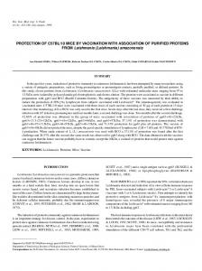
3099(03)005176. PubMed PMID: 12560194. Zijlstra, E. E. (2014). PKDL and other dermal lesions in HIV coinfected patients with leishmaniasis: review of clinical presentation in relation to immune responses. PLoS Negl Trop Dis 20, 8:e3258. doi: 10.1371/journal.pntd.0003258. PubMed PMID: 25412435. Figure/Table Captions Maximum of 2 figures and 2 tables Figure 1. Case I: Leishmania species amastigotes in Giemsa-stained bone marrow smears under oil immersion (×1000) (a) amastigotes in extracellular area, (b) amastigotes in extracellular area Figure 2. Case II: Leishmania species amastigotes in Giemsa-stained bone marrow smears under oil immersion (×1000) (a) intracellular amastigotes in macrophage, (b) amastigotes in extracellular area Abbreviations
Abbreviations: VL
visceral leishmaniasis
CCS
Clinical Centre of Serbia
BMS
bone marrow smear(s)
PKDL
post-kala-azar dermal leishmaniasis
ITS
internal transcribed spacer
CITD
Clinic for Infectious and Tropical Diseases
IHA
indirect hemagglutination analysis
SSI
Statens Serum Institut
12
Downloaded from www.microbiologyresearch.org by IP: 181.215.111.6 On: Thu, 09 Mar 2017 13:03:37
VP
ventriculoperitoneal
Ethical Statement Maximum of 50 words The study was approved by the Clinical Centre of Serbia Ethics Committee. Acknowledgements/Declaration of Interest Maximum of 50 words Acknowledgement The collaboration between our two labs under the auspices of the COST Action TD1302 (CYSTINET) is established. Declaration of Interest The authors have no dual or conflicting interests to declare. The results of this paper have been presented in part at the 25th ECCMID, Copenhagen, Denmark, 25–28 April 2015. Signature:
Date: August 10, 2016
*Licence to Publish forms are provided during submission through Editorial Manager. **Authors are responsible for attaining patient consent and will be asked to confirm this during submission. Read our ethical guidelines here: http://jmmcr.sgmjournals.org/site/misc/ifora.xhtml#req-ethics.
13
Downloaded from www.microbiologyresearch.org by IP: 181.215.111.6 On: Thu, 09 Mar 2017 13:03:37
Figure
Click here to download Figure Fig. 1(a).jpg
Downloaded from www.microbiologyresearch.org by IP: 181.215.111.6 On: Thu, 09 Mar 2017 13:03:37
Figure
Click here to download Figure Fig. 1(b).jpg
Downloaded from www.microbiologyresearch.org by IP: 181.215.111.6 On: Thu, 09 Mar 2017 13:03:37
Figure
Click here to download Figure Fig. 2(a).jpg
Downloaded from www.microbiologyresearch.org by IP: 181.215.111.6 On: Thu, 09 Mar 2017 13:03:37
Figure
Click here to download Figure Fig. 2(b).jpg
Downloaded from www.microbiologyresearch.org by IP: 181.215.111.6 On: Thu, 09 Mar 2017 13:03:37