RESEARCH ARTICLE
Lethality of mice bearing a knockout of the Ngly1-gene is partially rescued by the additional deletion of the Engase gene Haruhiko Fujihira1, Yuki Masahara-Negishi1, Masaru Tamura2, Chengcheng Huang1, Yoichiro Harada1, Shigeharu Wakana2, Daisuke Takakura3, Nana Kawasaki3, Naoyuki Taniguchi4, Gen Kondoh5, Tadashi Yamashita6, Yoko Funakoshi1, Tadashi Suzuki1*
a1111111111 a1111111111 a1111111111 a1111111111 a1111111111
1 Glycometabolome Team, Systems Glycobiology Research Group, RIKEN-Max Planck Joint Research Center, Global Research Cluster, RIKEN, Saitama, Japan, 2 Technology and Development Team for Mouse Phenotype Analysis, Japan Mouse Clinic, BioResourse Center, RIKEN, Ibaraki, Japan, 3 Graduate School of Medical Life Science, Yokohama City University, Kanagawa, Japan, 4 Disease Glycomics Team, Systems Glycobiology Research Group, RIKEN-Max Planck Joint Research Center, Global Research Cluster, RIKEN, Saitama, Japan, 5 Laboratory of Integrative Biological Science, Institute for Frontier Life and Medical Sciences, Kyoto University, Kyoto, Japan, 6 Laboratory of Biochemistry, School of Veterinary Medicine, Azabu University, Kanagawa, Japan *
[email protected]
OPEN ACCESS Citation: Fujihira H, Masahara-Negishi Y, Tamura M, Huang C, Harada Y, Wakana S, et al. (2017) Lethality of mice bearing a knockout of the Ngly1gene is partially rescued by the additional deletion of the Engase gene. PLoS Genet 13(4): e1006696. https://doi.org/10.1371/journal.pgen.1006696 Editor: Robert S. Haltiwanger, University of Georgia, UNITED STATES Received: October 5, 2016 Accepted: March 15, 2017 Published: April 20, 2017 Copyright: © 2017 Fujihira et al. This is an open access article distributed under the terms of the Creative Commons Attribution License, which permits unrestricted use, distribution, and reproduction in any medium, provided the original author and source are credited. Data Availability Statement: All relevant data are within the paper and its Supporting Information files. Funding: This work was supported in part by PRESTO/CREST, Japan Science and Technology Agency (JST) (to TS), Center of Excellence Program, Osaka University (to NT and TS), Yamada Science Foundation, Mochida Memorial Foundation for Medical and Pharmaceutical Research, Toray Science Foundation, Grace Science Foundation (to TS), a Grant-in-Aid for
Abstract The cytoplasmic peptide:N-glycanase (Ngly1 in mammals) is a de-N-glycosylating enzyme that is highly conserved among eukaryotes. It was recently reported that subjects harboring mutations in the NGLY1 gene exhibited severe systemic symptoms (NGLY1-deficiency). While the enzyme obviously has a critical role in mammals, its precise function remains unclear. In this study, we analyzed Ngly1-deficient mice and found that they are embryonic lethal in C57BL/6 background. Surprisingly, the additional deletion of the gene encoding endo-β-N-acetylglucosaminidase (Engase), which is another de-N-glycosylating enzyme but leaves a single GlcNAc at glycosylated Asn residues, resulted in the partial rescue of the lethality of the Ngly1-deficient mice. Additionally, we also found that a change in the genetic background of C57BL/6 mice, produced by crossing the mice with an outbred mouse strain (ICR) could partially rescue the embryonic lethality of Ngly1-deficient mice. Viable Ngly1deficient mice in a C57BL/6 and ICR mixed background, however, showed a very severe phenotype reminiscent of the symptoms of NGLY1-deficiency subjects. Again, many of those defects were strongly suppressed by the additional deletion of Engase in the C57BL/6 and ICR mixed background. The defects observed in Ngly1/Engase-deficient mice (C57BL/ 6 background) and Ngly1-deficient mice (C57BL/6 and ICR mixed background) closely resembled some of the symptoms of patients with an NGLY1-deficiency. These observations strongly suggest that the Ngly1- or Ngly1/Engase-deficient mice could serve as a valuable animal model for studies related to the pathogenesis of the NGLY1-deficiency, and that cytoplasmic ENGase represents one of the potential therapeutic targets for this genetic disorder.
PLOS Genetics | https://doi.org/10.1371/journal.pgen.1006696 April 20, 2017
1 / 23
Generation of a viable mouse model of an NGLY1-deficiency
Scientific Research (grant no. 26110725) from the Ministry of Education, Culture, Sports, Science and Technology of Japan (to TS) and a grant for Incentive Research Project, RIKEN (to HF). We also would like to express sincere gratitude to Mr. Hiroshi Mikitani (Rakuten Inc.; Tokyo, Japan) for his financial support for Ngly1 research. CH is supported by the RIKEN Foreign Postdoctoral Researcher Program. The funders had no role in study design, data collection and analysis, decision to publish, or preparation of the manuscript. Competing interests: The authors have declared that no competing interests exist.
Author summary Ngly1 is a cytoplasmic de-N-glycosylating enzyme that is ubiquitously found in eukaryotes. This enzyme is involved in a process referred to as endoplasmic reticulum-associated degradation (ERAD), one of the quality control mechanisms for newly synthesized proteins. A genetic disorder, NGLY1-deficiency, caused by mutations in the NGLY1 gene has recently been discovered. However, the precise mechanism for the pathogenesis of this devastating disease continues to remain unclear. We report herein that Ngly1-deficient mice are embryonically lethal in a C57BL/6 background. Surprisingly, the lethality was suppressed by crossing the mice with an outbred mouse strain (ICR), suggesting that the phenotypic consequence of Ngly1 is greatly influenced by their genetic background. In both cases, the additional deletion of Engase in Ngly1-deficient mice could strongly mitigate the phenotypes. Interestingly, the remaining defects in Ngly1-deficient or Ngly1/ Engase-deficient mice were reminiscent of the symptoms of subjects with an NGLY1-deficiency. Our results clearly point to the importance of Ngly1 in mammals and show that the inhibition of ENGase represents an effective therapy for treating an NGLY1-deficiency. Most importantly, the mice described herein could serve as valuable viable model mice for studies related to the pathophysiology of an NGLY1-deficiency.
Introduction The endoplasmic reticulum (ER) is the organelle responsible for the biosynthesis of proteins that pass through the secretory pathway. This organelle has an efficient protein quality control or homeostasis system, in which abnormal or excess proteins are targeted for destruction [1– 3]. In such systems, cytoplasmic proteasomes play a major role in degrading proteins, and the degradation system is often referred to as ER-associated degradation, or ERAD in short. Ngly1 is a highly conserved deglycosylating enzyme that is involved in the ERAD process [4–6]. Ngly1 cleaves N-glycans from misfolded glycoproteins during the ERAD process, and is thought to play an important role in the efficient degradation of misfolded glycoproteins [7– 11]. N-glycans released by Ngly1 are known to be processed by cytoplasmic glycosidases such as ENGase and the α-mannosidase, Man2C1 (Fig 1A, upper scheme) [12–14]. Cytoplasmic ENGase is another deglycosylating enzyme that acts directly on N-glycans that are attached to glycoproteins but leaves a single N-acetylglucosamine (GlcNAc) residue attached to the protein (Fig 1A, lower scheme). This enzyme belongs to the Glycosyl Hydrolase (GH) family 85, and while conserved widely in eukaryotes, some yeast, including S. cerevisiae and S. pombe, do not produce this enzyme. Several studies dealing with the biological functions of Ngly1 and/or ENGase have been reported. Mutants of Ngly1 and its orthologues have been analyzed in various organisms [7, 15–19]. Interestingly, the phenotypic consequences were found to be quite distinct between species; for example, budding yeast show no significant viability/growth defects [7], while mutant flies exhibited severe growth delay/arrest [15]. The molecular details behind the phenotypes, however, remain poorly understood. As of this writing, mutants of Engase have been analyzed in A. thaliana [20] and C. elegans [21] but, in both cases, no significant phenotypes were observed. In 2012, the first patient harboring mutations in NGLY1 alleles (NGLY1-deficiency) was identified through an exome analysis [22] and, since then, clinical cases involving humans have been reported [23–26]. These patients show very severe systemic symptoms such as
PLOS Genetics | https://doi.org/10.1371/journal.pgen.1006696 April 20, 2017
2 / 23
Generation of a viable mouse model of an NGLY1-deficiency
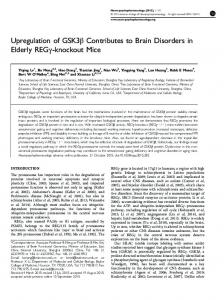
Fig 1. Generation of Ngly1- and Engase-deficient mice. (A) Schematic views of the non-lysosomal degradation of free oligosaccharides (upper scheme) and the action of ENGase on glycoproteins (lower scheme). (B, C) A diagram of the targeting construct used to generate Ngly1-deficient mice (B) and Engase-deficient mice (C) (Figures Reproduced from ref. 27). In the Ngly1−/− allele, from exon 11 to 12 were flanked by loxP site (filled triangles). Open triangles indicate FRT sites. In Engase−/− allele, exon 5 was replaced with a PGK-Neor cassette. (D) Cytoplasmic fraction of MEF cells derived from Ngly1−/− or Ngly1+/+ (wild type, WT) embryos were immunoblotted with an antibody against mouse Ngly1. GAPDH was used as a loading control. Allow indicates Ngly1 and asterisks indicate non-specific bands. https://doi.org/10.1371/journal.pgen.1006696.g001
delayed global development, movement disorders, hypotonia and hypo/alacrima [24, 26]. These reports point to the biological significance of Ngly1 in the normal development of mammals. More recently, using a model ERAD substrate, we reported that the ablation of Ngly1 causes a disruption in the ERAD process in mouse embryonic fibroblast (MEF) cells [27]. Interestingly, this ERAD disruption was found to be caused by an unexpected deglycosylating activity of ENGase, and the direct action of this enzyme towards the model substrate was shown to result in the formation of aggregation-prone N-GlcNAc proteins [27]. Moreover, the disruption of ERAD in Ngly1-deficient cells was restored by the additional deletion of the Engase gene. While this result using a model substrate suggests that an ENGase inhibitor could be a potential therapeutic target for treating an NGLY1-deficiency, the issue of how Engasedeletion affects mice phenotypes lacking Ngly1 remains unknown.
PLOS Genetics | https://doi.org/10.1371/journal.pgen.1006696 April 20, 2017
3 / 23
Generation of a viable mouse model of an NGLY1-deficiency
Results Ngly1−/− mice in C57BL/6 are embryonically lethal The goal of this study was to clarify the details of the biological function of the deglycosylating enzymes, Ngly1 and ENGase, at the individual level in mice. To this end, we used mice (C57BL/6 background mice) that had been generated in a previous study, in which the Ngly1 and Engase genes had been knocked out [27]. The knockout constructs of Ngly1 and Engase are shown in Fig 1B and 1C, respectively. While the generation of these KO mice has been described previously [27], a detailed phenotypic analysis of those mice has not been reported. The targeted genomic disruption of Ngly1 and ENGase was confirmed by PCR using 8 sets of primers (Fig 1B and 1C; S1 Table for primer sequences). The loss of Ngly1 activity was confirmed by an activity assay [27]. We further examined the expression of the Ngly1 protein by western blot analysis using cytoplasmic fractions from MEF cells. As shown in Fig 1D, the loss of Ngly1 in MEF cells from Ngly1−/− mice was confirmed. In the case of ENGase, we confirmed the loss of the ENGase activity through an activity assay in a previous study [27] as well as by conducting a detailed structural analysis of the free oligosaccharides (fOSs) in the cytoplasm of MEF cells [28]. The Ngly1 heterozygous (Ngly1−/+) mice were fertile and did not show any obviously recognizable phenotypes. However, viable homozygous Ngly1−/− pups were not produced, despite the repeated crossing of the Ngly1−/+ mice (Table 1), suggesting that the deletion of the Ngly1 allele results in a lethal condition in C57BL/6 mice. To delineate the timing of the Table 1. Results of the genotyping of pups/embryos by crossing of Ngly1−/+, or Ngly1−/+;Engase−/− mice in the C57BL/6 background. Stage E14.5
E16.5
Genotype Ngly1
18 (21.95) [25.00]
0
50 (60.98) [50.00]
0
Ngly1−/−
14 (17.07) [25.00]
0 0
+/+
Ngly1
56 (28.00) [25.00]
Ngly1−/+
113 (56.50) [50.00]
0
Ngly1−/−
31 (15.50) [25.00]
0
Ngly1+/+;Engase−/−
20 (22.73) [25.00]
0
Ngly1−/+;Engase−/−
52 (59.09) [50.00]
0
Ngly1
E18.5
P0
2 wks
Number of inviable embryos/pups
Ngly1−/+
−/−
E17.5
Number of viable embryos/pups
+/+
−/−
16 (18.18) [25.00]
0
Ngly1+/+
;Engase
21 (28.77) [25.00]
0
Ngly1−/+
38 (52.05) [50.00]
1
Ngly1−/−
14 (19.18) [25.00]
7
+/+
Ngly1
23 (31.08) [25.00]
0
Ngly1−/+
38 (51.35) [50.00]
1
Ngly1−/−
13 (17.57) [25.00]
5
+/+
Ngly1
22
−
Ngly1−/+
35
−
Ngly1−/−
0
−
+/+
Ngly1
49
−
Ngly1−/+
84
−
Ngly1−/−
0
−
E: Embryonic day, P: Postnatal day, wks: weeks, V: Viable, IV: Inviable. (number): birth rates (%), [number]: expected (%). https://doi.org/10.1371/journal.pgen.1006696.t001
PLOS Genetics | https://doi.org/10.1371/journal.pgen.1006696 April 20, 2017
4 / 23
Generation of a viable mouse model of an NGLY1-deficiency
lethality, Ngly1−/+ mice were crossed and embryos were collected at several stages of gestation. The viability of collected embryos was confirmed by checking their heart beat and their genotypes were analyzed using genomic DNA extracted from the amnion. The results of this genotyping are summarized in Table 1. The Ngly1−/− embryos were viable, even at embryonic day 18.5 (E18.5), one day prior to birth. At the same time, however, about 30% of Ngly1−/− embryos were inviable at later stages of development (E17.5–18.5). When embryos were collected early in the morning of the day of birth, only the Ngly1−/+ and Ngly1+/+ embryos were alive when we revived them by gentle massaging, but no Ngly1−/− mice could be revived. We also analyzed the genotype of pups within a few hours after their birth (P0) and confirmed the absence of Ngly1−/− pups (Table 1). At E14.5 and E16.5, however, only viable Ngly1−/− embryos were observed. Therefore, the lethality caused by the Ngly1 deficiency appears to occur between E16.5 and before birth.
A ventricular septal defect is observed in Ngly1−/− embryos To investigate the defects in Ngly1−/− embryos in more detail, X-ray micro-computed tomography (μ-CT) analyses were carried out on Ngly1−/− or Ngly1+/+ embryos at E16.5 (Fig 2A and 2B). As shown in Fig 2B, the Ngly1−/− embryos showed a ventricular septal defect (VSD) (5 out of 5 embryos (5/5)). Histological analyses also confirmed the occurrence of a VSD in Ngly1−/− embryos (3/3) (Fig 2C and 2D). VSD is one of the most frequently-observed cardiovascular phenotypes in embryonic/perinatal lethal mice [29]. We also found that some Ngly1−/− embryos showed anemia (12/28 [42.86%], Fig 2E, left panel) or edema (4/28 [14.29%], Fig 2F, left panel), which were not observed in Ngly1+/+ embryos (0/44, Fig 2E and 2F, right panel).
The additional deletion of the Engase gene partially rescues the lethality caused by the loss of Ngly1 In sharp contrast to the case of Ngly1−/− mice, the Engase−/− mice showed normal behavior/values in several tests as follows: behavior test (open field), morphology/behavioral/sensory test (RIKEN modified-SHIRPA), hematology/clinical chemistry test (hematology, urinalysis, clinical blood chemistry), pathology test (body weight), cardiovascular test (blood pressure, electrocardiogram) and neurology/psychiatry test (light/dark transition, home-cage activity, tail suspension, hot plate, tail flick) (the number of tested mice are described in Materials and Methods). According to our previous cell-based study using an ERAD model substrate [27], we hypothesized that ENGase could have the ability to function as a deglycosylating enzyme and that its deletion could rescue the defects caused by the lack of Ngly1. In this study, we attempted to verify the effect of the additional Engase deletion of Ngly1−/− in mice at an individual level. To this end, we crossed Ngly1−/+;Engase−/+ or Ngly1−/+;Engase−/− mice and found that surviving Ngly1−/− mice were produced upon the crossing (Table 2). Upon further examination, all of the surviving mice were found to be Ngly1−/−;Engase−/− double-knockout mice, strongly indicating that the additional deletion of Engase partially rescued the lethality caused by the defect of Ngly1 at an individual level.
VSD appears to be one of the critical causes of lethality in Ngly1−/− mice We next investigated the issue of whether VSD was present in Ngly1−/−;Engase−/− embryos at E16.5. If VSD was to be critical for the lethality of Ngly1−/− embryos, then this phenotype should be suppressed by the additional deletion of Engase. As we expected, VSD was not observed in the Ngly1−/−;Engase−/− embryos, as evidenced by μ-CT (0/2) and histological analyses (0/2) (Fig 2G and 2H). It is therefore possible that VSD is at least one of the critical phenotypes that cause the lethality of Ngly1−/− mice/embryos and the deletion of the Engase gene, for
PLOS Genetics | https://doi.org/10.1371/journal.pgen.1006696 April 20, 2017
5 / 23
Generation of a viable mouse model of an NGLY1-deficiency
Fig 2. Loss of Ngly1 causes ventricular septal defects (VSD) and the additional Engase deletion rescues the VSD phenotypes. (A, B, G) Maximum intensity projection of heart μ-CT images of E16.5 embryo of wild-type (A), Ngly1−/− (B), and Ngly1−/−;Engase−/− (G). White arrows in (B) indicates VSD. (C, D, H) Transverse section of wild-type (C), Ngly1−/− (D), and Ngly1−/−;Engase−/− embryo (H) at E16.5 were stained with H&E. White arrows in (D) indicate VSD. Shown are representative sections (n = 3). Scale bar in (C), (D) and (H) indicate 200 μm. RA: right atrium, RV: right ventricle, LA: left atrium, LV: left ventricle. (E) Anemia was observed in Ngly1−/− embryo at E16.5 (left panel). Right panel shows Ngly1+/+ embryo at E16.5 (littermate of the left panel). (F) Edema was observed in Ngly1−/− embryo at E16.5 (left panel). Right panel shows Ngly1+/+ embryo at E16.5 (littermate of the left panel). Black arrowhead indicates edema. (I) Anemia was observed in Ngly1−/−;Engase−/− embryo at E16.5 (left panel). Right panel shows Ngly1+/+;Engase−/− embryo at E16.5 (littermate of the left panel). Representative images were shown. https://doi.org/10.1371/journal.pgen.1006696.g002
unknown reasons, rescues this phenotype. We also examined the gross morphology of embryos at E16.5 and found that some Ngly1−/−;Engase−/− embryos showed anemia (5/16 [31.25%], Fig 2I, left panel) while no Ngly1+/+;Engase−/− embryo showed this phenotype (0/20, Fig 2I, right panel). Additionally, Ngly1−/−;Engase−/− embryos did not show edema (0/16). Unexpectedly, there is no significant difference of the appearance ratio between Ngly1−/− embryos and Ngly1−/−;Engase−/− embryos at E16.5 (Table 1). These results suggest that Engase
PLOS Genetics | https://doi.org/10.1371/journal.pgen.1006696 April 20, 2017
6 / 23
Generation of a viable mouse model of an NGLY1-deficiency
Table 2. Genotyping results of pups by crossing of Ngly1−/+;Engase−/+ mice or Ngly1−/+;Engase−/− mice in C57BL/6 background. Ngly1−/+;Engase−/+ × Ngly1−/+;Engase−/+ Genotype +/+
Ngly1
;Engase
Number of pups (%) −/−
Ngly1−/+;Engase−/−
Expected %
11 (9.40)
6.25
20 (17.09)
12.50
Ngly1−/−;Engase−/−
3 (2.56)*
6.25
Ngly1+/+;Engase+/−
20 (17.09)
12.50
Ngly1−/+;Engase+/−
34 (29.06)
25.00
Ngly1−/−;Engase+/−
0 (0.00)***
12.50
Ngly1+/+;Engase+/+
8 (6.84)
6.25
21 (17.95)
12.50
0 (0.00)***
6.25
−/+
Ngly1
;Engase
+/+
Ngly1−/−;Engase+/+
Ngly1−/+;Engase−/− × Ngly1−/+;Engase−/− Genotype +/+
Ngly1
;Engase
Number of pups (%) −/−
Expected %
73 (28.52)
25.00
Ngly1−/+;Engase−/−
160 (62.5)***
50.00
Ngly1−/−;Engase−/−
23 (8.98)***
25.00
Statistic difference to expected value was examined by Chi-squared test *p