© 2000 Nature America Inc. • http://genetics.nature.com
letter
The human Rhesus-associated RhAG protein and a kidney homologue promote ammonium transport in yeast
© 2000 Nature America Inc. • http://genetics.nature.com
Anne-Marie Marini1, Giorgio Matassi2,3, Virginie Raynal2, Bruno André1, Jean-Pierre Cartron2 & Baya Chérif-Zahar2
The Rhesus blood-group antigens are defined by a complex association of membrane polypeptides that includes the nonglycosylated Rh proteins (RhD and RhCE) and the RHag glycoprotein, which is strictly required for cell surface expression of these antigens1. RhAG and the Rh polypeptides are erythroidspecific transmembrane proteins belonging to the same family (36% identity)2,3. Despite their importance in transfusion medicine, the function of RhAG and Rh proteins remains unknown, except that their absence in Rhnull individuals leads to morphological and functional abnormalities of erythrocytes, known as the Rh-deficiency syndrome. We recently found significant sequence similarity between the Rh family proteins, especially RhAG, and Mep/Amt ammonium transporters4,5. We show here that RhAG and also RhGK, a new human homologue expressed in kidney cells only, function as ammonium transport proteins when expressed in yeast. Both specifically complement the growth defect of a yeast mutant deficient in ammonium uptake. Moreover, ammonium efflux assays and growth tests in the presence of toxic concentrations of the analogue methylammonium indicate that RhAG and RhGK also promote ammonium export. Our results provide the first experimental evidence for a direct role of RhAG and RhGK in ammonium transport. These findings are of high interest, because no specific ammonium transport system has been characterized so far in human.
Ammonium transporters of the Mep/Amt family are polytopic membrane proteins highly conserved in eubacteria, archaebacteria, fungi, plants and invertebrates6,7,8. Although no Mep/Amt protein has yet been found in vertebrates, a definite sequence relationship (20–27% identity) exists between the Mep/Amt transporters and Rh family proteins, especially RhAG proteins4,5. Furthermore, RhAG homologues are found in slime mold4,
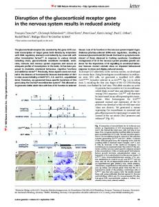
a
b
c
d
sponge9, nematode4 and fruit fly, indicating that their function is not confined to erythrocytes. RhAG proteins probably derived from Mep/Amt proteins, Rh proteins most likely originating later in evolution from an RhAG-like ancestor10,11. To test whether human RhAG is capable of ammonium transport activity, we performed functional complementation assays in a yeast mutant with deletions in the three endogenous ammonium-transporter genes (mep1∆ mep2∆ mep3∆, henceforth named triple-mep∆). The RhAG cDNA was cloned, placed under the control of the yeast methionine-repressible MET25 promoter and expressed in wild type and triple-mep∆ yeast cells. Westernblot analysis led to the detection of an approximately 38–43-kD signal corresponding to RhAG (Fig. 1a,b). RhAG does not appear to be N-glycosylated in yeast, because the electrophoretic mobility of the detected signal was unaffected by treatment of extracts with N-glycosidase and by neo-synthesis of RhAG in the presence of tunicamycin (data not shown). Expression of RhAG is not toxic to yeast cells, because the growth rate of transformed strains was the same whether methionine was added or not (data not shown). We first studied the ability of RhAG to promote ammonium transport in yeast by testing the growth of triple-mep∆ strains, expressing or not expressing the RhAG protein, on media containing ammonium at various concentrations (Fig. 2). This triplemep∆ mutant strain cannot grow on minimal media containing less than 5 mM ammonium (pH 6.1; ref. 8). After a 5-day incubation, a clear complementation of the growth defect was observable at ammonium concentrations down to 3 mM for the triple-mep∆ mutant expressing RhAG. After a longer incubation period (7 days), growth was also detectable at 1 mM ammonium. The transport function restored by RhAG in yeast appears ammonium-specific, because the RhAG protein could not repair the growth defects
Fig. 1 Immunodetection of the RhAG and RhGK proteins expressed in yeast. a,c, Wild-type cells (23344c) transformed with the p426-MET25 vector and triple-mep∆ (called mep∆) cells (31019b) transformed with p426-MET25, p426MET25-HsRhAG, or p426-MET25-HsRhGK were grown on minimal glutamine medium. Equal amounts of protein from membrane-enriched fractions were subjected to SDS–PAGE. Proteins were probed with an anti-RhAG antiserum (a) or an anti-RhGK antiserum (c). A signal at 38–43 kD (arrow), composed of at least three closely migrating bands, the band corresponding to the highest molecular mass being the most intense, appeared specifically with extracts of triple-mep∆ cells transformed with the p426-MET25-HsRhAG plasmid. A nonspecific signal (•), detected at about 48 kD, was used as a loading control. Similarly, a specific signal appeared at 51 kD (arrow) in extracts of triple-mep∆ cells transformed with the p426-MET25-HsRhGK plasmid. Surrounding non-specific signals (•), at 46, 52 and 55 kD, were used as loading controls. b,d, To confirm that the signals at 38–43 kD and 51 kD correspond to the RhAG and RhGK proteins respectively, we tested the production of these proteins in mep∆ cells (31019b) transformed with p426-MET25-HsRhAG or p426-MET25-HsRhGK, growing exponentially on glutamine medium in the presence of methionine (t=0 min) and after its removal (t=60–240 min). Proteins were probed with an anti-RhAG antiserum (b) or an anti-RhGK antiserum (d).
1Laboratoire de Physiologie Cellulaire, Université Libre de Bruxelles, Institut de Biologie et de Médecine Moléculaires, Gosselies, Belgium. 2Unité INSERM U76, Institut National de la Transfusion Sanguine Paris, France. 3Laboratorio di Evoluzione Molecolare, Stazione Zoologica “Anton Dohrn”, Naples, Italy.
Correspondence should be addressed to B.A. (e-mail:
[email protected]) or B.C.-Z. (e-mail:
[email protected]). nature genetics • volume 26 • november 2000
341
letter
© 2000 Nature America Inc. • http://genetics.nature.com
© 2000 Nature America Inc. • http://genetics.nature.com
Fig. 2 RhAG and RhGK proteins complement a yeast mutant deficient in ammonium transport. Wild-type cells (23344c) transformed with the p426-MET25 vector and triple-mep∆ cells (31019b) transformed with p426-MET25, p426-MET25HsRhAG, or p426-MET25-HsRhGK were grown on minimal medium containing 3 mM ammonium (5 d) or 1 mM ammonium (7 d) as the sole nitrogen source. The triple-mep∆ growth defect observed on ammonium at concentrations below 5 mM was partially repaired by both the RhAG and the RhGK protein.
of the gap1∆ agp1∆ double mutant deficient in amino-acid transport (Fig. 3a), nor those of the trk1∆ trk2∆ double mutant deficient in potassium transport (Fig. 3b). This latter observation is noteworthy because potassium is similar in size and charge to ammonium. The triple-mep∆ strain expressing RhAG did not grow as fast as mep∆ cells expressing any one of the three endogenous Mep ammonium transporters8, suggesting that the ammonium uptake activity restored by RhAG is low. Accordingly, we detected no ammonium (or radiolabelled methylammonium) uptake activity in transport assays (data not shown). Unexpectedly, however, expression of RhAG confers to triple-mep∆ cells an enhanced resistance to a toxic concentration of methylammonium (200 mM; Fig. 4), a result consistent with RhAG promoting export of the ammonium analogue. To test the role of RhAG in ammonium export, we grew the cells on arginine, a nitrogen source whose catabolism leads to ammonium production. On induction of RhAG expression, ammonium was excreted by the cells at a distinctly higher rate compared with cells not expressing the human protein (Fig. 5), indicating that RhAG mediates ammonium export in yeast. Blast similarity searches12 in GenBank revealed that the PDRC2 human protein is a new RhAG homologue (∼50% identity). This protein (renamed RhGK for Rh glycoprotein kidney) also shares sequence homology (∼24% identity) with the Mep/Amt ammonium transporters. Analysis of the human EST database with the RhGK sequence produced the highest-score matches with tags mainly from kidney. In northern-blot analysis, consistently, among the tissues examined, only kidney cells displayed the 2.2-kb RhGK mRNA (Fig. 6). The RhGK cDNA, amplified from human kidney RNA, was expressed in yeast as a 51-kD protein (Fig. 1c,d). RhGK also specifically repairs the growth defects of the triple-mep∆ strain on ammonium (Figs 2, 3 and 4). The results obtained in the presence of 1 mM ammonium even suggest that RhGK functions slightly more efficiently than RhAG. Moreover, RhGK confers to triplemep∆ cells both enhanced resistance to methylammonium (Fig. 4) and increased ammonium excretion (Fig. 5). Together with the sequence similarity between the RhAG homologues and Mep/Amt
proteins, our results suggest that RhAG and RhGK could be ammonium transport system components in erythrocytes and kidney cells, respectively. Although the importance of ammonium transport in red blood cells is not well documented, the ammonium concentration is reported to be three times higher inside erythrocytes than in plasma13. A tentative hypothesis would be that RhAG, in the Rh complex, promotes export of ammonium accumulated into erythrocytes. RhAG might also promote erythrocytemediated retention of ammonium from the plasma and its release to detoxifying organs such as the liver and brain14. Our results will certainly lead to new studies of human Rhnull patients, such as the analysis of ammonium concentrations in plasma and erythrocytes and of possible perturbation in labile nitrogen metabolism. In the human kidney, excretion of excess ammonium ions is crucial to maintaining the systemic acid-base balance15. Although participation of potassium transport systems in the transport of ammonium in the kidneys is well documented16, our data on RhGK suggest that specific ammonium transporters could also be involved. Notably, organisms such as worm and fruit fly contain both RhAG and Mep/Amt homologues, suggesting that these categories of proteins differ somehow in function. The stoichiometry of ammonium transport could be different and adapted to distinct electrochemical gradients specific to certain cell types. As recently suggested for the yeast Mep2 transporter17, RhAG homologues might be involved in sensing external ammonium11. Alternatively, RhAG homologues might have evolved from ammonium transporters to proteins more specialized in ammonium efflux. Interestingly, pulses of ammonia excretion have recently been involved in the regulation of growth of neighbouring yeast colonies18. Our results can be regarded as a crucial step towards unravelling the function of Rh family proteins and the molecular bases of ammonium transport in humans.
a
b
342
Fig. 3 RhAG and RhGK proteins do not complement yeast mutants deficient in amino acid or potassium transport. To test whether the complementation of the ammonium growth impairment could be due to an non-specific leak of the yeast plasma membrane, we tested the ability of the RhAG and RhGK proteins to complement mutations causing other transport defects in yeast. a, The p426MET25, p426-MET25-HsRhAG and p426-MET25-HsRhGK plasmids were used to transform a yeast mutant (30633c) bearing deletions in the genes encoding the general amino-acid permease Gap1 and the inducible amino-acid permease Agp1 (ref. 24). Wild-type cells (23344c) were transformed with the p426-MET25 vector. The growth defect of the double gap1∆ agp1∆ mutant on minimal medium containing leucine (1 mM) as the sole nitrogen source was not repaired by expression of the RhAG or RhGK protein. Similar results were obtained using 1 mM citrulline or 1 mM tryptophan (not shown). b, Because ammonium and potassium are very similar in size and charge, we tested whether the RhAG and RhGK proteins might be involved in potassium transport. For this purpose, the same constructs as in (a) were used to transform the trk1∆ trk2∆ mutant (CY162). This strain bears deletions in two related potassium transporters, Trk1 and Trk2, and shows a severe growth defect compared with its wild type (S288c) at potassium concentrations below or equal to 7 mM (at pH 5.9) (refs 25,26). Growth tests were performed on minimal media (with glutamine as the sole nitrogen source) containing potassium at various concentrations. Neither of the RhAG and RhGK proteins was able to restore growth of the mutant.
nature genetics • volume 26 • november 2000
© 2000 Nature America Inc. • http://genetics.nature.com
letter
© 2000 Nature America Inc. • http://genetics.nature.com
Fig. 4 RhAG and RhGK proteins confer resistance to methylammonium to yeast cells. Wild-type cells (23344c) transformed with the p426-MET25 vector and triple-mep∆ cells (31019b) transformed with p426-MET25, p426-MET25HsRhAG or p426-MET25-HsRhGK were grown on minimal medium containing proline 0.1% as sole nitrogen source and supplemented with 200 mM methylammonium. Expression of RhAG or RhGK in the triple-mep∆ strain conferred resistance to methylammonium, whereas resistance to other toxic compounds such as D-histidine 0.05% was not altered (not shown).
Methods Construction of yeast expression plasmids. We inserted human RhAG (nt –22 to nt 1,243) and RhGK (nt –6 to 1,521) cDNAs (+1 is the A of the ATG start codon) into the HindIII/XhoI and SmaI sites, respectively, of the highcopy expression vector pRS426 (ref. 19). The vector contains the MET25 promoter causing expression of RhAG and RhGK to be repressed upon addition of methionine to the medium. We obtained two constructs, p426MET25-HsRhAG and p426-MET25-HsRhGK. We detected no PCR-introduced mutations by sequencing of both cDNAs in the constructs. Strains, growth conditions, methods. S. cerevisiae strains 23344c (ura3), 31019b (mep1∆ mep2∆ mep3∆ ura3) and 30633c (gap1∆ agp1∆ ura3) are isogenic with Σ1278b. Strain CY162 (trk1∆ trk2∆ ura3) (R. Gaber) is isogenic with S288c. We grew cells in minimal buffered (pH 6.1) medium20, with 3% glucose as carbon source. The nitrogen sources used were 0.1% glutamine, 1–10 mM (NH4)2SO4, 1 mM leucine, 1 mM citrulline, 1 mM tryptophan, 0.1% proline, 0.1% arginine. Methylammonium was used at 200 mM. We added methionine (1 mM) to the medium, to repress synthesis of RhAG and RhGK. For testing complementation of the mutation causing the potassiumrelated growth defect, potassium salts were replaced with sodium salts in the above-mentioned medium and KCl was added at a concentration ranging from 0.2–100 mM. Yeast cells were transformed as described21. Antibody production. We obtained antibody to the carboxy terminus of the RhAG protein by immunizing rabbits with a synthetic peptide (aa 389–408) coupled to keyhole limpet haemocyanin (Neosystem). We tested its specificity on membrane proteins isolated from normal and Rh null erythrocytes. We obtained a signal in the expected size range (from about 45 to 75 kD) for normal red blood cells, this being characteristic of the heterogeneously glycosylated RhAG protein1. No signal was detected with Rhnull erythrocytes (data not shown). We obtained antibody raised to the amino terminus of the RhGK protein (aa 31–48) as above. Western immunoblotting. For membrane-enriched preparations, 108 yeast cells were filtered, washed with cold water and resuspended in 0.2 ml lysis buffer (0.1 M Tris-HCl, pH 7.5; 0.15 M NaCl; 5 mM EDTA) containing proteinase inhibitors: 100 µg/ml phenylmethylsulfonyl fluoride (PMSF); 1 µg/ml leupeptin; 1 µg/ml pepstatin; 50 mM N-ethyl maleimide. We added an equal volume of glass beads and cells were lysed at 4 °C by vortex mixing for 3 min. We diluted extracts with one volume of lysis buffer and centrifuged for 5 min at 3,000 r.p.m. We obtained the membrane-enriched fraction from the supernatant as described22. For westernblot analysis, we loaded 10 µl of solubilized proteins on a 10% SDS–polyacrylamide gel in a Tricine system23. After transfer to a nitrocellulose membrane, we probed proteins with rabbit antiserum raised against the C-terminal region of RhAG (1:250) or the N terminus of RhGK (1:250). We detected primary antibodies with horseradish-peroxidase-conjugated (HRP-conjugated) anti-rabbit-IgG secondary antibody, followed by measurement of chemoluminescence (Lumi-LightPLUS, Roche). nature genetics • volume 26 • november 2000
Fig. 5 RhAG and RhGK promote excretion of ammonium. Triple-mep∆ cells (31019b) transformed with p426-MET25 (❍), p426-MET25-HsRhAG (❐), or p426-MET25-HsRhGK (+) were grown on minimal liquid medium containing arginine plus methionine. At time 0, cells were transferred to arginine medium without methionine to induce RhAG and RhGK expression. At time intervals, samples of supernatant were withdrawn and assayed for ammonium concentration. Triple-mep∆ strain is defective in re-uptake of ammonium that on arginine medium tends to leak out of the cells thereby leading to detectable excretion8. Expression of RhAG or RhGK in these cells clearly enhances the rate of ammonium excretion in the medium.
PNGase F treatment. We prepared a membrane-enriched fraction from wild-type glutamine-grown cells (108 cells) as described in the preceding section. We resuspended membranes in 100 µl incubation buffer as recommended (Boehringer), with or without 10 U of peptide-N-glycosidase F (PNGase F). We incubated the samples for 20 h at 37 °C. Tunicamycin treatment. We grew cells on glutamine plus methionine medium to repress RHAG expression, then transferred the cells to and incubated them for 3 h in glutamine medium with tunicamycin (5 µg/ml) or with an equal volume of the corresponding solvent (95% ethanol). We prepared membrane-enriched fractions from samples collected before and after transfer to glutamine medium. Northern-blot analysis. After reverse transcription of 1 µg of total human kidney RNA performed with the first strand cDNA kit (Pharmacia), we amplified by PCR a 1,487-bp cDNA fragment, corresponding to the entire
Fig. 6 Northern-blot analysis of RhGK mRNA. The entire coding region (1,487 bp) of the RhGK cDNA was PCRamplified from human kidney RNA and used on northern blots to probe poly(A)+ RNAs (2 µg) from various human tissues. A transcript of ∼2.2 kb was detected only in kidney. This mRNA was not detected in lymph node, bone marrow, fetal liver and testis, even after overexposure (not shown). A weaker signal at ∼2.7 kb was revealed in brain, heart, skeletal muscle, kidney, liver (arrow-head) but also in fetal liver, bone marrow and testis (not shown).
kb
343
letter
© 2000 Nature America Inc. • http://genetics.nature.com
RhGK coding region, using the following oligonucleotides (derived from GenBank entry AF081497): 5´–TGCAGCATGGCCTGGAACACCAACC–3´ and 5´–AGGACAGTCTGTGGAGCCTGCTCCTCA–3´. PCR conditions were: 94 °C for 1 min followed by 30 cycles at 94 °C for 30 s and 68 °C for 3 min, using the Advantage cDNA PCR Kit (Clontech). The amplified cDNA fragment was used as a hybridization probe on the following multiplehuman-tissue northern blots: 12-lane (brain, heart, skeletal muscle, colon, thymus, spleen, kidney, liver, small intestine, placenta, lung, peripheral blood leukocyte); Immune System II (spleen, lymph node, thymus, peripheral blood leukocyte, bone marrow, fetal liver); Endocrine System (pancreas, adrenal medulla, thyroid, adrenal cortex, testis, thymus, small intestine, stomach) (Clontech). We carried out hybridizations for 1 h at 68 °C with the ExpressHybTM hybridization solution and 1×10⁶ cpm/ml of the cDNA probe labelled with 32P, according to the manufacturer’s instructions. Ammonium excretion assay. Cells growing exponentially on arginine plus methionine were transferred to and incubated for 6 h in arginine medium
1.
© 2000 Nature America Inc. • http://genetics.nature.com
2.
3. 4.
5.
6.
7. 8. 9.
10.
11.
12. 13. 14.
Chérif-Zahar, B. et al. Candidate gene acting as a suppressor of the RH locus in most cases of Rh-deficiency. Nature Genet. 12, 168–173 (1996). Cartron, J.P. Red cell membrane and its disorders. in Baillière’s Clinical Haematology (eds Tanner, M.J.A. & Anstee, D.J.) 655–689 (Harcourt, London, 1999). Avent, N.D. & Reid, M.E. The Rh blood group system: a review. Blood 95, 375–387 (2000). Marini, A.M., Urrestarazu, A., Beauwens, R. & Andre, B. The Rh (rhesus) blood group polypeptides are related to NH4+ transporters. Trends Biochem. Sci. 22, 460–461 (1997). Matassi, G., Chérif-Zahar, B., Raynal, V., Rouger, P. & Cartron, J.P. Organization of the human RH50A gene (RHAG) and evolution of base composition of the RH gene family. Genomics 47, 286–293 (1998). Marini, A.M., Vissers, S., Urrestarazu, A. & Andre, B. Cloning and expression of the MEP1 gene encoding an ammonium transporter in Saccharomyces cerevisiae. EMBO J. 13, 3456–3463 (1994). Ninnemann, O., Jauniaux, J.C. & Frommer, W.B. Identification of a high affinity NH4+ transporter from plants. EMBO J. 13, 3464–3471 (1994). Marini, A.M., Soussi-Boudekou, S., Vissers, S. & Andre, B. A family of ammonium transporters in Saccharomyces cerevisiae. Mol. Cell. Biol. 17, 4282–4293 (1997). Seack, J., Pancer, Z., Muller, I.M. & Muller, W.E. Molecular cloning and primary structure of a Rhesus (Rh)-like protein from the marine sponge Geodia cydonium. Immunogenetics 46, 493–498 (1997). Kitano, T., Sumiyama, K., Shiroishi, T. & Saitou, N. Conserved evolution of the Rh50 gene compared to its homologous Rh blood group gene. Biochem. Biophys. Res. Commun. 249, 78–85 (1998). Matassi, G., Chérif-Zahar, B., Pesole, G., Raynal, V. & Cartron, J.P. The members of the RH gene family (RH50 and RH30) followed different evolutionary pathways. J. Mol. Evol. 48, 151–159 (1999). Altschul, S.F. et al. Gapped BLAST and PSI-BLAST: a new generation of protein database search programs. Nucleic Acids Res. 25, 3389–3402 (1997). Huizenga, J.R., Tangerman, A. & Gips, C.H. Determination of ammonia in biological fluids. Ann. Clin. Biochem. 31, 529–543 (1994). Dejong, C.H., Deutz, N.E. & Soeters, P.B. Ammonia and glutamine metabolism
344
(6.106 cells/ml). After transfer, we withdrew samples of supernatant and assayed for ammonium concentration as described6. Accession numbers. Fruit fly RhAG, AE003482; human PDRC2, AF081497. Acknowledgements
We thank C. Hattab for help in preparing rabbit antibodies; R. Gaber for yeast strains; and C. Jauniaux and S. Lecomte for technical contributions. This research was supported by The Commission of the European Communities and the Communauté Française de Belgique, Direction de la Recherche Scientifique. A.-M.M. is Chargé de recherches du Fonds National belge de la Recherche Scientifique. G.M. is currently a fellow of the International Centre for Genetic Engineering and Biotechnology. Received 7 April; accepted 6 September 2000.
15. 16. 17.
18. 19.
20.
21. 22.
23.
24.
25. 26.
during liver insufficiency: the role of kidney and brain in interorgan nitrogen exchange. Scand. J. Gastroenterol. Suppl. 218, 61–77 (1996). Good, D.W. & Knepper, M.A. Ammonia transport in the mammalian kidney. Am. J. Physiol. 248, F459–471 (1985). Knepper, M.A., Packer, R. & Good, D.W. Ammonium transport in the kidney. Physiol. Rev. 69, 179–249 (1989). Lorenz, M.C. & Heitman, J. The MEP2 ammonium permease regulates pseudohyphal differentiation in Saccharomyces cerevisiae. EMBO J. 17, 1236–1247 (1998). Palkova, Z. et al. Ammonia mediates communication between yeast colonies. Nature 390, 532–536 (1997). Mumberg, D., Muller, R. & Funk, M. Regulatable promoters of Saccharomyces cerevisiae: comparison of transcriptional activity and their use for heterologous expression. Nucleic Acids Res. 22, 5767–5768 (1994). Jacobs, P., Jauniaux, J.C. & Grenson, M. A cis-dominant regulatory mutation linked to the argB-argC gene cluster in Saccharomyces cerevisiae. J. Mol. Biol. 139, 691–704 (1980). Ito, H., Fukuda, Y., Murata, K. & Kimura, A. Transformation of intact yeast cells treated with alkali cations. J. Bacteriol. 153, 163–168 (1983). Galan, J.M., Moreau, V., Andre, B., Volland, C. & Haguenauer-Tsapis, R. Ubiquitination mediated by the Npi1p/Rsp5p ubiquitin-protein ligase is required for endocytosis of the yeast uracil permease. J. Biol. Chem. 271, 10946–10952 (1996). Schagger, H. & von Jagow, G. Tricine-sodium dodecyl sulfate-polyacrylamide gel electrophoresis for the separation of proteins in the range from 1 to 100 kDa. Anal. Biochem. 166, 368–379 (1987). Iraqui, I. et al. Amino acid signaling in Saccharomyces cerevisiae: a permease-like sensor of external amino acids and F-Box protein Grr1p are required for transcriptional induction of the AGP1 gene, which encodes a broad-specificity amino acid permease. Mol. Cell. Biol. 19, 989–1001 (1999). Ko, C.H. & Gaber, R.F. TRK1 and TRK2 encode structurally related K+ transporters in Saccharomyces cerevisiae. Mol. Cell. Biol. 11, 4266–4273 (1991). Nakamura, R.L., Anderson, J.A. & Gaber, R.F. Determination of key structural requirements of a K+ channel pore. J. Biol. Chem. 272, 1011–1018 (1997).
nature genetics • volume 26 • november 2000