© 1999 Nature America Inc. • http://genetics.nature.com
letter
Disruption of the glucocorticoid receptor gene in the nervous system results in reduced anxiety François Tronche1*, Christoph Kellendonk1*, Oliver Kretz2, Peter Gass1, Katrin Anlag1, Paul C. Orban3, Rudolf Bock2, Rüdiger Klein3 & Günther Schütz1
© 1999 Nature America Inc. • http://genetics.nature.com
*These authors contributed equally to this work.
The glucocorticoid receptor (Gr, encoded by the gene Grl1) controls transcription of target genes both directly by interaction with DNA regulatory elements and indirectly by cross-talk with other transcription factors1,2. In response to various stimuli, including stress, glucocorticoids coordinate metabolic, endocrine, immune and nervous system responses and ensure an adequate profile of transcription. In the brain, Gr has been proposed to modulate emotional behaviour, cognitive functions and addictive states3–5. Previously, these aspects were not studied in the absence of functional Gr because inactivation of Grl1 in mice causes lethality at birth6 (F.T., C.K. and G.S., unpublished data). Therefore, we generated tissue-specific mutations of this gene using the Cre/loxP-recombination system7. This allowed us to generate viable adult mice with loss of Gr function in selected
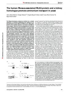
a
Grl1loxP allele
Grl1– allele
b hGH poly(A)
c
e
g
i
d
f
h
j
tissues. Loss of Gr function in the nervous system impairs hypothalamus-pituitary-adrenal (HPA)–axis regulation, resulting in increased glucocorticoid (GC) levels that lead to symptoms reminiscent of those observed in Cushing syndrome. Conditional mutagenesis of Gr in the nervous system provides genetic evidence for the importance of Gr signalling in emotional behaviour because mutant animals show an impaired behavioural response to stress and display reduced anxiety.
To disrupt Grl1 selectively in the nervous system, we developed two mouse lines. Using homologous recombination in embryonic stem (ES) cells, we generated a modified Grl1 allele (Grl1tm2Gsc, hereafter referred to as Grl1loxP; Fig. 1a) in which exon 3, encoding the first zinc finger of the Gr DNA-binding domain, was flanked by loxP sites. This allele encodes an active Gr protein, but is sensitive to Cre recombinase, which binds to loxP sites and deletes the intervening DNA sequence. Grl1loxP will therefore be inactivated in any cell expressing the recombinase. Mice homozygous for Grl1loxP appeared normal and expression of the Gr protein was identical to that of wild-type mice (data not shown). We generated a mouse expressing Cre recombinase in neuronal and glia cell precursors by expressing Cre under the control of the rat nestin (Nes) promoter and enhancer8 (Fig. 1b). Fig. 1 Generation of mice deficient for Gr in the nervous system. a, Organization of Grl1 encompassing exons 3 and 4. We flanked the third exon with loxP sites in two steps. First, we generated the modified allele by homologous recombination in ES cells. Second, transient expression of Cre recombinase leads to removal of the selection cassette, generating Grl1loxP and Grl1– alleles. A scheme of the wild-type gene locus, the targeting vector and the resulting alleles is depicted (open triangles, loxP; S, SacI; P, PstI). b, Structure of the nestin-Cre transgene. Cre recombinase was expressed under the control of the promoter and the nervous system-specific enhancer present in the second intron of the rat nestin gene8. hGH poly(A), human growth hormone polyadenylation signal. c–j, Expression of Gr is selectively lost in the nervous system of GrNesCre mice. Immunohistochemical analysis of Gr protein distribution in control (c,e,g,i) and GrNesCre (d,f,h,j) mice. c,d, ×25, 50 µm hippocampal section; e,f, ×200, 10 µm hypothalamus; g,h, pituitary (top, whole pituitary, ×50; bottom, magnification of the anterior lobe, ×1,000; 6 µm); i,j, kidney and adrenal gland. Note that the Gr is expressed in nuclei of the pituitary corticotrope cells of GrNesCre mice that also express Acth. We double stained these cells using antibodies directed against Gr (brown) and Acth (blue; ×200, 6 µm). CA, cornu ammonis; DG, dentate gyrus; PVN, paraventricular nucleus; k, kidney; cort, adrenal cortex; med, adrenal medulla.
1Molecular Biology of the Cell I, Deutsches Krebsforschungzentrum, Im Neuenheimer Feld 280, 69120 Heidelberg, Germany. 2Institute of Anatomy, Universität des Saarlandes Homburg, Germany. 3EMBL, Heidelberg, Germany. Correspondence should be addressed to G.S. (e-mail:
[email protected]).
nature genetics • volume 23 • september 1999
99
letter
© 1999 Nature America Inc. • http://genetics.nature.com
© 1999 Nature America Inc. • http://genetics.nature.com
Fig. 2 Mice lacking Gr in the nervous system display symptoms characteristic of Cushing syndrome including reduced size, altered fat distribution and reduced bone density. a, Histological analysis of serial Nissl-stained sections did not reveal any gross abnormalities in the central nervous system between control (left) and GrNesCre (right) mice. b, GrNesCre mutant mice (right) were reduced in size compared with control littermates (left). Adult GrNesCre males (6–14 weeks) were 70±3% of the weight (n=10) and displayed 82±0.4% of the length (n=6) of control littermates. c, GrNesCre mice have altered fat distribution. Fat tissue is visualized in white on digitalized pictures obtained by nuclear magnetic resonance tomography. Note the displacement of fat tissue toward the head and neck in mutants (bottom) compared with control mice (top). Mutant mice are not obese as often observed in Cushing syndrome patients. d, Osteoporosis was present in mutant mice as revealed by measuring bone density by Xray tomography in the skull or pelvis of control (n=6) and mutant (n=6) littermates.
a
b
We mated mice expressing the nestin-Cre transgene (NesCre) with mice carrying Grl1loxP. Specificity c and efficiency of Grl1 inactivation via recombination was determined by immunostaining using an antibody directed against the amino-terminal part of the Gr protein. We did not detect Gr in brain of mutant mice (Grl1loxP/loxPNesCre, abbreviated GrNesCre; Fig. 1d,f), but observed normal Gr expression in control mice (Grl1loxP/loxP; Fig. 1c,e). In contrast, the distribution of Gr protein observed in anterior pituitary, kidney, adrenal glands and other organs of control (Fig. 1g,i) and mutant mice were similar (Fig. 1h,j, and data not shown). Ubiquitous inactivation of Gr resulting from deletion of exon 3 leads to perinatal lethality1, whereas deletion of exon 3 in the nervous system did not impair development or post-natal survival (Fig. 2a). Mutant mice displayed several symptoms characteristic of patients suffering from Cushing syndrome9, a disease characterized by elevated levels of circulating GCs (Fig. 2b–d). Morning levels of circulating GCs were elevated in mutant mice but circadian rhythm was maintained (Fig. 3a). The alterations observed in mutant mice can be explained as a consequence of the elevated levels of GCs activating Gr present in cells outside the nervous system. GCs are synthesized by the cortex of the adrenal gland under the stimulus of adrenocorticotropic hormone (Acth) released by the anterior lobe of the pituitary9–11. Acth expression and release are under the control of corticotropin-releasing hormone (Crh)
a
corticosterone levels
GrNesCre
GrNesCre
b
d P