Plant Physiol. (1986) 80 ... Photocontrol of Chloroplast and Mitochondrial Polypeptide. Levels in ... in total cellular protein and without a chae in the size of the cellular .... tochondria will be referred to as cytoplasmic, a number of these .... roplasts are intact and contain both solubleand membrane ... glycolate metabolism.
Plant Physiol. (1986) 80, 618-622 0032-0889/86/80/06 18/05/$01.00/0
Photocontrol of Chloroplast and Mitochondrial Polypeptide Levels in Euglenal Received for publication August 12, 1985 and in revised form November 2, 1985
ANTONIO F. MONROY, BENITO GOMEZ-SILVA2, STEVEN D. SCHWARTZBACH*, AND JEROME A. SCHIFF3
School of Biological Sciences, University of Nebraska, Lincoln, Nebraska 68588 (A.F.M., S.D.S.), and Photobiology Institute, Brandeis University, Waltham, Massachusetts 02254 (B.G., J.A.S.) ABSTRACT Two dimensional polyacrylme gel electrophoresis resolved protein from intact chkroplasts of wild type Eugkxagrailis Klebs var. bacillaris Cori into 185 polypeptides of which 55 were lli on the whole cell polypeptide map. Of these chloroplast polypeptides, the relative amounts of 49 inased, the relative amounts of two decreased, and the relative amounts of four polypeptides were unaltered by exposure of dark grown resting cells to Light for 72 hours. Proteins from intact purified mitochondria obtined from a bleached mutant (W,.BSmL) lacking plastids were resolved into 193 polypeptides of which 44 were lalized on the whole cell polypeptide map from wild type cells. Of these mitochondrial polypeptides, the relative amount of one increased, the relative amounts of 12 were unaltered, and the relative amounts of 31 decreased after exposure of the dark grown rest cells to light. Since it is known that the development of the choroplast in Eagkxa occurs without a net increase in total cellular protein and without a chae in the size of the cellular amino acid pools, the degradation of mitochondrial polypeptides represents a major source of amino acids for the synthesis of chloroplast polypeptides.
Light acting through a blue absorbing nonchloroplast photoreceptor, a red absorbing nonchloroplast photoreceptor, and a blue-red absorbing chloroplast photoreceptor which may be
Pchl(ide) controls the polypeptide con;position of Euglena (24).
The most obvious result of exposure of dark grown resting cells to light is the transformation of the proplastid into a photosynthetically competent chloroplast. Many proteins which are induced by light are chloroplast constituents, although light also induces the formation of a number of nonchloroplast enzymes such as the microbody localized isozyme of glycolate dehydrogenase (1 1, 27). Other enzymes are transiently induced by light (1, 2, 12). Their levels increase during the first 12 h of light exposure and then decline. The induction of these enzymes appears to represent a response of starving cells to a utilizable carbon source, a nutritional shift up (12, 24). Both mitochondrial enzymes, such as fumarase (12) and citrate synthase (1), as well as cytoplasmic enzymes, such as NADPH-glutamate dehydrogenase (2), are regulated in this manner. The relative amounts of a third group of proteins decrease upon light exposure and these proteins include a number of glycolytic enzymes (5) and
mitochondrial Cyt (23). Based on electron microscopic (13, 22, 23) and autoradiographic studies (23), mitochondrial structure and proteins appear to be degraded during chloroplast development. In nondividing carbon starved resting cells, the carbon skeletons and energy required for chloroplast development are obtained through the degradation of preexisting molecules. Light promotes the degradation of the storage carbohydrate paramylum (25) and the degradation of RNA (3). Chloroplast proteins are synthesized in the absence of a net increase in total cellular proteins (23, 24). Since the size of the amino acid pools remains constant during chloroplast development (23), the amino acids used for the synthesis of chloroplast proteins must be derived through the degradation of preexisting proteins. In photosynthetic cells, the chloroplast can provide most of the ATP, fatty acids, storage carbohydrates, and carbon skeletons required for growth. Enzymes required for the synthesis of these compounds during organotrophic growth are at least partially gratuitous during phototrophic growth and they are likely to be degraded to provide the amino acids for the synthesis of chloroplast proteins. Although it is clear that light selectively alters the polypeptide composition of the cell, the extent to which these alterations are localized in specific cellular compartments remains unclear. Studies of changes in the specific activity of individual enzymes are time consuming and they provide information regarding only a single enzyme whose regulation may not be charcteristic of the regulation of the majority of the enzymes within a specific intracellular compartment. Changes in activity which are found can be the consequence of enzyme activation or inactivation rather than changes in the amount of enzyme protein. Two dimensional gel electrophoresis is a technique which is free from the problems associated with studies of specific marker enzymes and which rapidly provides a global picture of changes in cellular polypeptide levels. Polypeptides are chosen for study based on their physical properties (charge and molecular weight) rather than their function; this ensures that the polypeptides studied are a representative sample of the cellular proteins. Changes in the staining intensity of a polypeptide are proportional to changes in the amount of the polypeptide (15). Two dimensional gel electrophoresis resolves total cellular protein from Euglena into 640 polypepfides detectable by silver staining (15). Light exposure reproducibly induces the accumulation of 79 polypeptides and lowers the relative amounts of 72 polypeptides (15). Most of these changes are catabolite sensitive; they are inhibited when cells are exposed to light in the presence of ethanol (16), a carbon source which specifically inhibits chloroplast development (16, 24) while inducing the formation of enzymes of the glyoxylate cycle (11). The light induced accumulation of many polypeptides is dependent on the developmental status of the chloroplast while the disappearance of most
'Supported by National Science Foundation grant PCM 8202472 to S. D. S. and by National Institutes of Health grant GM 14595 and National Science Foundation grant PCM 7918142 to J. A. S. 2Goodman Graduate Fellow. 3Abraham and Etta Goodman Professor of Biology. 618
COMPARTMENTATION OF PHOTOREGULATED POLYPEPTIDES
polypeptides is independent of chloroplast development (18). In a small number of cases, a nonchloroplast photoreceptor controls the accumulation or disappearance of specific polypeptides ( 15). To determine the intracellular localization of those polypeptides whose levels increase or decrease after light exposure, proteins isolated from intact functional chloroplasts and mitochondria purified on Percoll gradients have been separated by two dimensional gel electrophoresis and localized on the whole cell polypeptide map in the present work. A brief report of this work was presented at the 1984 meeting of American Society of Plant Physiologists ( 14).MATERIALS AND METHODS Euglena gracilis Klebs var. bacillaris Cori and the plastidless mutant, W,OBSmL (21), derived from this stain were used throughout this work. For the isolation of chloroplasts, wild type cells were grown in the light with limiting concentrations of vitamin B12 (10). For the isolation of mitochondria, W1OBSmL was grown in the dark on limiting concentrations of vitamin B12 (9). Chloroplasts (8, 10) and mitochondria (9) were isolated by modifications of the trypsin digestion method and purified by Percoll density gradient centrifugation. Based on marker enzyme distributions and EM, the isolated chloroplasts (8, 10) and mitochondria (9) were free of contamination by other cellular organelles. The isolated chloroplasts were intact as evidenced by high rates of light dependent CO2 fixation and protein synthesis as well as by the retention of Cyt-c552 (8). The isolated mitochondria were intact based on measurements of substrate dependent 02 uptake, coupled phosphorylation, and amino acid incorporation into protein (7, 9). Proteins were extracted with phenol, precipitated with methanol, separated by two dimensional gel electrophoresis, and stained with silver as described previously (15). The intracellular loalization of polypeptides resolved by two dimensional gel electrophoresis of total cellular protein was determined in the following manner. A set of three gels containing 20 tg of organelle protein, a mixture of 20 gg of organelle protein and 40 Mg of total cellular protein, or 40 ,ug of total cellular protein were subjected to electrophoresis and stained at the same time. Polypeptides found in the organelle fraction were identified on the whole cell map based on their relative position on the gel and their increased staining intensity on a gel containing a mixture of organelle polypeptides and total cell protein relative to their intensity on a gel containing total cellular protein alone. Each set of gels was analyzed independently by two observers. The results presented are those polypeptides which were consistently identified by both observers on four sets of gels produced from two independent isolated chloroplast or mitochondrial fractions. In some cases polypeptides were localized in an organelle in some but not all ofthe analyses and these polypeptides are not reported. Although polypeptides not localized within chloroplasts or mitochondria will be referred to as cytoplasmic, a number of these may actually be chloroplast or mitochondrial proteins which were not positively identified in all of the analyses. The set of cytoplasmic polypeptides is also composed of polypeptides found in the cytoplasm, nucleus, and other organelles; compartments whose polypeptide composition has not been separately analyzed. Polypeptides are referred to by an alphanumeric system (15) consisting of a letter (A-D; A being the most acidic) corresponding to the isoelectric focusing sector of the gel in which the polypeptide is found and a number corresponding to the apparent mol wt of the polypeptide rounded to the nearest 1000. In the case of polypeptides within a sector having the same apparent mol wt, a decimal is added starting with the most acidic polypeptide.
619
RESULTS AND DISCUSSION Proteins extracted from isolated chloroplasts were resolved by two dimensional gel electrophoresis into approximately 185 polypeptides detectable by silver staining (Fig. 1). By comparing gels of chloroplast proteins (Fig. 1, top), whole cell proteins extracted from dark grown resting cells exposed to light for 72 h (Fig. 1, middle) and a mixture of whole cell proteins and chloroplast proteins (Fig. 1, bottom), 55 polypeptides found in isolated chloroplasts were localized on the whole cell polypeptide map (Fig. 1; squares). Proteins extracted from isolated mitochondria were resolved by two dimensional gel electrophoresis into 193 polypeptides detectable by silver staining (Fig. 2). By comparing gels of mitochondrial proteins (Fig. 2, top), whole cell proteins extracted from dark grown wild type cells (Fig. 2, middle), and a mixture of whole cell proteins and mitochondrial proteins (Fig. 2, bottom), 44 polypeptides found in isolated mitochondria were localized on the whole cell polypeptide map (Fig. 2; circles). A composite map indicating the relative positions and shapes of all of the polypeptide spots resolved from 60 Mg of total cellular protein extracted from dark grown resting cells maintained in the dark or exposed to light is shown in Figure 3. The polypeptides found in isolated mitochondria are distinctly different from those found in isolated chloroplasts (compare Figs. 1 and 2). Of the 44 mitochondrial and 55 chloroplast polypeptides localized on the whole cell map, only 3 polypeptides, B89, D43.2, and C25, were common to both organelle fractions (Fig. 3; squares within circles). Polypeptide B89 appears as a circular spot on gels of proteins extracted from chloroplasts (Fig. 1, top) and as a teardrop on gels of proteins extracted from mitochondria (Fig. 2, top). On gels of total protein extracted from dark grown cells, polypeptide B89 appears as a teardrop shaped spot (Fig. 2, middle), while on gels of total protein extracted from chloroplast containing cells, polypeptide B89 appears as a teardrop with three circular ridges on the top (Fig. 1, middle). These variations in the shape of the spot corresponding to polypeptide B89 suggest that chloroplasts and mitochondria contain different polypeptides which migrate to the same position on the two dimensional gel. It cannot be determined whether the other polypeptides found in both organelles represent comigrating polypeptides or whether they truly represent polypeptides found in both chloroplasts and mitochondria. Taken together with the biochemical and electron microscopic characterization of the isolated chloroplasts and mitochondria, the low numbers of comigrating polypeptides found in both the chloroplast and mitochondrial fractions indicate that the organelles purified on Percoll gradients are free of contamination by other cellular constituents. Exposure of dark grown resting cells to light increases the relative amounts of 79 polypeptides and decreases the relative amounts of 72 polypeptides (15; summarized in Fig. 3). A 2- to 3-fold change in the relative amount of a polypeptide is the minimum change in polypeptide levels which produces a visually detectable change in staining intensity (15). Of the 55 chloroplast polypeptides localized on the whole cell map, the relative amount of most chloroplast polypeptides, 49, is increased by light exposure (Fig. 3, squares). The relative amount of two chloroplast polypeptides, C25 (which was also found in the mitochondrial fraction) and D28, is decreased by light exposure (Fig. 3, squares). The relative amount of four chloroplast polypeptides, A 100, C2 1, D43.2 (which was also found in the mitochondrial fraction), and D18. 1, is unaltered by light exposure (Fig. 3, squares). The accumulation of 11 light induced chloroplast localized polypeptides, A24.1, A24.3, A23, A 18.2, B27.2, B22.2, B22.3, B21, C19, D2 1, and D17, appears to be correlated with the synthesis of Chl (18). Polypeptides of similar mol wt have been isolated from pigment protein complexes (4, 19), suggesting that these polypeptides are components of these complexes. Six polypeptides are presumed to be chloroplast translation products (I18) and as
620
MONROY ET AL. p--
Plant Physiol. Vol. 80, 1986
0 -
og -@t6 .~~~~~ ~~~~~
.
GL
IV
---W' ~ ~
~
*R N7-eG a- II8®
.04., (t
4
_
_
_
_
,
46
v_. W l
s B
I
.
8i8I
ie
~
t
0-
_% i
-
_
a&
.. Mm -W
W
.-
.s
.- -
.~~~~~~~~. f4 Si e 0- 0 1
(ad;
a ew .~ ~
S0
' 0 4._
*
r0 0W
*.1W
I
_ S_...
f-'
L .0, "A .."
p.
11
.4sbgse
A...
4 S:
w
As _
Pr0,
k4 hS
__
i
0
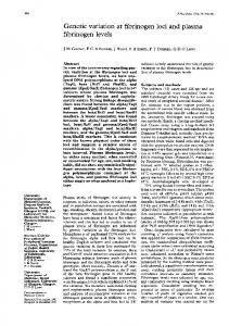
FIG. 1. Silver stained two dimensional gel of 20 Mg of protein extracted from Percoll-purified chloroplasts (top), 40 Mg of total cellular protein extracted from dark grown resting cells exposed to light for 72 h (middle), and a mixture of 20 Mg chloroplast protein and 40 Mg total cellular protein (bottom). Those polypeptides which are found in chloroplasts and have been localized on the whole cell polypeptide map are enclosed in squares. The acidic end of the gel is on the right.
FIG. 2. Silver stained two dimensional gel of 20 Ag of protein extracted from Percoll-purified mitochondria (top), 40 ,g of total cellular protein extracted from dark grown resting cells (middle), and a mixture of 20 Mg mitochondrial protein and 40Mgg total cellular protein (bottom). Those polypeptides which are found in mitochondria and have been localized on the whole cell polypeptide map are enclosed in circles. The acidic end of the gel is on the right.
expected, five of these polypeptides, A51, A17, B51, C47, and D54, were found in the isolated chloroplasts. Polypeptide D54 is the large subunit of ribulose-bisphosphate carboxylase (15) and its presence in the isolated chloroplasts indicates that the chloroplasts are intact and contain both soluble and membrane bound (Chl binding) polypeptides. As found for the induction of
COMPARTMENTATION OF PHOTOREGULATED POLYPEPTIDES
621
FIG. 3. Composite diagram indicating the photoregulation of those chloroplast and mitochondrial polypeptides localized on the whole cell polypeptide map. The relative positions of all of the polypeptides detected on silver stained two dimensional gels of 60 ,ug of total cellular protein of Euglena are shown. Solid and striped spots indicate those polypeptides whose relative amounts are increased and decreased, respectively, upon light exposure. Squares and circles indicate those polypeptides which are found in isolated chloroplasts and mitochondria, respectively.
the microbody localized isozyme of glycolate dehydrogenase (1 1, 27), a number of polypeptides (six) are induced by a product of photosynthetic CO2 fixation, presumably glycolate, rather than directly by light exposure (18). Five ofthese polypeptides, A22.2, A22.3, B44. 1, B1 5, and B29, were not found in the chloroplasts or mitochondria, suggesting that, like the microbody localized isozyme of glycolate dehydrogenase which is a microbody enzyme (27), these polypeptides represent enzymes involved in glycolate metabolism. W3BUL is a bleached mutant which lacks Pchl(ide), the chloroplast photoreceptor, but it has retained a proplastid remnant which undergoes limited development when exposed to light (20). Twelve polypeptides accumulate when dark grown resting W3BUL are exposed to light (15) and eight of these, Al 1O, A76, A42.1, A42.2, A28.2, A22.1, D93, and D26, are chloroplast proteins. Of the 44 mitochondrial polypeptides localized on the whole cell map, the relative amount of one polypeptide, B89, a polypeptide comigrating with a chloroplast polypeptide, is increased by light exposure (Fig. 3, circles). The relative amount of 12 mitochondrial polypeptides, B72. 1, B72.2, B72.3, B6 1, B43, C54, C38, C24. 1, D43. 1, D43.2 (which was also found in the chloroplast fraction), D23. 1, and D23.2, appear unaltered by light exposure (Fig. 3, circles). The relative amount of the remaining 31 polypeptides is decreased by light exposure (Fig. 3, circles). The levels of 14 polypeptides decreased when the bleached mutant W3BUL was exposed to light (15); 10 of these polypeptides A38.1, A25.1, C56, C32.1, C32.2, D120, D104, D65.1, D65.2, and D56, are mitochondrial proteins. Since protein turnover increases in resting cells exposed to light (24, 26), the selectivity of the light dependent decrease in the relative amounts of specific mitochondrial and cytoplasmic proteins can be due to selective protein degradation or to continued degradation through turnover with a light dependent decrease in the rate at which specific proteins are resynthesized. Pulse labeling studies have shown that light exposure decreases the relative rate of synthesis of many polypeptides, including mitochondrial polypeptides whose levels decrease after light exposure ( 17). It appears that light exposure decreases the relative amounts of cytoplasmic
polypeptides and the relative amounts of a subset (70%) of the most abundant mitochondrial polypeptides by inhibiting their resynthesis while their degradation through turnover continues. Since total cellular protein content (23, 24) and the size of the amino acid pools (23) are unaltered during chloroplast development, the amino acids required for the synthesis of chloroplast proteins are derived from cytoplasmic polypeptides and a majority of the most abundant mitochondrial proteins. Action spectra and studies with mutants have shown that at least three photoreceptors function independently and synergistically to control the polypeptide composition of Euglena (1, 24). One of these, a red-blue receptor which may be identical with Pchl(ide) appears to be present in the chloroplast and seems to control the synthesis of proteins on both chloroplast and cytoplasmic ribosomes (6, 24). Although the increase in the levels of a large number of chloroplast localized polypeptides and the decrease in the levels of a large number of mitochondrial polypeptides is independent of Chl synthesis or chloroplast protein synthesis (18), the bleached mutant W3BUL has lost the ability to photoregulate the levels of many, but not all, proteins (15) which are found in the chloroplast, cytoplasm, and mitochondria. The simplest explanation of this loss of photoresponsiveness is that the levels of these polypeptides are controlled by the chloroplast photoreceptor, Pchl(ide), and that this receptor is absent from the mutant W3BUL. In addition to inducing the synthesis of many chloroplast proteins, the chloroplast photoreceptor appears to inhibit the synthesis of many mitochondrial proteins. Studies with the bleached mutant W3BUL indicate that the accumulation of a few chloroplast, cytoplasmic, and mitochondrial proteins is also regulated by a nonchloroplast photoreceptor (15). Two nonchloroplast photoreceptors have been identified. A red only photoreceptor controls the transient induction of a number of mitochondrial and cytoplasmic enzymes in carbon starved resting cells but not in growing cells (1, 2, 12). These enzymes are induced in a similar manner when resting cells are provided with a utilizable carbon source (12), suggesting that the red receptor allows the cell to respond to light in the same manner as it would respond to any utilizable carbon source.
622
MONROY ET AL.
Two dimensional gel electrophoresis has failed to identify polypeptides which are induced by both light and organic carbon ( 15). It is therefore unlikely that the red photoreceptor regulates the levels of the cytoplasmic, mitochondrial, and chloroplast polypeptides detectable on silver stained two dimensional gels. The blue absorbing nonchloroplast photoreceptor is also present in bleached mutants (3, 12, 20, 24-26). Events specifically required for chloroplast development, such as cytoplasmic rRNA synthesis (3), the synthesis of proteins required for rapid Chl synthesis (6), and the mobilization and aerobic metabolism of storage carbohydrates (25), appear to be controlled by the blue photoreceptor. Like Chl synthesis, all of the light dependent changes in polypeptide levels detected by two dimensional gel electrophoresis are catabolite sensitive; they are inhibited when cells are provided with specific carbon sources (16). Thus it is probably the blue photoreceptor which is responsible for the changes in the levels of chloroplast, mitochondrial, and cytoplasmic polypeptides observed in the bleached mutant W3BUL. LITERATURE CITED 1. CANNONS AC, MJ MERRErT 1984 Regulation of synthesis of citrate synthase in regreening Euglena gracilis. Eur J Biochem 142: 597-602 2. CHAUDHARY MF, AC CANNONS, MJ MERRErr 1984 Photoregulation of
3. 4. 5.
6.
7.
8. 9.
NADPH-glutamate dehydrogenase in regreening cultures of Euglenagracilis. Plant Sci Lett 34: 89-94 COHEN D, JA SCHIFF 1976 Events surrounding the early development of Euglena chloroplasts. Photoregulation ofthe transcription of chloroplast and cytoplasmic ribosomal RNAs. Arch Biochem Biophys 177: 201-216 CUNNINGHAM FX JR, JA SCHIFF 1986 Chlorophyll-protein complexes.from Euglena grailis and mutants deficient in chlorophyll b. II. Polypeptide composition. Plant Physiol 80: 231-238 DOCKERTY A, MJ MERRETT 1979 Isolation and enzymic characterization of Euglena proplastids. Plant Physiol 63: 468-473 EGAN JM, D DORSKY, JA SCHIFF 1975 Events surrounding the early development of Euglena chloroplat VI. Action spectr for the formation of chlorophyll, lagelimination in chlorophyll synthesisand appearance of TPNdependent triose-phosphate dehydrogenase and alkaline DNase activities. Plant Physiol 56: 318-323 GOMEZ-SILVA B, E DELORME, AI STERN, JA SCHIFF 1984 Protein synthesis in organello by purified, intact, functional mitochondria from Euglena gracilis var. Bacillaris. Plant Physiol 75: S-196 GoMEZ-SILVA B, JA SCHIFF 1985 The light requirement for protein synthesis and carbon dioxide fixation in highly purified intact Euglena chloroplast PlantSci 39: 111-120 GoMEZ-SILVA B, AI STERN, T SAIDHA, JA SCHIFF 1985 Oxidative phosphoryl-
Plant Physiol. Vol. 80, 1986
ation coupled to respiration in highly purified intact Euglena mitochondria. J Plant Physiol 120: 431-40 10. GOMEZ-SILVA B, MP TIMKO, JA SCHIFF 1985 Chlorophyll biosynthesis from glutamate or 5-aminolevulinic acid in intact Euglena chloroplasts. Planta 165: 12-22 1 1. HORRUM MA, SD SCHWARTZBACH 1981 Nutritional regulation of organelle biogenesis in Euglena. Induction of microbodies. Plant Physiol 68: 430434 12. HORRUM MA, SD SCHWAR1ZRACH 1981 Induction of fumaase in resting Euglena. Biochim Biophys Acta 714: 407-414 13. LEFORT M 1964 Modifications du chondriome dans les cellules etiolees de 1'Euglena gracilis (klebs). C R Hebd Seances Acad Sci 258: 4318-4321. 14. MONROY AF, B GomEZ-SILVA, SD SCHWARTZBACH, JA SCHIFF 1984 Compartmentation of photoregulated polypepties in Euglena gracilis var. bacillaris. Plant Physiol 75: S-156 15. MONROY AF, SD SCHWARTZBACH 1983 Photocontrol of the polypeptide composition of Euglena. Analysis of two-dimensional gel electrophoresis. Planta 158: 249-258 16. MONROY AF, SD SCHWARTZBACH 1984 Catabolite r on of chloroplast development in Euglena. Proc Natl Acad Sci USA 81: 2786-2790 17. MONROY AF, SD SCHWARTZBACH 1984 Photoregulation of polypeptide synthesis in Euglena. Plant Physiol 75: S- 141 18. MONROY AF, SD SCHWARTZBACH 1985 Influence of photosynthesis and chlorophyll synthesis on polypeptide accumulation in greening Euglena. Plant Physiol 56: 313-317 19. ORI7Z W, E STUTZ 1980 Synthesis of polypeptides of the chlorophyll-protein complexes in isolated chloroplasts of Euglena gracilis. FEBS Lett 1 16: 298302 20. OSAFUNE T, JA SCHIFF 1980 Events surrounding the early development of Euglena chloroplasts. 17. Light induced changes in a proplastid remnant in mutant W3BUL. J Ultratuct Res 73: 64-76 21. OSAFUNE T, JA SCHIFF 1983 W10BSmL, a mutant of Euglena gracilis var. bacillaris lacking plastids. Exp Cell Res 148: 530-535 22. PELLEGRINI, M 1980 Three-dimensional reconstruction of organelles in Euglena grailis Z. I. Qualitative and quantitative changes of chloroplasts and mitochondrial reticulum in synchronous photoautotrophic culture. J Cell Sci43: 137-166 23. SCsAN1Z R, ML SCHANrz, H DURANTON 1975 Changes in amino acid and peptide composition of Euglena gracilis cells during chloroplast development. Plant Sci Lett 5: 313-324 24. ScHIFF JA, SD SCHWARTZBACH 1982 Photocontrol of chloroplast development in Euglena. In DW Buetow, ed, The Biology of Euglena, Vol III. Physiology. Academic Press, New York, pp 313-352 25. SCHWARTZBACH SD, JA SCHIFF, NH GOLDSTEIN 1975 Events surrounding the early development of Euglena chloroplasts. V. Control of paramylum degradation. Plant Physiol 56: 313-317 26. SCHWARTZBACH SD, JA ScHIFF 1979 Events surrounding the early development of Euglena chloroplasts. 13. Photocontrol of protein synthesis. Plant Cell Physiol 20: 827-838 27. YOKOTA A, Y NAKANO, S KfTAOKA 1978 Different effects of some growing conditions on glycolate dehydrogenase in mitochondria and microbodies in Euglena gracilis. Agric Biol Chem 42: 115-120