Nacional, Heredia,4 and International Centerfor Medical Research and Training, Louisiana State ... erinaria, Universidad Nacional, Heredia, Costa Rica. Phone: ...
CLINICAL AND DIAGNOSTIC LABORATORY IMMUNOLOGY, Mar. 1994, 107 l-412X/94/$04.00+0 Copyright © 1994, American Society for Microbiology
p.
206-213
Vol. 1, No. 2
Immunochemical Identification of Brucella abortus Lipopolysaccharide Epitopest NORMAN ROJAS, 2 ENRIQUE FREER,2'3 ANDREJ WEINTRAUB,2 MARGARITA RAMIREZ,4,5 SIV LIND,2 AND EDGARDO MORENO45* Facultad de Microbiologia' and Departamento de Fisiologia, Facultad de Medicina Universidad de Costa Rica, San Jose, Programa de Investigaci6n en Enfermedades Tropicales (PIET), Escuela de Medicina Veterinaria, Universidad Nacional, Heredia,4 and International Center for Medical Research and Training, Louisiana State University, INCIENSA, Tres Rios, Cartago,5 Costa Rica, and Karolinska Institute, Department of Clinical Bacteriology, Huddinge Hospital, Huddinge, Sweden2 Received 2 July 1993/Returned for modification 6 August 1993/Accepted 16 November 1993
Sera from Brucella abortus-infected and -vaccinated bovines recognized four lipopolysaccharide (LPS) determinants: two in the O-polysaccharide (A and C), one in the core oligosaccharide from rough Brucella LPS (R), and one in lipid A (LA). From 46 different hybridomas secreting monoclonal antibodies (MAbs) against various LPS moieties, 9 different specificities were identified. Two epitopes, A and C/Y, were present in the O-polysaccharide. Two epitopes were found in the core oligosaccharide (RI and R2) of rough Brucella LPS. MAbs against Rl and R2 epitopes reacted against LPS from different rough Brucella species; however, MAbs directed to the R2 epitope also reacted against enterobacterial LPS from deep rough mutants. Three epitopes (LAI, LA2, and LA3) were located in the lipid A backbone. Different sets of MAbs recognized two epitopes in the lipid A-associated outer membrane protein (LAOmp3-1 and LAOmp3-2). LPS preparations from smooth brucellae had small amounts of rough-type LPS. Although LPS from rough brucellae did not show smooth-type LPS in Western blots (immunoblots), two hybridomas generated from mice immunized with rough B. abortus produced antibodies against smooth B. abortus LPS. Results are discussed in relation to the structure and function of B. abortus LPS and to previous findings on the epitopic density of the molecule.
antigen predominating in B. melitensis (48). Diaz et al. (11) demonstrated that the A and M determinants were located in the O-polysaccharide. Further studies showed that the crossreaction between Brucella species and Yersinia enterocolitica 0:9 was due to common determinants located in the 0polysaccharide (18). The demonstration by Douglas and Buchanan (13) that the cross-reaction between B. abortus (biotype 1) and B. melitensis (biotype 1) was due to the existence of a "common" epitope(s), and not to "different proportions of A and M antigenic determinants," challenged the prevailing interpretation of Wilson and Miles supported by others (4, 27). Later, the presence of at least two common (C and C/Y) epitopes in LPS from smooth strains was established (13, 40). In recent investigations, Bundle et al. (4) and Meikle et al. (27) showed that the A, M, and C/Y epitopes resided in different linkages of N-formylperosamine residues along the O-polysaccharide. In this work, we describe the distribution of nine different epitopes defined in B. abortus LPS. The results are discussed in relation to the structure of Brucella LPS and to previous findings on the epitopic density of this molecule.
Brucella abortus S19 (biotype 1) lipopolysaccharide (LPS) is composed of an O-polysaccharide consisting of an unbranched linear homopolymer of 10 to 100 cx-1,2-linked 4-formamido4,6-dideoxy-D-mannose (N-formylperosamine) residues (6). The O-polysaccharide is covalently linked to a core oligosaccharide composed of mannose, glucose, 2-amino-2,6-dideoxyglucose (quinovosamine), 2-amino-2-deoxyglucose (GlcN), 3-deoxy-D-manno-2-octulosonic acid (KDO), and unidentified sugars (3, 35). The core is linked to the lipid A, which in contrast to most LPS studied, contains both GlcN and 2,3diamino-2,3-dideoxyglucose (GlcN3N) as backbone sugars and amide- and ester-linked long-chain saturated (Ci6:0 to C18:0) and hydroxylated (3-OH-C12:0, to 29-OH-C30:0) fatty acids (37). The long aliphatic chains of the lipid A have been proposed to span the outer membrane of the bacteria (2, 37). The thermotropic phase behavior of lipid A from B. abortus S19 and B. melitensis 16M suggests a disaccharide backbone molecule linked in a rll->6 configuration (43). Ethanolamine, neutral sugars, and ester-linked acyl oxyacyl fatty acids are not found in B. abortus lipid A (31, 37). Phosphate is absent or present in reduced quantities in the lipid A moiety (7, 37). Furthermore, B. abortus S19 LPS contains an outer membrane protein(s) (Omp) strongly bound to the lipid A moiety, not removed by conventional procedures used to release the lipid A-associated protein of enterobacterial LPS (1, 30). In 1932, Wilson and Miles designated two different determinants: the A antigen predominating in B. abortus, and the M
MATERIALS AND METHODS Antigens. The Brucella strains, the extraction procedure, and purification methods for the various LPS have been described elsewhere (1, 32, 35, 37). The enterobacterial and photosynthetic strains and the methods for extraction and purification of their LPS have been extensively described (18, 26, 32, 35, 37). The chemical and physical characterization of LPS from smooth and rough strains, the conditions for LPS hydrolysis, and preparation of the different lipid A moieties and 0polysaccharide have been summarized in several publications (2, 23, 24, 26, 30, 32-37). Purification of lipid A was carried out by high-performance
* Corresponding author. Mailing address: Programa de Investigacion en Enfermedades Tropicales (PIET), Escuela de Medicina Veterinaria, Universidad Nacional, Heredia, Costa Rica. Phone: (506) 37-3004. Fax: (506) 38-1298. t Part of project 801-90-565, Vicerrectoria de Investigaci6n, Universidad de Costa Rica.
206
VOL. 1, 1994
thin-layer chromatography (HPTLC) according to the method of Freer (15). Briefly, a continuous line containing 150 ,ug of lipid A was spotted and chromatographed on HPTLC precoated silica gel 60 plates (Merck, Darmstadt, Germany) in a mobile phase of chloroform-methanol-ammonia-water (25:14: 1:2); then a strip of the plate was stained with 10% sulfuric acid in ethanol. With the visible bands as a reference, the unstained fractions on the silica plate were removed and collected by carefully scraping the silica with a scalpel. Finally, the lipid A was eluted by stirring the silica for 4 h at 25°C in the same solvent as that used for chromatography. The supernatants were centrifuged at 2,000 x g, and the organic solvent was removed under a stream of nitrogen. The lipid A was stored dry for further analysis or dissolved in 7 mM triethylamine (Sigma, St. Louis, Mo.) for immunochemical assays. Purification of O-polysaccharide was done as previously described (34, 36). B. abortus 45/20 core oligosaccharide was purified from LPS as described by Suarez et al. (47). Deacylated lipopolysaccharide (LPS-OH) and lipid A (lipid A-OH) were prepared according to formerly described methods (26). Salmonella minnesota R595 tetrasaccharide and Haemophilus influenzae 169 Rd-lb+ dephosphorylated trisaccharide were prepared as described previously (24). Brucella blebs (Omp-rich fractions) and purified Omp preparations were obtained from I. Moriyon (Department of Microbiology, University of Navarra, Navarra, Spain). B. abortus fatty acids were obtained and purified as described elsewhere (37). Periodate oxidation of the B. abortus 45/20 LPS was carried out according to the methods of Mayer et al. (26) and Woodward et al. (50). GlcN3N was obtained from H. Mayer (Max Planck Institute for Immunobiology, Freiburg, Germany). GlcN was purchased from Sigma. The chemical composition and biological properties of the different preparations have been published elsewhere (7, 30-37). Antibodies. The following alkaline phosphatase conjugates (Sigma) were used in enzyme immunoassays (EIA), dot blot, or Western blot (immunoblot): rabbit immunoglobulin G (IgG) anti-mouse Ig, IgG, IgA, and IgM; goat IgG anti-rabbit Ig; and rabbit IgG anti-bovine Ig. Polyclonal antibodies against B. melitensis 16M LPS were produced in rabbits and ensured to be monospecific to B. melitensis (M) serotypes as described previously (34, 37). Sera from B. abortus field-infected and -vaccinated bovines were obtained from herds in Costa Rica. Rabbit IgGs were purified by using protein A-Sepharose as described in the Pharmacia manual (Pharmacia, Uppsala, Sweden). Absorption of bovine serum with LPS fractions was carried out as previously described (36). Monoclonal antibodies (MAbs) against Brucella 0-polysaccharide common (C) and abortus (A) epitopes, Lipid A, lipid A-associated Omp, and B. abortus 45/20 core oligosaccharide (R) were produced according to the protocol of St. Groth and Scheidegger (46). Six- to ten-week-old female BALB/c mice were immunized by intraperitoneal injections of 0.2 ml of an emulsion in Freund's complete adjuvant (Sigma) containing 5 x 10⁸ acetone-killed B. abortus S19 cells, acetic acid-treated B. abortus S19, or acetic acid-treated B. abortus 45/20 postcoated with lipid A from B. abortus S19 (25). One milligram of B. abortus 45/20 LPS per ml was administered, using a similar protocol of immunization (200 ,ug per mouse). A second dose with the same quantity of bacterial preparation or LPS in incomplete Freund's adjuvant was injected intraperitoneally 10 to 15 days later. A booster in phosphate-buffered saline (PBS), pH 7.2, was injected by the same route 3 days before the fusion. Hybridizations were performed with spleen cells from immunized mice with myeloma cell line P3X63-AG656 in supplemented RPMI 1640 medium (GIBCO, Grand Island, N.Y.) or with the Sp2/0 myeloma cell line in RPMI 1640 medium with
IDENTIFICATION OF B. ABORTUS LPS EPITOPES
207
supplements (GIBCO). The hybridomas were selected in medium containing 6 x 10-' M hypoxanthine, 5 x 10'7 M aminopterin, and 2 x 10⁻⁵ M thymidine, all from GIBCO. The hybridoma supernatants from the different fusions were screened for antibody production by EIA 10 days after fusion, using the following antigens: B. abortus S19 LPS and B. melitensis 16M LPS; B. abortus 45/20 LPS and B. abortus S19 lipid A. The selected clones were expanded and cloned by limiting dilution, using maintenance medium with 104 peritoneal macrophages per well as feeder cells. MAbs containing supernatants from hybridomas grown in either complete RPMI 1640 (10% fetal bovine serum) or supplemented serum-free medium (GIBCO) were concentrated with 50% (vol/vol) ammonium sulfate-saturated solution, dialyzed, and lyophilized. In addition, cloned hybridoma cells (2 x 10⁷) were injected intraperitoneally into Pristane (2,6,8,14-tetramethylpentadecane; Aldrich Chemical Co., Milwaukee, Wis.)-primed BALB/c mice. Ascites fluid was collected 12 or 22 days after hybridoma inoculation. Antibody isotypes were determined by immunodiffusion, using rabbit anti-mouse Igs (Litton Bionetics, Kensington, Md.). Serological assays. Microtiter polystyrene plates (Nunc, Roskilde, Denmark) were coated with 100 ,ul of LPS (10 ,ug/ml) in 0.1 M sodium carbonate buffer (pH 9.6) overnight at 37°C. The coating of lipid A at 10 ,ug/ml was performed as described by Freudenberg et al. (16). Indirect EIA was carried out as described previously (24, 34). EIA inhibition was performed as follows: MAbs were diluted in PBS containing 0.05% Tween 20 to a concentration equivalent to an optical density of 0.90 ± 0.08 at 405 nm after 100 min, which corresponds to 30 to 50% saturation of antibody bound to the coating antigen. Then, 1 volume of the diluted antibody was mixed with 1 volume (in PBS-Tween 20) of the corresponding inhibitor diluted at concentrations ranging from 250 to 0.04 ,ug/ml. After 1 h of incubation at 22°C, an indirect EIA was performed. The inhibitory value was recorded as the amount of inhibitor needed to obtain a 50% lowering of the optical density at 405 nm after 100 min compared with controls with no inhibitor added. Sodium dodecyl sulfate-polyacrylamide gel electrophoresis (SDS-PAGE) for separation of LPS and lipid A was carried out in 10, 12, or 15% gels, as described by Krauss et al. (21). Western and dot blotting of the preparations was carried out as described by Douglas and Palmer (14). A radial immunodiffusion test for polysaccharide preparations was carried out as previously described (10). RESULTS
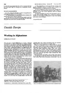
Immunoreactivity of bovine polyclonal antibodies. Sera from B. abortus-infected bovines recognized at least four antigenic determinants in LPS: two (A, abortus; and C, common) present in the O-polysaccharide, one (R) present in the core oligosaccharide from rough strains, and one (IA) present in the lipid A (Fig. 1). Serum from a B. abortus (biotype 1)-infected cow reacted in the radial immunodiffusion test against B. melitensis 16M polysaccharide native hapten (NH), while serum from a vaccinated cow did not react against this material (not shown). Absorption of serum from the B. abortus-infected cow with homologous LPS resulted in elimination of antibodies against both B. abortus S19 and B. melitensis 16M LPS. However, when the same serum was absorbed with B. melitensis 16M LPS, the titer against this antigen decreased almost to the background level but maintained a significant reactivity against B. abortus S19 LPS (optical density at 405 nm of 0.46). The absorption experiments are consistent with the
208
ROJAS ET AL.
CLIN. DIAGN. LAB. IMMUNOL.
B
A co
cn v
and ori-
-,
No
Co t ,
-H _ _
TABLE 1. EIA reactivity' of MAbs against LPS and LPS-derived antigens
C
n _>
A
v A n 1l L
mi
LI L2
ofnor
| t
Reactivity
Antigen"
Ss
-
-
28K
-22K i
CCAfRRRRRRLPPC FIG. 1. Western blot of different Brucella antigen preparations. (A) Western blots of Brucella antigen preparations against different MAbs and rabbit antibodies. Lanes: a, B. abortus; m, B. melitensis; o, B. ovis; c, B. canis. C, Baps-C/Y; A, Baps-A; M, monospecific rabbit IgG anti-M epitope; R, Baro-1; L, Bala-3; P, left (one band), Baomp3-1; P, right (two bands), Baomp3-2. LPS are from B. abortus S19 and B. melitensis 16M smooth strains (S-LPS) and from rough B. abortus 45/20, B. melitensis B15, B. ovis, and B. canis strains (R-LPS). Lipid A is from B. abortus S19. Omps are from B. abortus 45/20, and Omp-rich fractions (blebs) are from B. abortus S19. (B) Western blots of Brucella and Yersinia antigen preparations against serum from a B. abortusinfected bovine. Lanes: Y, Y enterocolitica 0:9 LPS; A, B. abortus S19 LPS; M, B. melitensis 16M LPS; R, B. abortus 45/20 LPS; L, B. abortus S19 lipid A. (C) Western blots of B. abortus S19 lipid A preparations against Baps-C/Y MAb. Lanes: L,, lipid A after hydrolysis with 2% acetic acid for 5 h at 100°C; L2, lipid A purified by HPTLC. Twelve percent polyacrylamide gels were used. The following negative Western blot reactions are not shown: Baro MAbs tested against 104treated B. abortus 45/20 LPS, Y enterocolitica 0:9 LPS, and purified lipid A's; Baps-C/Y tested against purified lipid A's, purified Omp, and rough Brucella LPS; Baps-A tested against B. melitensis 16M LPS, purified lipid A's, purified Omp, and rough Brucella LPS; Bala MAbs tested against Brucella LPS and purified Omp; monospecific rabbit IgG anti-M epitope against B. abortus S19 LPS, Y enterocolitica 0:9 LPS, and purified lipid A.
of
Baro- I
Baro-2
2.18
1.90
B. abortus 45/20 LPS B. abortus 45/20 LPS 104 treated B. melitensis B115 LPS B. canis LPS B. ovis Reo LPS B. abortus S19 LPS B. melitensis 16M LPS B. abortus S19 lipid A (20%
2.70 1.10 1.18
protein) E. coli J5 (Re) LPS S. minnesota R595 (Re) LPS S. typhimurium SL1181 (Rd2) LPS S. typhimurium SL1032 (Rdl) LPS S. typhimurium Rc to Ra LPSs S. typhimurium SH4809 LPS
2.34 1.37 1.51
0.11
0.14
0.05 0.07
0.40 0.33 0.23
given
MAb
(OD)
Baps-A
Baps-C/Y
ND
0.12 ND
0.06
0.15
0.10 1.67 0.13 0.37
1.27 0.94 0.56
0.08 0.07
0.11 ND
ND
a Indirect EIA values are duplicate averages of a representative assay expressed as optical density (OD) at 405 nm - optical density at 405 nm of the background. A blank space indicates optical density of 1
~3
W:
0
Q-
-)
-ci0
i
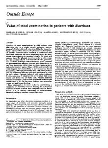
i% FIG. 2. Western
blotting
reactions of Baro-2 MAbs
against
differ-
enterobacterial and Brucella LPS. Lanes: R, B. abortus 45/20 LPS; Re, S. minnesota R595 LPS; Rd2, typhimurium S1181 LPS; Rd,, S. typhimurium SL1032 LPS; Rb3, S. typhimurium TV148; S, S. typhient
R2
.;
o0
0
_
0)
:Kdo.,
0
£'
murium SH4809 LPS. Fifteen percent
polyacrylamide gels
were
used.
LA3
smooth-type LPS. None of the polysaccharide antigens penetrated the polyacrylamide gels, indicating the absence of an SDS-binding moiety such as LPS, lipid A, or protein. B. abort us S19 LPS present in Omp-rich fractions was recognized as a long smear, revealing the presence of protein ghosts along the gel. Lipid A before purification showed a diffuse band with Baps-C/Y, indicating the presence of 0-polysaccharide epitopes in this preparation. After purification of lipid A, reaction with Baps-C/Y was not observed (Fig. 1C). Core KDO and quinovosamine were not detected in any of the lipid A preparations, demonstrating the absence of LPS. Baro-2 MAbs reacted against LPS of Salmonella deep core mutants but not against LPS harboring "large" or complete core oligosaccharide or possessing 0-polysaccharide antigen (Fig. 2). Baro-2 could recognize several bands in the LPS of the deepest rough mutant of S. minnesota Re but fewer bands in the LPS from Salmonella Rdl and Rd2. The intensity of the Western blot reactions agreed with the ELA results.
DISCUSSION
Our
previous
rabbits
(32-37) has demonstrated that antibod(biotype 1)-infected or -hyperimmunized
work
ies from B. abortus
recognized
of Brucella strains: of smooth strains,
determinants in the LPS
at least six distinct a common an
charide of B. abortus,
determinant
(C)
in the 0 chain
A determinant present in the one
R determinant in the
0-polysacoligosac-
LA1
LAOmp3-1 LAOmp3-2 Associated Omp3 peptide
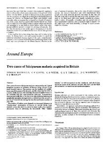
FIG. 3. Illustration of the most likely location and distribution of the B. abortus (biotype 1) LPS epitopes (A, C/Y, RI, R2, LAI, LA2, LA3, LAOmp3-1, and LAOmp3-2). To the right, the three regions of the smooth-type LPS are indicated in black, gray, and white, respectively. A scheme of the three LPS moieties corresponding to the three regions is presented in the center. For simplicity, the three moieties are not linked to demonstrate the epitopes. The triangles, rectangles, and trapezoids in the model indicate the dominant interactions between the epitope and the cognate MAb, corresponding to Baps-A (IgG3), Baps-C/Y (IgG3), Baro-1 (IgG3), Baro-2 (IgM), Bala-3 (IgM), Bala-5 (IgM), Bala 13 or Bala 15 (IgM), Baomp3-1 (IgGl), and Baomp-3-2 (IgGI). The less reactive region is located towards the narrower vertex, whereas increasing reactivity is illustrated by the wider areas of the geometric figures. The circles represent the a-1,2-linked N-formylperosamine residues of the 0-polysaccharide, the gray diamonds represent the sugars of the core oligosaccharide, and the black ovals represent the lipid A backbone aminosugars (GlcN3N, diaminoglucose; GlcN, glucosamine).
core
rough strains, two GIcN and GlcN3N determipresent in the lipid A backbone of all strains, and one
charide from nants
determinant in the
present work,
we
lipid
A-associated
have
Omp
determined
the
of all strains. In the
presence
of
nine
specificities in LPS preparations of B. abortus, and their proposed epitopes have been assigned. Two epitopes (A and C/Y) previously defined by MAbs (14, 40) are present in the O-polysaccharide, two (Rl and R2) are found in the LPS core oligosaccharide from rough strains, three (LA1, LA2, and LA3) are in lipid A, and two ([AOmp3-1 and LAOmp3-2) are in lipid A-associated protein (Fig. 3). It has been proposed that the A epitope resides in four to five oL-1,2-linked N-formylperosamine residues, while the necessary element of the M epitope is a disaccharide of ot-1,3linked N-formylperosamine residues that requires adjacent oa-1,2-linked residues for full activity (4). Douglas and Palmer (1 4, 40) demonstrated the presence of two common epitopes in the LPS of different smooth Brucella species. These authors produced MAbs reacting with one epitope (C/Y) shared with
the LPS of Y enterocolitica 0:9, while the other epitope (C) was only present in smooth Brucella strains. Since nuclear magnetic resonance analysis revealed that the chemical structure of Y enterocolitica 0:9 LPS is identical to that of the B. abortus S19 LPS (6), then the C/Y epitope must reside in 2 to 4 units of a-1,2-linked N-formylperosamine residues (4, 40). The location of the C epitope remains uncertain. Interestingly, lipid A preparations obtained after acid hydrolysis of LPS of smooth B. abortus or B. melitensis before purification also reacted with anti-C/Y MAbs, suggesting at first glance the existence of nonhydrolyzed LPS in the lipid A preparations. However, the absence of KDO and quinovosamine, core oligosaccharide markers, is consistent with the following alternative: the binding of anti-C/Y to lipid A is due to the presence of a lipid-bound polysaccharide (lipid-NH) that is antigenically related to the 0-polysaccharide but in essence is different from the LPS, as previously proposed (7, 31). Furthermore, the
VOL. 1, 1994
absence of a KDO linkage would make the lipid-NH resistant to the hydrolysis conditions used to release O-polysaccharide from the lipid A (42), and due to its hydrophobic domain, the lipid-NH might migrate in SDS-PAGE and be detected in immunoblots (Fig. 1C). Since the description of the A and M determinants by Wilson and Miles in 1932 (48), the interpretation that B. abortus biotype 1 and B. melitensis biotype 1 LPS possess only two antigenic epitopes and that the cross-reaction between the abortus and melitensis serotypes is due to different proportions of A and M determinants has prevailed and consequently dominated the scientific literature on brucellosis for the last 70 years. Though we recognize this pioneering work as a major contribution to the field of brucellosis, in light of the present knowledge on the antigenic and chemical structures of Brucella LPS, this concept should be modified. Assuming the presence of significant quantities of A epitope in the M serotype and vice versa, absorption of anti-A with M-serotype bacteria and anti-M sera with A-serotype bacteria should eventually remove antibodies against both determinants. However, sera against O-polysaccharide exhaustively absorbed with B. abortus (A serotype) or B. melitensis (M serotype) always reacted in lower titer (compared with the original antiserum) against the homologous but not the heterologous bacteria. This implies the absence or, ultimately, the presence of nonsignificant quantities of heterologous epitopes, that is, A in melitensis serotype and M in abortus serotype, respectively (13, 14, 27, 40). Furthermore, that different proportions and expression of A and M determinants may be regulated by plasmids or bacteriophages, as in epitopes of other bacteria (4, 27), is unlikely since several extensive analyses have failed to reveal natural plasmids or lysogenic phages in Brucella species (28). In conclusion, our results and interpretation are in close agreement with those presented by Douglas and Palmer (14, 40). Therefore, the proper understanding regarding the extensive cross-reactivity between B. abortus (biotype 1) and B. melitensis (biotype 1) that is explained by the existence of immunodominant common determinants in the LPS molecule should prevail over the interpretation that this event is due to different proportions of A and M determinants. Several authors have reported the presence of 0-polysaccharide determinants in rough B. abortus 45/20 (11, 45) and B. melitensis B115 (9) strains. In this work, we obtained two hybridomas from mice immunized with B. abortus 45/20 cells reacting against smooth Brucella LPS. The fact that 0-polysaccharide epitopes are detected in rough Brucella spp. does not necessarily imply the presence of smooth-type LPS in these strains since there are other molecules (e.g., NH and poly B) which share antigenic determinants with smooth-type LPS. Therefore, it would be of interest to further investigate the expression of LPS epitopes in those brucellae that have been regarded as rough mutant strains. None of the anti-core oligosaccharide MAbs reacted against O-polysaccharide preparations, suggesting that core epitopes are not accessible in the LPS from smooth Brucella spp. Alternatively, the rough-type LPS core oligosaccharide could be structurally different from that of the smooth-type core LPS. On the basis of limited chemical analysis (7, 32) and comparative immunochemical studies shown in this paper, we propose that one of the epitopes defined in the Brucella core oligosaccharide includes common determinants with an enterobacterial core (R2) whereas the other does not (R1). Therefore, Baro-1 was highly specific for Brucella rough-type oligosaccharide. The RI epitope most likely includes mannose, glucose, and GlcN sugars but not quinovosamine or KDO. The R2 epitope is most likely located within the KDO region and
IDENTIFICATION OF B. ABORTUS LPS EPITOPES
211
may be masked by additional sugars present in the Salmonella core mutants Rc to Ra or the O-polysaccharide of smooth bacteria. In addition, the reactivity of the Baro-2 MAb suggests that although KDO is found in fewer amounts in Brucella LPS, its conformation might be the same or very similar to the KDO linked to the lipid A backbone of the deep enterobacterial rough mutants (e.g., unsubstituted KDO). This hypothesis is supported by the fact that the Baro-2 MAb was inhibited by lower quantities of LPS isolated from S. minnesota Re than LPS from rough mutants with increasing core sugars (Table 2). Since the order of reactivity against LPS was rough brucellae > Re > Rd2 > Rdl > Rc = Ra = smooth strains, we concluded that Baro-2 antibody, in addition to binding KDO, needs other sugars for full recognition. On the basis of inhibitory analysis, the lipid A displayed three different epitopes. One epitope (LA1), which is preferentially present in Brucella spp., seems to be a disaccharide aminosugar molecule d-ifferent from that of enterobacteria (LA2) and photobacteria (LA3) studied. The other two epitopes are shared by lipid A preparations from enterobacteria or photobacteria. It has been shown that enterobacterial LPS contains "free" lipid A capable of migrating in HPTLC and being detected by anti-lipid A MAbs (15). Bala MAbs were unable to react against lipid A epitopes in the complete LPS molecule, indicating the absence of free lipid A in Brucella LPS. Because of the contamination with polysaccharides and proteins, purification of lipid A by HPTLC was performed. Neither anti-O-polysaccharide nor anti-Omp MAbs bound to these lipid A preparations. Anti-Omp3 MAbs bound to LPS and to the lipid A fraction before HPTLC purification, indicating that Omp group 3 epitopes are accessible for antibodies. The fact that Baomp3 MAbs react against different bands in immunoblots in the region corresponding to Omp group 3 agrees with the proposal that this group of proteins is a family of closely related proteins associated with the LPS (17). It has been suggested that Omp group 3 might be the counterpart of OmpA of E. coli (49). We do not know to which peptide region the LAOmp3-1 and LAOmp3-2 epitopes correspond. Attempts to isolate lipid A-associated protein from LPS or lipid A have failed, suggesting that after proteinase K and acid treatment the protein is destroyed and only small, strongly bound immunodominant peptides remain. It must be stressed that antibody responses of infected or immunized animals to LPS are directed not only to epitopes localized in the O-polysaccharide (A, M, C/Y, or C) but also to epitopes found in the rough-type LPS, lipid A, lipid A-associated Omp, and NH moieties. Although simple, these observations are remarkable. With a few exceptions (22, 23, 33), works involving diagnosis, passive protection, or immune responses against Brucella antibodies to LPS determinants different from those present in O-polysaccharide have been disregarded (5, 8, 12, 20, 29, 38, 39). For instance, LPS antigens containing strongly bound proteins considerably influence the interpretation of serological immunoassays due to the presence of different immunoglobulins directed against protein epitopes (22, 33). This is relevant in light of extensively used EIAs for the diagnosis of bovine brucellosis (19) performed with smooth-type LPS antigens containing variable quantities of LPS-associated proteins and rough-type LPS, as determined by one of us (E.M.).
ACKNOWLEDGMENTS We thank Alf Lindberg (Karolinska Institute) for criticism of the manuscript, Ignacio Moriy6n (University of Navarra, Navarra, Spain) for help in the characterization of anti-Omp MAbs, J. Weckesser
212
ROJAS ET AL.
(Albert-Ludwigs-Universitat, Freiburg, Germany) for the Rhodopseudomonas LPS, and Edgar Bolafios for technical assistance. N. Rojas received support from the Costa Rican National Council for Scientific and Technological Research (CONICIT). This work was supported by a grant to the Karolinska International Research Training (KIRT) Program from the Swedish Agency for Research Cooperation with Developing Countries (SAREC), a grant from the Swedish Medical Research Council (16X-656), and a grant from the Third World Academy of Sciences (TWAS 92-108 RG/BIO/LA). REFERENCES 1. Berman, D. T., and R. Kurtz. 1987. Relationship of biological activities to structures of Brucella abortus endotoxin and LPS. Ann. Inst. Pasteur Microbiol. 138:96-101. 2. Bhat, U. R., R. W. Carlson, M. Busch, and H. Mayer. 1991.
Distribution and phylogenetic significance of 27-hydroxy-octacosanoic acid in lipopolysaccharides from bacteria belonging to the alpha-2 subgroup of Proteobacteria. Int. J. Syst. Bacteriol. 41:213217.
CLIN. DIAGN. LAB. IMMUNOL.
18. 19.
20. 21. 22. 23.
3. Bowser, D. V., R. W. Wheat, J. W. Foster, and D. Leong. 1974.
Occurrence of quinovosamine in lipopolysaccharides of Brucella species. Infect. Immun. 9:772-774. 4. Bundle, D. R., J. W. Cherwonogrodzky, M. A. J. Gidney, P. J. Meikle, M. P. Perry, and T. Peters. 1989. Definition of Brucella A
and M epitopes by monoclonal typing reagents and synthetic oligosaccharides. Infect. Immun. 57:2829-2836. 5. Canning, P. C., B. L. Deyoe, and J. A. Roth. 1988. Opsonin-
dependent stimulation of bovine neutrophil oxidative metabolism by Brucella abortus. Am. J. Vet. Res. 49:160-163. 6. Caroff, M., D. R. Bundle, M. B. Perry, J. W. Cherwonogrodzky, and J. R. Duncan. 1984. Antigenic S-type lipopolysaccharide of Brucella abortus 1119-3. Infect. Immun. 46:384-389. 7. Cherwonogrodzky, J. W., G. Dubray, E. Moreno, and H. Mayer. 1990. Antigens of Brucella, p. 19-64. In K. Nielsen and B. Duncan
(ed.), Animal brucellosis. CRC Press, Inc., Boca Raton, Fla. 8. Cloeckaert, A., I. Jacques, P. de Wergifosse, G. Dubray, and J. N. Limet. 1992. Protection against Brucella melitensis or Brucella abortus in mice with immunoglobulin G (IgG), IgA, and IgM monoclonal antibodies specific for a common epitope shared by the Brucella A and M smooth lipopolysaccharides. Infect. Immun. 60:312-315. 9. Cloeckaert, A., M. S. Zygmunt, J. Nicolle, G. Dubray, and J. L. Limet. 1992. 0 chain expression in the rough Brucella melitensis strain B1 15: induction of O-polysaccharide-specific monoclonal antibodies and intracellular localization demonstrated by immunoelectron microscopy. J. Gen. Microbiol. 138:1211-1219. 10. Diaz, R., P. Garatea, L. M. Jones, and I. Moriy6n. 1979. Radial immunodiffusion test with a Brucella polysaccharide antigen for differentiating infected from vaccinated cattle. J. Clin. Microbiol. 10:37-41. 11. Diaz, R., L. M. Jones, D. Leong, and J. B. Wilson. 1968. Surface
antigens of smooth brucellae. J. Bacteriol. 96:893-901. 12. Diaz, R., and M. A. Oyeledun. 1977. Studies of some biological activities of Brucella endotoxin in normal and infected animals and the role of hypersensitivity factor. Ann. Sclavo 19:117-130. 13. Douglas, J. T., and T. M. Buchanan. 1980. Quantitation of Brucella lipopolysaccharide antigen by enzyme-linked immunosorbent assay. Abstr. Annu. Meet. Am. Soc. Microbiol. 1980, B29, p. 22. 14. Douglas, J. T., and D. A. Palmer. 1988. Use of monoclonal antibodies to identify the distribution of A and M epitopes on smooth Brucella species. J. Clin. Microbiol. 26:1353-1356. 15. Freer, E. 1990. Production and characterization of monoclonal antibodies against Brucella lipid A and demonstration of heterogeneity of Brucella lipid A by high performance thin-layer chromatography (HPTLC). M.S. thesis. Karolinska Institute, Stockholm. 16. Freudenberg, M., A. Fomsgaard, I. Mitov, and C. Galanos. 1989. ELISA for antibodies to lipid A, lipopolysaccharides and other hydrophobic antigens. Infection 17:322-328. 17. Gamazo, C., A. J. Winter, I. Moriy6n, J. I. Riezu-Boj, J. M. Blasco, and R. Diaz. 1989. Comparative analysis of proteins extracted by
24.
25.
26. 27. 28. 29.
30. 31. 32.
33. 34. 35.
36. 37.
38.
hot saline or released spontaneously into outer membrane blebs from field strains of Brucella ovis and Brucella melitensis. Infect. Immun. 57:1419-1426. Hurvell, B., and A. A. Lindberg. 1973. Serological cross reactions between different Brucella species and Yersinia enterocolitica. Acta Pathol. Microbiol. Scand. Sect. B 81:113-119. Joint FAO/IAEA Division. 1992. Animal production and health: brucellosis indirect ELISA kit. Animal Production and Health Section, Joint FAO/IAEA Division, International Atomic Energy Agency, Vienna. Jones, L. M., R. Diaz, and A. G. Taylor. 1973. Characterization of allergens prepared from smooth and rough strains of Brucella melitensis. Br. J. Exp. Pathol. 52:492-508. Krauss, J. H., J. Weckesser, and H. Mayer. 1988. Electrophoretic analysis of lipopolysaccharides of purple nonsulfur bacteria. Int. J. Syst. Bacteriol. 38:157-163. Kurtz, R. S., and D. T. Berman. 1986. Influence of endotoxinprotein in immunoglobulin G isotype responses of mice to Brucella abortus lipopolysaccharide. Infect. Immun. 54:728-734. Lamb, V. L., L. M. Jones, G. G. Schurig, and D. T. Berman. 1979. Enzyme-linked immunosorbent assay for bovine immunoglobulin subclass-specific response to Brucella abortus lipopolysaccharides. Infect. Immun. 26:240-247. Lind, S., L. Kenne, and A. A. Lindberg. 1990. Mapping of the antibody combining site of five monoclonal antibodies recognizing 3-deoxy-D-manno-octulosonic acid (Kdo) in bacterial lipopolysaccharides. J. Immunol. 146:3864-3870. Luk, J. M. C., N. A. Nalue, and A. A. Lindberg. 1990. Efficient production of mouse and rat monoclonal antibodies against 0 antigens of Salmonella serogroup C1, using LPS-coated bacteria as immunogen. J. Immunol. Methods 129:243-250. Mayer, H., R. N. Tharanathan, and J. Weckesser. 1985. Analysis of lipopolysaccharides of gram-negative bacteria. Methods Microbiol. 18:157-207. Meikle, P. J., M. B. Perry, J. W. Cherwonogrodzky, and D. R. Bundle. 1989. Fine structure of A and M antigens from Brucella biovars. Infect. Immun. 57:2822-2828. Meyer, M. E. 1990. Current concepts in the taxonomy of the genus Brucella, p. 1-17. In K. Nielsen and B. Duncan (ed.), Animal brucellosis. CRC Press, Inc., Boca Raton, Fla. Montaraz, J. A., A. J. Winter, D. M. Hunter, B. A. Sowa, A. M. Wu, and L. G. Adams. 1986. Protection against Brucella abortus in mice with O-polysaccharide-specific monoclonal antibodies. Infect. Immun. 51:961-963. Moreno, E., D. T. Berman, and L. A. Boetcher. 1981. Biological activities of Brucella abortus lipopolysaccharides. Infect. Immun. 30:363-370. Moreno, E., D. Borowiak, and H. Mayer. 1987. Brucella lipopolysaccharides and polysaccharides. Ann. Inst. Pasteur Microbiol. 138:102-105. Moreno, E., L. M. Jones, and D. T. Berman. 1984. Immunochemical characterization of rough Brucella lipopolysaccharides. Infect. Immun. 43:779-782. Moreno, E., R. S. Kurtz, and D. T. Berman. 1984. Induction of immune and adjuvant immunoglobulin G responses in mice by Brucella lipopolysaccharide. Infect. Immun. 46:74-80. Moreno, E., H. Mayer, and I. Moriy6n. 1987. Characterization of a native polysaccharide hapten from Brucella melitensis. Infect. Immun. 55:2850-2853. Moreno, E., M. W. Pitt, L. M. Jones, G. G. Schurig, and D. T. Berman. 1979. Purification and characterization of smooth and rough lipopolysaccharides from Brucella abortus. J. Bacteriol. 138:361-369. Moreno, E., S. L. Speth, L. M. Jones, and D. T. Berman. 1981. Immunochemical characterization of Brucella lipopolysaccharide and polysaccharides. Infect. Immun. 30:214-222. Moreno, E., E. Stackebrandt, M. Dorsch, J. Wolters, M. Busch, and H. Mayer. 1990. Brucella abortus 16S rRNA and lipid A reveal phylogenetic relationship with members of the alpha-2 subdivision of the class Proteobacteria. J. Bacteriol. 172:3569-3576. Nielsen, K., J. W. Cherwonogrodzky, J. R. Duncan, and D. R. Bundle. 1989. Enzyme-linked immunosorbent assay for differentiation of the antibody response of cattle naturally infected with
VOL. 1, 1994
39. 40. 41.
42. 43.
44.
Brucella abortus or vaccinated with strain 19. Am. J. Vet. Res. 50:5-9. Nielsen, K., P. F. Wright, J. Cherwonogrodzky, J. R. Duncan, and B. Stemshorn. 1987. Enzyme immunoassay for diagnosis of bovine brucellosis. Ann. Inst. Pasteur Microbiol. 138:75-79. Palmer, D. A., and J. T. Douglas. 1989. Analysis of Brucella lipopolysaccharide with specific and cross-reacting antibodies. J. Clin. Microbiol. 27:2331-2337. Perera, V. Y., A. J. Winter, and B. Gnem. 1984. Evidence for covalent binding of native hapten proteins complex to smooth lipopolysaccharide of B. abortus. FEMS Microbiol. Lett. 21:263266. Raetz, C. R. 1990. Biochemistry of endotoxins. Annu. Rev. Biochem. 59:129-170. Ramos-Sanchez, M. C., A. Orduia-Domingo, A. RodriguezTorres, F. J. Martin-Gil, and J. Martin-Gil. 1992. Investigations on thermotropic phase behavior of lipid A from Brucella and other Gram-negative bacteria. Thermochim. Acta 144:299-305. Redfearn, M. S. 1960. An immunochemical study of antigens of Brucella extracted by the Westphal technique. Ph.D. thesis. Uni-
IDENTIFICATION OF B. ABORTUS LPS EPITOPES
213
versity of Wisconsin-Madison, Madison. 45. Schurig, G. G., R. M. Roop II, T. Bagchi, S. Boyle, D. Buhrman, and N. Sriranganathan. 1991. Biological properties of RB51: a stable rough strain of Brucella abortus. Vet. Microbiol. 28:171188. 46. St. Groth, F., and D. Scheidegger. 1980. Production of monoclonal antibodies: strategy and tactics. J. Immunol. Methods 35:1-21. 47. Suarez, C. E., G. A. Pacheco, and A. Vigliocco. 1990. Immunochemical studies of oligosaccharides obtained from lipopolysaccharide of Brucella ovis. Vet. Microbiol. 22:335-340. 48. Wilson, G. S., and A. A. Miles. 1932. The serological differentiation of smooth strains of the Brucella group. Br. J. Exp. Pathol. 13:1-13. 49. Winter, A. J. 1987. Outer membrane proteins of Brucella. Ann. Inst. Pasteur Microbiol. 138:87-89. 50. Woodward, M. P., Jr., W. W. Young, and R. A. Bloodgood. 1985. Detection of monoclonal antibodies specific for carbohydrate epitopes using periodate oxidation. J. Immunol. Methods 78:143153.