Mg2, 25 mmol/l HEPES, pH 7.4; Gibco), antibiotics and fungizone as above; 300 IU/ml Collagenase Type IV (Worthington, Freehold,. NJ, USA) and 22.5 IU/ml ...
Molecular Human Reproduction vol.5 no.8 pp. 757–766, 1999
Localization of regulatory protein binding sites in the proximal region of human myometrial connexin 43 gene
Clement O.Echetebu, Mariam Ali, Michael G.Izban, Lynnete MacKay and Robert E. Garfield1 Department of Obstetrics and Gynecology, The University of Texas Medical Branch, Division of Reproductive Sciences, Galveston, TX 77555, USA 1To
whom correspondence should be addressed
Parturition is preceded by a large increase in gap junctions between myometrial smooth muscle cells. Connexin 43 is the major structural protein of myometrial gap junctions. To explore transcriptional regulation of the myometrial Cx43 gene, we used DNase I footprinting, electrophoretic mobility shift and transient transfection assays to examine a 312 bp promoter region (–164 to 1148) of the gene, utilizing human myometrial cell cultures and nuclear extracts. The DNase I studies showed four regions of nucleoprotein interactions. Protection of region 1 (–80 to –31) encompassed an Activator Protein 1 (AP1) (–44 to –36) and two Specificity Protein 1 (Sp1) (–77 to –69 and –59 to –48) consensus sequences. Regions 2 to 4 included the transcription initiation site (–10 to 125), an Ets/NF-kB consensus sequence (147 to 174) and a TA-rich region (181 to 1101) respectively. Gel mobility shift and supershift assays demonstrated c-Jun and Sp1 binding at the AP1 and Sp1 sites respectively. Promoter mutagenesis and transient transfection analyses combined with Sp1 and c-Jun/c-Fos over-expression studies indicate that both Sp1 and c-Jun are required for maximal promoter activity and, therefore, may positively regulate transcription of myometrial Cx43 during the initiation of labour. Key words: AP1/connexin-43/myometrium/promoter/Sp1
Introduction Gap junctions are specialized channels that connect adjacent cells and enable metabolic and electrical co-ordination between them (Pitts and Simms, 1977; Bennet and Goodenough, 1978). The major structural protein of gap junctions is connexin. There are many different kinds of connexins encoded by a multigene family dispersed on numerous chromosomes (Fishman et al., 1991). Connexin 43 (Cx43) is one of the most widely expressed members of this gene family, and is found in numerous tissues such as the uterus, heart, lens epithelium, kidney, mammary gland, ovary, placenta and intestine (Beyer et al., 1987; Risek and Gilula, 1991; Lee et al., 1992; Li et al., 1993). The onset of labour is preceded by a dramatic increase of gap junctions in uterine smooth muscle, the myometrium (Garfield et al., 1977; Miller et al., 1989; Hendrix et al., 1992; Chow and Lye, 1994). This increase is believed to enable the development of co-ordinated and intense uterine contractions necessary for the delivery of the fetus, whereas its suppression before term is believed to aid uterine quiescence. Evidence to support this concept comes from many functional studies showing that propagation of electrical activity of the myometrium is increased significantly during labour when gap junctions/Cx43 protein concentrations are increased (Garfield et al., 1977, 1988; Sims et al., 1982; Cole et al., 1985; Miller et al., 1989; Miyoshi et al., 1996). Cx43 mRNA and protein concentrations in the myometrium are low during pregnancy, but rise markedly at term, remain high throughout labour, then decline rapidly following © European Society of Human Reproduction and Embryology
delivery in rat (Risek et al., 1990; Hendrix et al., 1992; Tabb et al., 1992; Lye et al., 1993; Winterhager et al., 1993), sheep (McNutt et al., 1994) and human (Garfield and Hayashi, 1981). While the expression of Cx43 is up-regulated by oestrogens (Garfield et al., 1977; MacKenzie and Garfield, 1985) and down-regulated by progesterone (Garfield et al., 1977; MacKenzie and Garfield, 1985; Petrocelli and Lye, 1993; Zhao et al., 1996) the molecular events that regulate transcriptional activation remain unclear. Rat (Yu et al., 1994), mouse (Chen et al., 1995), and human (Geimonen et al., 1996) Cx43 promoters have been cloned and sequenced. In these promoters, extensive homology exists between –175 to 1143, relative to the start site of transcription (Chen et al., 1995). Transient expression studies (DeLeon et al., 1994; Chen et al., 1995; Geimonen et al., 1996) indicated that this region is important for regulating Cx43 promoter activity. It is likely, therefore, that similar regulatory elements may be operational within this promoter region in these species. In the mouse, genetic and biochemical studies have demonstrated both ‘repressor’ (–100 to –91) and ‘activator’ (–68 to –59) promoter elements (Chen et al., 1995). These regions are .90% conserved in the human Cx43 promoter. Transient transfection experiments in human cardiac myocytes have revealed the presence of negative regulatory element(s) between –2400 and –360 and a positive element between –175 and –50 of Cx43 gene (DeLeon et al., 1994). Transcription of the human Cx43 gene can be induced in primary human myometrial cell cultures after activation of 757
C.O.Echetebu et al.
protein kinase C by tetradecanoyl phorbol acetate (TPA) treatment (Geimonen et al., 1996). Human Cx43 promoter region from –697 to 1170 has been used in mutation analysis and transient expression studies to demonstrate that TPAinduced activation required a consensus Activator Protein 1 (AP1) site located between positions –44 to –36. The AP1 site is conserved between rodent and human promoters but distinct from the mouse ‘activator’ region (Chen et al., 1995). In addition, these studies demonstrated that TPA induced a dramatic rise in the protein concentrations of both c-Jun and c-Fos, further supporting the notion that transcriptional activation occurred through the promoter proximal AP1 site. However, over-expression of c-Jun and or c-Fos in the absence of TPA treatment had no effect on promoter activity, suggesting that TPA functioned by increasing and activating c-Jun/c-Fos. In this report, we used a combination of DNase I footprinting, electrophoresis mobility shift (EMSA) and transient transfection assays to begin a systematic study of the human Cx43 promoter. We confirm previous studies (Geimonen et al., 1996) and extend them by showing that the Ap1 site functionally interacts with c-Jun. In addition, we demonstrate that the Cx43 promoter contains tandem Specificity Protein 1 (Sp1) binding sites and that the proximal site, which is encompassed within a promoter segment homologous to the mouse ‘activator’ element functionally activates transcription. Furthermore, we show that over-expression of c-Jun and Sp1 either alone or in combination up-regulates Cx43 transcription in untreated primary cells in culture. Thus, both Sp1 and c-Jun are capable of and necessary for positively regulating human myometrial Cx43 transcription.
Materials and methods Cell culture Approval for the use of human tissue was obtained from the University of Texas Medical Branch Committee on Research Involving Human subjects. To obtain human myometrial factors that bind the Cx43 promoter at term when the levels of Cx43 mRNA and gap junctions are high, nuclear extracts should be made from myometrium obtained from biopsies or Caesarean section at these times. The quantity of tissue obtained from biopsies is insufficient for making amounts of nuclear proteins needed for our experiments. Moreover, it is difficult to homogenize human myometrium to liberate intact nuclei without first freezing the tissue or using harsh disintegration procedures that also break the nuclei and freezing is not recommended for preparing factors active in transcription (M.G.Izban, personal communication). Crude nuclear extracts were prepared from primary cultures of human myometrial cells initiated and grown as follows: the tissue from Caesarian sections or biopsies (from patients, gestational age, 37–39 weeks and not in labour) was cleaned of blood vessels, fat and endometrium. It was washed several times in wash buffer, Hank’s balanced salt solution (HBSS; Gibco Life Technologies, Grand Island, NY, USA), supplemented with 100 IU/ml penicillin G; 100 µg/ml streptomycin sulphate and 25 µg/ml amphotericin B as fungizone (Gibco); then minced and rinsed three times in the same buffer. It was then digested for ~75 min at 37°C with three times its volume of dispersion solution (HBSS, containing 1 mmol/l Ca21, 1 mmol/l Mg21, 25 mmol/l HEPES, pH 7.4; Gibco), antibiotics and fungizone as above; 300 IU/ml Collagenase Type IV (Worthington, Freehold, NJ, USA) and 22.5 IU/ml DNase I (Worthington). The dispersion
758
process included trituration every 3 min with decreasing bore-size pipettes, collection of supernatants by centrifugation (20 g for 5 min) at room temperature every 10 min and addition of fresh dispersion solution. The pooled supernatants were centrifuged at 430 g for 10 min. The separated cells were washed twice with the wash buffer and once with the growth medium (Minimal Essential Medium; Gibco) supplemented with 10% (v/v) fetal calf serum, 2 mmol/l Lglutamine, 1 mmol/l sodium pyruvate; antibiotics and fungizone as above. They were then resuspended in adequate volume of growth medium, plated and grown at 37°C in a 5% CO2/95% humidified air incubator. Medium was changed every 3 days (Andersen et al., 1993; Zhao et al., 1996).
Preparation of nuclear extract To obtain sufficient quantity of nuclear proteins, the cultures were passaged four times. A total of 50 culture dishes (150315 mm) of cells (~33109 cells) were used per experiment. After change of media (Geimonen et al., 1996), the cells were treated for 6–8 h with phorbol ester, 12-O-tetradecanoyl-phorbol-13-acetate (TPA), (CalbiochemNovobiochem Corporation, La Jolla, CA, USA) at 100 ng/ml, harvested and nuclear extract made by a modified method (Shapiro et al., 1988) using a hand-held Dounce homogenizer. The modification included the addition of protease inhibitors: leupeptin and aprotinin, at 2 µg/ml; pepstatin-A (1 µg/ml), and phosphatase inhibitor (β-glycerophosphate) (10 mmol/l) to all buffers except the dialysis buffer which contained only β-glycerophosphate as an addition. Extracts were divided into small volume aliquots, flash-frozen in liquid nitrogen and stored at –70°C till used. Plasmid constructs A 312 bp fragment (–164 to 1148) was amplified using the polymerase chain reaction (PCR) from a 6 kb EcoRI human Cx43 gene 59-region that was cloned into pGF10 and kindly supplied by Dr G.I.Fishman (Fishman et al., 1991). The PCR primers contained KpnI and XbaI sites, which were used to insert the 312 bp fragment into a pUC19 polycloning site to create pCx312. The primers were: Sense: 59-GCGCGGTACCGATCCCACTGCTCTTTG-39 Antisense: 59-GCGCTCTAGACCTTGGAGGATGAAGTAAAATG-39. A 171 bp fragment (–164 to 17), was excised from the 312 bp fragment and inserted into KpnI/HindIII sites of pUC19 and is hereafter referred to as pCx171. The sequences of the fragments were confirmed by the dideoxy chain termination method. In-vitro mutagenesis Seven independent 10 bp replacement mutants (seriatim linker scan spanning –99 to –30 of the promoter) were made using PCR from pCx312 (Vallette et al., 1989; Vanden Heuvel et al., 1993). A 10 bp sequence 59-ATCTCGAGAT-39 bearing a unique XhoI site was used for each replacement. The mutants ∆–99/–90, ∆–89/–80, ∆–79/–70, ∆–69/–60, ∆–59/–50, ∆–49/–40, ∆–39/–30 were inserted into pGL3 basic vector in the KpnI/BglII cloning site. Specific confirmatory 2 or 3 bp replacement mutants ∆–93/–91 [CCC→TTT; ∆–74/–72, (CTC→TGT); ∆–55/–53, (CTC→TGT); and ∆ –59/–58 (GC→TT)] were also constructed as additional checks for the ∆–99/–90, ∆–79/– 70 and ∆–59/–50 mutations. Briefly, the 312 bp fragment was excised from pCx312 and inserted into PGL3 basic vector in the KpnI/BglII cloning site and mutagenesis was performed directly in this vector using transformer site-directed mutagenesis kit (Clontech Labs Inc, Palo Alto, CA, USA) and the following primers: for ∆–93/–91: 59-CAAAGGGAAAGGCTAAAAGGAGGTAGGGAG-39, ∆–74/–72: 59-GCTAGAAGAAAGGGACAGGCAAAGGGAAAGGC-39;
AP1 and Sp1 regulate human myometrial Cx43 gene expression ∆–59/–58: 59-CAACTGGGAGGAGGGAATAGAAGAAAGGGGAGG-39, ∆–55/–53: 59-CTGACTCAACTGGGAGACAGGGCTAGAAGAAAGGGG-39, and a selection primer, 59-GCCGGCCGCCCCGACTCGCGAATTACACGGCGATC-39 were used to change XbaI (TCTAGA) to NruI (TCGCGA) site. Sequences were confirmed by the ABI PRISM Automatic Sequencer Model 373, Version 3.0.
Labelling of DNA fragments: The 312 and 171 bp fragments used in DNaseI protection assays were 39-end labelled at EcoRI, XbaI or HindIII sites of the plasmids pCx312 and pCx171 with [α-32P]dNTPs (specific activity, 3000 Ci/ mmol; Amersham Life Science, Arlington Heights, IL, USA) and the Klenow fragment of DNA Polymerase I (Promega, Madison, WI, USA). High specific activity was achieved by employing double labels [α-32P]dATP and [α-32P]dTTP for EcoRI and HindIII 39-ends and [α-32P]dCTP and [α-32P]dTTP for XbaI 39-end. Labelled inserts were separated from the vector by restriction with appropriate enzymes and purified by 5% non-denaturing polyacrylamide gel electrophoresis (PAGE), ion exchange chromatography (NACS column; Gibco Life Technologies) and ethanol precipitation. Double-stranded oligonucleotides used in EMSA were 59-end labelled with [γ-32P]ATP (Amersham) and T4 Polynucleotide kinase, then purified by gel filtration (Pharmacia, Piscataway, NJ, USA) using G-50 spin columns or PAGE. They include oligonucleotides for AP1, AP2 and Sp1, purchased from Santa Cruz Biotechnology (Santa Cruz, CA, USA) and custom oligonucleotides made by Genosys Biotechnologies (The Woodlands, TX, USA). DNase I footprinting DNase I footprint assay (Angel et al., 1987) was performed in a 25 µl reaction mixture containing (final concentration); 10 mmol/l HEPES (pH 8.0), 1.6 mmol/l dithiothreitol (DTT), 5 mmol/l MgCl, 0.1 mmol/l EDTA, 25 mmol/l KCl, 1 µg [poly (dI-dC). poly (dIdC)], (Pharmacia), 12% glycerol, 5 µg bovine serum albumin (BSA), nuclear proteins (0.0 to 60 µg) and 13105 cpm (0.1 pmoles) of labelled DNA. After incubation on ice for 60 min, samples were equilibrated for 3 min at room temperature and digested for 1 min. with 0.5–6 µl of DNase I (Worthington) diluted to 6.25 µg/ml in buffer (10 mmol/l HEPES, pH 7.6, and 25 mmol/l CaCl2). The reaction was terminated with 200 µl of stop buffer (10 mmol/l Tris– HCl, pH 7.6, 20 mmol/l EDTA, 3 µg/ml sheared calf thymus DNA, 0.2 mg/ml proteinase K and 0.2% sodium dodecyl sulphate), then incubated for 45 min at 37°C. After phenol–chloroform extraction, the DNA was ethanol-precipitated, resuspended in formamide dye mixture (95% formamide, 0.05% Bromophenol Blue and 0.05% Xylene Cyanol FF) and fractionated on 8% polyacrylamide sequencing gel along with a G1A chemical sequencing reaction lane (Maxam and Gilbert, 1980) and visualized by autoradiography. Electrophoresis motility shift assay (EMSA) Standard EMSA were performed with [γ-32P]ATP-labelled doublestranded oligonucleotides. Binding reactions (15 µl) contained 10 mM Tris–HCl (pH 7.6), 50 mmol/l KCl, 5 mmol/l DTT, 0.5 mmol/ l EDTA, 1 mmol/l MgCl2 and varying amounts of nuclear extract and poly (dI-dC) as indicated in the figure legends. The mixture was incubated on ice for 10 min, then 5.0–7.53104 cpm of label was added and the incubation was continued for 25 min at 25°C. Samples were then fractionated on 4% non-denaturing polyacrylamide gel with 0.53 Tris/Borate/EDTA buffer. Protein–DNA bands were visualized by autoradiography. For supershift and competition assays, antibodies to specific proteins (Santa Cruz Biotech Santa Cruz, CA,
USA; and Oncogene Research Products, Cambridge, MA, USA) or unlabelled mutant or non-specific oligonucleotides were added to the incubation mixtures. Incubations involving Sp1 antibody contained additional 1.6 mmol/l ZnSO4. Double-stranded oligonucleotides used (showing only the coding strand sequences) contain either AP1 (double underline), AP2 consensus (bold-faced), ‘CCTCC’ elements (single underline) or Ets/NF-kB consensus (dotted underline) are as follows: AP1 (–52/–29): 59-CTCCCAGTTGAGTCAGTGGCTTGA-39. CCTCC/ proximal AP2: (–100/–80): 59-ACCTCCTCCCAGCCTTTCCCT-39. AP2 distal (–139/–119): 59-TCTCCCCCCAGCCCTTCCACA-39. –70/–48: 59-CCTTTCTTCTAGCCCCTCCTCCCA-39 –100/–50: 59-ACCTCCTCCCAGCCTTTCCCTTTGCCCTTTCTTCTAGCCCCTCCTTCCCCC-39 –83/–65: 59-CCTTTGCCCTCCCCTTTC-39. 150/173: 59-GGCGTGAGGAAAGTACCAAACAGC-39. (Ets/ reversed NF-kB motif) –32/–9: 59-TTGAAACTTTTAAAAGCTCTGTGC-39 bA-4: 59-TCTTCGCATCCCTCCTCCAGT-39, motif found in chicken adult β-globin gene (Emerson et al., 1985).
Transient transfections Human myometrial primary culture cells between second and fourth passages were plated to ~50–60% confluence in 60 mm diameter culture dishes. They were transfected with 20 µg total DNA (15.0 µg of test plasmids, 1.0 µg of internal control pCMVlacZ plasmid and 4.0 µg of Salmon sperm DNA) by the calcium phosphate procedure. At 4–6 h post-transfection, the cells were glycerol-shocked for 1 min, washed with phosphate-buffered saline (PBS) and supplemented with fresh medium. Cells were harvested 48 h later, lysed and assayed for luciferase using the Promega Luciferase Assay System (Promega). β-Galactosidase activity was measured by the standard colorimetric method and used for normalizing luciferase activity. Co-transfection with c-Jun, c-Fos and Sp1 expression plasmids The following plasmids were used: pCMV Sp1, which over-expresses Sp1, was generously provided by R.Tjian; pCMV c-Jun and pCMV c-Fos, which over-express c-Jun and c-Fos respectively, were gifts from A.Kopland. Wild-type Cx43 (5 µg; 312 bp) in pGL3 basic-luc reporter was mixed with either 1 µg of each expression plasmid or their combinations containing 1 µg of each plasmid (see legend to Figure 7 for details) and 1 µg of pCMVlacZ. The total DNA was adjusted to 20 µg with salmon sperm DNA. Transfections were performed as described above. Statistical analysis The transfection assays were performed four times. Samples in each group of experiments were in duplicates or triplicates. All data were expressed as mean 6 SE. One-factor analysis of variance (ANOVA) was used to test the overall hypothesis of no group differences, followed by two-sample t-tests for pairwise comparisons where the overall hypothesis was rejected. P , 0.05 was considered to be statistically significant.
Results Transcription factor binding sites within the human Cx43 promoter It has been demonstrated that TPA treatment of primary myometrial cells in culture leads to stimulation of Cx43 759
C.O.Echetebu et al.
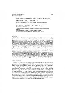
Figure 1. (A) DNase I footprint of 312 bp promoter region of human Cx43 gene. DNase I protection analysis of the coding strand of labelled DNA probe (–164 to 1148). Amounts of human myometrial crude nuclear extract proteins used are indicated at the tops of lanes (3, 4 and 5). Protected regions one through four are indicated on the right of the autoradiograph and sequence positions relative to transcription start site are on the left and right margins. G1A (lane 1) indicates Maxam & Gilbert chemical sequence ladder. Free DNA (lane 2) contains no proteins and shows natural sensitivity to DNase I. (B) DNaseI footprint of Cx43 gene region (–164 to 17). The 171 bp Sau3AI/HindIII Cx43 promoter (region –164 to 17), labelled at the HindIII ends was used as a probe. Amounts of extract proteins used are at the tops of lanes. Protected regions are indicated by lines along the right side of the autoradiograph. Positions relative to transcription start site are shown on the left side. The positions of Activator Protein (AP)1, Sp1 and AP2 consensus sequences are indicated. Free DNA (lanes 1 and 3) contain no proteins. Lane 2 shows chemical sequence ladder.
promoter (Geimonen et al., 1996). To define the regions within the promoter of the human Cx43 gene capable of interacting with transcription factors in similarly treated cells we carried out DNase I footprinting analysis. The results using a promoter fragment bearing (relative to the start site of transcription) from –164 to 1148 (Figure 1A) and from –164 to 17 (Figure 1B) demonstrated several protected regions separated by sites of enhanced DNase I sensitivity. Protection on the coding (Figure 1) and non-coding (data not shown) strands is indicated in Figure 2. Region 1 extends from –80 to –31 and encompasses a consensus AP1 site (–44 to –36) and a segment (–80 to –45) that envelops other potential protein binding sites summarized in Figure 2. The promoter sequence (–65 to –48) homologous to the mouse ‘activator’ element (Chen et al., 1995) is also located in this region. Three other regions considered further only in the discussion include the site of initiation, Region 2 (–5 to 115), Region 3 (147 to 170) which is coincident with an Ets/NFkB consensus site and Region 4 (180 to 1 100) that spans a TA-rich site (181 to 760
196). Note that transcription factor binding was not observed either within the CCTCC/AP2 sequence (–93 to –87) which corresponds to the mouse ‘repressor’ site (Chen et al., 1995), or at the AP2 consensus (–134 to –127).
c-Jun binds to the AP1 consensus site of the Cx43 promoter Previous studies (Geimonen et al., 1996) have suggested that Cx43 promoter activation through the proximal AP1 site following TPA treatment of primary myometrial cells relies on the concomitant increase in c-Jun and c-Fos protein concentrations. To test this directly, we performed mobility shift and supershift assays using a double-stranded oligonucleotide that encompassed the AP1 consensus site (–52 to –29). Incubation of the radiolabelled Cx43 AP1 motif with nuclear extracts derived from TPA-treated primary myometrial cell cultures (Figure 3 A,B, lane 2) or, as a control, from actively growing HeLa cell spinner cultures (Figure 3B, lane 6) resulted in a single shifted band. Formation of this nucleoprotein complex
AP1 and Sp1 regulate human myometrial Cx43 gene expression
Figure 2. Summary of human Cx43 promoter DNaseI footprint in the presence of human myometrial nuclear extract proteins. Lines above and below the sequences, marked off by numbers, indicate regions 1–4, protected from DNase1 digestion. The start site of transcription is at 11. Brackets indicate cis-elements and arrow above Ets shows direction of the incomplete Ets element.
was competed by inclusion of non-labelled self (Figure 3A, lane 8) or by a commercially available oligonucleotide containing the consensus AP1 binding motif (Figure 3A, lane 9; Figure 3B, lane 10). Competition was not observed with a mutated AP1 binding motif that does not support AP1 binding (Figure 3B, lane 11). These data support the notion that this region within the Cx43 promoter is an AP1-binding site. AP1 is a family of dimeric transcription factors comprised of either c-Jun homodimers or heterodimers of certain Jun/ Fos family members. In order to identity the Jun/Fos family members within the band-shifted nucleoprotein complex, we initially performed supershift analysis using a monoclonal c-Jun antibody specific for phosphorylated c-Jun (c-JunP, Figure 3A, lane 4,), and polyclonal antibodies raised against the aminoterminal regions of either the c-Jun (c-Jun, Figure 3A, lane 3) or c-Fos (c-Fos, Figure 3A, lane 5) proteins. In Figure 3A, a supershift for phosphorylated c-Jun was observed (lanes 4 and 7). However, supershifts were not observed with c-Jun (lanes 3 and 6) or c-Fos (lanes 5 and 6) either alone (lanes 3 and 5) or in combination (lane 6). It was surprising that c-Jun antibody had no effect on band mobility since this antibody should have recognized both the phosphorylated and non- phosphorylated forms of c-Jun. Therefore, we repeated these experiments using a polyclonal antibody (pan Jun) that recognizes a highly conserved region of the DNA binding domain of Jun family members c-Jun, Jun B and Jun D. As expected, this antibody disrupted Jun binding when either myometrial (Figure 3B, lane 3) or HeLa cells (Figure 3B, lane 7) were used as a protein source. The supershift assay performed with the c-JunP antibody demonstrated that phosphorylated c-Jun was present within the myometrial-derived complex (Figure 3B, lane 5) but not within the nucleoprotein complex formed using the HeLa cell nuclear extract (Figure 3B, lane 9). This latter result was not entirely unexpected since the HeLa cell extracts were prepared using buffers that lacked phosphatase inhibitors. In contrast, preincubation of myometrial (Figure 3B, lane 4) or HeLa cell nuclear extracts (lane 8) with pan Fos (which recognizes the amino terminus of Fos family members c-Fos, Fos B, Fra-1 and Fra-2) prior to addition of the labelled oligonucleotide had no effect on
the mobility shift. These data suggest that the AP1 motif within the Cx43 promoter was recognized either by a Jun homodimer or a heterodimer comprised of Jun and either an undefined Fos family member or the CREB/ATF family that often heterodimerize with Jun.
Identification of Sp1 binding sites in Cx43 promoter We next sought to identify the factor(s) that bind to the rest of Region 1 encompassing from –80 to –45. Based on the extent of DNase I protection, this region probably contains tandem transcription factor binding sites. A portion of this region (–65 and –48) is homologous to the mouse ‘activator’ element (Chen et al., 1995). This element overlaps a sequence motif (–59 to –48) that resembles the ‘CCTCC’ elements, reported to bind the ubiquitous transcriptional activator protein Sp1 and found within the promoters of rat growth hormone (Schaufele et al., 1990), low density lipoprotein receptor promotors (Dawson et al., 1988) and CDC11c promotors (Noti et al., 1996). The upstream portion of Region 1 (–77 to –69) contains another CCTCC motif. Electrophoretic mobility shift assay was performed using a double-stranded oligonucleotide spanning the Cx43 promoter from –100 to –50 (Figure 4). We included the DNase I sensitive promoter sequence from (–100 to –81) because the sequence (–93 to –87) corresponds to the mouse ‘repressor’ element (Chen et al., 1995). Note also that the sequence from –99 to –89 (59-CCTCCTCCCAG) is homologous to a portion of the CCTCC element within the downstream portion of Region 1 (–56 to –46). The results in Figure 4 show two major (a and c) and a minor (b) complex formed using extracts from the TPA-treated primary myometrial cell cultures (lane 2). The complexes are competed out at 200-fold excess of unlabelled oligonucleotides for self –100/–50 (lane 3) indicating site specificity. We also performed competition analysis using selected subregions of the –100/–50 segment. All three bands were efficiently competed by addition of an unlabelled oligonucleotide derived from Region 1 containing either the distal CCTCC motif (–83/–65, lanes 11 and 12) or the proximal CCTCC motif and overlapping mouse activator element (–70/–48 lanes 5 and 6). Competition was also demonstrated (data not shown) with a 761
C.O.Echetebu et al.
Figure 4. Competition gel shift assay of region –100/–50 of human Cx43 promoter. Binding of myometrial nuclear factors to a labelled probe spanning the –100 to –50 region (lane 2) was challenged with excess 100-fold (odd-numbered lanes) and 200-fold (evennumbered lanes) of specific competitors for Activator Protein 2 (AP2) (lanes 9–10) or other CCTCC-containing oligonucleotides (lanes 3–8, 11 and 12) and are non site specific oligonucleotide (lanes 13 and 14); a, b, c indicate the complexes.
Figure 3. (A). c-Jun binds to the Activator Protein 1 (AP1) site in human Cx43 promoter. [γ-32]P 59-end-labelled promoter-sequence specific AP1 double-stranded oligonucleotide –52/–29 was incubated without (lane 1) or with 5 µg of tetradecanoyl phorbol acetate (TPA)-treated myometrial extract protein (lanes 2–9). Antibodies (10 µg each) were added as indicated. Lanes 3 and 6 contained polyclonal c-Jun antibody (Sc-1694X); lanes (4 and 7) contained c-Jun monoclonal antibody specific for phosphorylated c-Jun (Sc-822X); lanes (5, 6 and 7) contained polyclonal antibody to c-Fos (Sc-52X). Lanes 8 and 9 contained 100-fold excess unlabelled AP1 oligonucleotides. Arrows indicate c-Jun and its phosphorylated supershifted complex. (B) Binding of c-Jun proteins from myometrial and HeLa extracts to the Cx43 AP1 site. Radiolabelled oligonucleotide –52/–29 was incubated as described in (A) without added protein (lane 1), or with 5 µg of myometrial extract (lanes 2–5), or HeLa extract (lanes 6–11). Polyclonal pan-Jun antibody (Sc-44X) was added to lanes 3 and 7; plyclonal pan Fos (Sc-413X) was added to lanes 4 and 8 and monoclonal antiphosphorylated c-Jun (Sc-822X) was added to lanes 5 and 9. Lanes 10 and 11 contained 100-fold excess AP1 consensus and mutant oligonucleotides respectively.
consensus SP1 oligonucleotide and an oligonucleotide that contained the CCTCC motif found within the chicken adult β-globin promoter (Emerson et al., 1985). Competition was not observed using oligonucleotide (–139/–119) that contained a consensus AP2 site, (lanes 9 and 10) or the non-related Region 4 sequence (150/173, lanes 13, 14). The –80/–100 oligonucleotide (which encompasses the homologous mouse ‘repressor’ region and contains an overlapping AP2 consensus and CCTCC-like elements) partially competed for complex formation (lanes 7 and 8). We attribute this to the CCTCC motif within this sequence. These data indicate that all three nucleoprotein complexes contain factors bound at the CCTCC motifs. The CCTCC motif has been reported to bind the ubiquitous 762
Figure 5. Sp1 binds to the ‘CCTCC’ motifs of the region –100/–50. Labelled oligonucleotides –100/–50 was incubated with myometrial extract and probed with polyclonal antibodies or Ets consensus oligonucleotide as indicated. Only Sp1 is supershifted (lane 6). a, b, c indicate three different complexes binding to the oligonucleotide.
transcriptional activator protein Sp1 (Dawson et al., 1988; Noti et al., 1996). To test this prediction we performed supershift analysis using an anti Sp1 antibody (Figure 5, lane 6). We conclude from these and other similarly performed experiments (data not shown) that the nucleoprotein complexes a and b contain Sp1. The identity of the protein(s) within complex c remains to be determined. It is possible that this nucleoprotein complex contains the ‘activator’ protein that appears not to be Sp1 (Chen et al., 1995). In this regard, recall that all three complexes were effectively competed for by either the –70/–48 or–83/–64 oligonucleotides. These sequences also contain the motif 59-TCC-39 identified as the core binding bases for most Ets family of transcription factors (Batchelor et al., 1998; Graves, 1998). The Ets family of transcription factors comprises more than 20 distinct proteins found in different organisms. Preliminary studies, however, indicated that complex c was unaffected by competition with a consensus oligonucleotide recognized by a subset of Ets family members (lane 7). Additionally, no effect was observed when supershift assays were performed with antibodies that recognize the Ets family members PU1 (lane 3), Ets1/Ets2 (lane 4) and GABPα/ β (lane 5). The identity of the protein(s) within complex c remains unresolved.
AP1 and Sp1 regulate human myometrial Cx43 gene expression
Figure 7. Co-transfection assay shows co-operation between c-Jun and Sp1. Promoter-less pGL3-luc (-control, lane 1), or wild-type Cx43-luc construct (WT, lanes 2 to 9), (5 µg) was co-transfected with 1 µg of β-galactosidase (transfection efficiency normalizer) and expression plasmids for c-Jun, c-Fos and Sp1 as follows: lane 3 WT 1 c-Jun; lane 4 WT 1 c-Fos; lane 5 WT 1 c-Jun 1c-Fos; lane 6 WT 1 Sp1; lane 7 WT 1 Sp1 1 c-Jun; lane 8 WT 1 Sp1 1 c-Fos; lane 9 WT 1 Sp1 1 c-Jun 1 c-Fos. Three experiments performed in triplicate and analysed as in Figure 6.
mutated. Minimal base substitution has previously demonstrated that the AP1 site is required for maximal minimal promoter activity (Geimonen et al., 1996). Using a similar strategy we constructed a series of 2 or 3 bp replacement mutants directed against core sequences within the ‘repressor’ (∆–93/–91) distal Sp1 (∆–74/–72) ‘activator’ (∆–59/–58) and proximal Sp1 (∆–55/–53) binding elements. The levels of expression determined by transient expression analyses (Figure 6B) support the conclusion that both the binding sites for AP1 (–44 to –36; Geimonen et al., 1996), and Sp1 (–56 to –49; this report), are required for maximal human myometrial Cx43 core promoter activity. Figure 6. Effect of replacement mutations in the region –100/–30 on Cx43 promoter activity. (A) 10 bp replacement mutant constructs of pCx312, and the wild-type (WT) pCx312 fused to pGL3 basic luciferase reporter. Luciferase activity was normalized against a co-transfected pCMV-β-galactosidase plasmid. The mean luciferase activities from triplicate experiments 6 SE (error bars) are indicated. (B) 2 or 3 bp replacement mutant replacement mutant constructs of pCx312, and WT fused to luciferase reporter. *P , 0.05 compared with wild-type.
AP1 and Sp1 binding sites are necessary for Cx43 promoter activity To determine the functional importance of the defined elements on Cx43 promoter activity, we performed transient expression assays using human myometrial primary cell cultures and Cx43 10 bp replacement mutant promoter constructs. The results (Figure 6A) show that systematic replacement mutations in the region –99 to –60 did not adversely affect promoter activity. However, sequential replacement mutations in the region –59 to –39, resulted in a dramatic reduction in promoter activity. In particular, a five-fold reduction in promoter activity was observed when either the proximal Sp1 (∆–59/–50, lane 4) or AP1 (∆–49/–40 and ∆–39/–30, lanes 5 and 6) sites were
Sp1 and c-Jun up-regulate Cx43 transcription To determine whether Sp1 and c-Jun can enhance Cx43 promoter activity, wild-type Cx43 promoter was co-transfected with expression vectors for c-Jun, c-Fos and Sp1 either alone or in combination. The results shown in Figure 7 indicate that over-expression of c-Jun (lane 3) or Sp1 (lane 6) or both (lane7) stimulated Cx43 promoter activity (compare with lane 2). c-Fos (lanes 4, 5, 8 and 9) was slightly inhibitory when over-expressed alone (compare lanes 2 and 4) or in combination with c-Jun (compare lanes 3 and 5), Sp1 (compare lanes 6 and 8) or c-Jun and Sp1 (compare lanes 7 and 9). The ability of c-Fos to inhibit promoter activity is consistent with supershift results (Figure 3) which failed to detect c-Fos within the AP1 complex bound within the –36 to –44 region of the Cx43 promoter. This result suggests that c-Jun and Sp1 either alone or in combination up-regulate Cx43 transcription.
Discussion Increase of myometrial gap junctions prior to and at term or preterm labour has been attributed to differential transcriptional and translational regulation. To understand the 763
C.O.Echetebu et al.
transcriptional control, we began systematic studies to identify the cis-elements in the human Cx43 promoter that interact with transcription factors from human myometrium. We used in-vitro DNase I footprinting to define potential regulatory elements within the promoter region (–164 to 1148) summarized in Figure 2. We identified four regions of protection: Region 1 encompassed ~50 bp of promoter sequence upstream of the RNA start site; Region 2 includes the start site; Regions 3 and 4 were localized to downstream sequences (Figure 1A). We then focused on Region 1 (–80 to –31) which contains an AP1 element (–44 to –36) and a region homologous to the ‘activator’ region found in the mouse Cx43 promoter (Chen et al., 1995). We have demonstrated by EMSA/supershift assays that c-Jun binds at the AP1 site (–44 to – 36) and that Sp1 binds at tandem sites (–59 to –48 and –77 to –69). Mutational and transient transfection studies indicated that the AP1 and the proximal Sp1 sites are important for maximal promoter function. Over-expression studies indicated that Sp1 and c-Jun either alone or in combination stimulate transcription. We were unable to demonstrate c-Fos binding to the AP1 site and over-expression of c-Fos either alone or in combination with c-Jun and or c-Fos slightly inhibited promoter activity. TPA treatment of primary myometrial cells in culture increases Cx43 expression and requires a functional AP1 site (Geimonen et al., 1996). TPA treatment of cells increases their levels of c-Jun and c-Fos proteins. Activation however, was not observed when c-Jun and or c-Fos were over-expressed in the absence of TPA-treatment. Thus it was postulated that TPA functioned by activating c-Jun/c-Fos via phosphorylation. In contrast, we performed similar over-expression experiments in cells not treated with TPA. The results (Figure 7) showed that over-expression of c-Jun stimulates promoter activity while over-expression of c-Fos either alone or in combination with c-Jun is slightly inhibitory. Figure 7 also showed that overexpression of Sp1 either alone or in combination with c-Jun stimulates promoter activity. Over-expression of c-Fos either alone or in combination with c-Jun and or Sp1 always resulted in slightly inhibited transient expression. The inhibitory effect of c-Fos and the inability to detect the Fos family in gel shifts (Figure 3A,B) lend support to the notion that the AP1 site is bound by c-Jun homodimers or heterodimers of c-Jun and either unidentified Fos family members or other factors known to interact with c-Jun (such as ATF/CREB). The Sp1 family of proteins includes Sp1, Sp2, Sp3 and Sp4 which bind to the canonical GC-box, (CCCGCCC; Kadonaga et al., 1986, 1988) and to the non-canonical CCTCC element found in several gene promoters: rat growth hormone (Schaufele et al., 1990), low density lipoprotein receptor (Dawson et al., 1988), HIV-1 LTR (Jones et al., 1986) CDC11c (Noti et al., 1996) and KDR (Hata et al., 1998). Our data (Figures 4 and 5) strongly indicates that Region 1 contains tandem Sp1 binding sites upstream of the AP1 element. In addition, this region promotes nucleoprotein complex formation (c) of unknown composition. Based on studies of the KDR promoter which contains an Sp1/Sp3 binding element identical to that found within the Cx43 promoter Region 1 – 60 to –51 (59-AGCCCCTCCTCC-39), this complex could 764
contain Sp3. This study also determined which residues were important (underline) or critical (bold-faced) for binding. Our data from promoter mutagenesis and transfection analyses demonstrated that only the proximal Sp1 site (–59 to –48) is functionally important and could correspond to the mouse ‘activator’ element. The mouse ‘activator’ was defined using an oligonucleotide (59-TTCTCCTAGCCCCTCCTT-39) that overlaps the human consensus Sp1 –67 to –50 (59TTCTTCTAGCCCCTCCTC-39and contains the critical Sp1 binding motif (bold). Mutations in the important region of the mouse activator (GC→TT, AGCCCCTCCTT) had a modest effect on activator protein binding. Paradoxically, this mutant also abolished promoter activity in transient expression studies. In contrast, our studies indicate that an identical mutation had essentially no effect on promoter activity. These discrepancies could be due to differences in promoter constructs used in the assays. The mouse study used a 59-deletion construct that removed the distal Sp1 binding motif whereas our strategy was to perform mutational analysis within the context of the entire minimal promoter sequence. Thus, it is possible that the GC→TT mutation in the human sequence may be recompensed by the distal Sp1 binding site which is absent in the mouse deletion construct (Chen et al., 1995). The mouse promoter also contains a ‘repressor’ element corresponding to a GC-box type (CCCGCC, –96 to–91) Sp1 site (Chen et al., 1995). These studies demonstrated that mutation ∆–96/–94 (CCC→TTT) in the ‘repressor’ abolished both protein binding and repressive ability. However susceptibility of this region in the human promoter to DNase I digestion in our foot print experiment (Figure 1A) and the minimal effect of identical mutational analysis on promoter activity (Figure 6A,B) strongly suggest that this region of the Cx43 promoter has diverged from the mouse promoter. Lack of repressor function is consistent with the A→C transversion within this region of the human CX43 promoter (CCCAGCC) which would be expected to abolish binding of Sp family members. High levels of transcriptional activation by Sp1 require multiple Sp1 binding sites close to the transcription start site (Courey and Tjian, 1988), although a single binding site appears to be sufficient for a promoter to be stimulated by Sp1 (Kadonaga et al., 1987). Cx43 promoter appears to contain tandem Sp1 binding sites (Figure 2). Our data however indicate that only the proximal Sp1 and the adjacent AP1 sites are required for maximal promoter activity. Sp1 recognition sequences are often found near binding sites for other transcription factors, CTF/NF-1 (Jones et al., 1985) and AP1 (Lee et al., 1987a; Angel et al., 1987). Moreover, synergy has been observed for adjacent AP1 and Sp1 sites in other promoters (Lee et al., 1987b; Noti et al., 1996; Ye et al., 1996). It is not clear how AP1 and Sp1 interact to regulate Cx43 promoter activity, but Sp1 has been shown to interact physically with a component, dTAFII110 at the transcription factor IID (TFIID) that binds to the TATA-box element (Gill et al., 1994). Cx43 promoter has a properly positioned TATA-like element (–25 to –19) that is not protected from DNase I digestion, yet transcription, initiates accurately at the start site (Yu et al., 1994; DeLeon et al., 1994; Chen et al., 1995; Geimonen
AP1 and Sp1 regulate human myometrial Cx43 gene expression
et al., 1996). Sp1 interaction with dTAFII110 could stimulate transcription by facilitating TFIID recruitment into the preinitiation complex. However, other elements might be used in establishing the level of promoter activity. Such other elements within the context of this study are the down-stream protected regions (2–4) which include the transcription initiation site, an Ets/NF-kB site and a TArich segment. The initiation site has the ascribed function of binding the RNA polymeraseII and other factor(s) such as the initiator factor (Zenzie-Gregory et al., 1992) required for accurate transcription initiation. The NF-kB family of proteins is induced as defensive response to stress, injury, viral as well as bacterial pathogens (Siebenlist et al., 1994). It is up-regulated by tumour necrosis factor (TNF)-α, lipopolysaccharides, interleukins and TPA, and itself regulates a number of stress as well as defensive genes. Labour is a stress process for both mother and fetus, and may require the intervention of NF-kB activity. The roles that these elements may play in uterine Cx43 transcriptional regulation are, as yet, unknown. In summary, our studies are the first to demonstrate by in-vitro DNase I foot printing potential regulatory protein binding sites in the region –164 to 1148 of the human myometrial Cx43 promoter. We show that c-Jun and Sp1 proteins bind to the promoter and independently up-regulate its activity but are both required for maximal promoter function. These data suggest that Sp1 and c-Jun may positively regulate transcription of myometrial Cx43 during the initiation of labour.
References Andersen, J., Grime, E.A., Eng, C.L.-Y. et al. (1993) Expression of connexin 43 in human myometrium and leiomyomas. Am. J. Obstet. Gynecol., 169, 1226–1276. Angel, P., Imagawa, M., Chiu, R. et al. (1987) Phorbol ester-inducible genes contain a common cis-element recognized by a TPA-modulated trans-acting factor. Cell, 49, 729–739. Batchelor, A.H., Piper, D.E., de la Brousse, F.C. et al. (1998) The structure of GABPα/β: an Ets domain-ankyrin repeat heterodimer bound to DNA. Science, 279, 1037–1041. Bennet, M.L.V and Goodenough, D.A. (1978) Gap junctions, electronic coupling and intercellular communication. Neurosci. Res. Bull., 16, 373– 386. Beyer, E.C., Paul, D.L. and Goodenough, D.A (1987) Connexin 43: a protein from rat heart homologous to a gap junction protein from liver. J. Cell Biol., 105, 2621–2629. Chen, Z.Q., Lefebvre, D., Bai, X.H. et al. (1995). Identification of two regulatory elements within the promoter region of the mouse connexin 43 gene. J. Biol. Chem., 270, 3863–3868. Chow, L. and Lye, S.J. (1994) Expression of the gap junction protein connexin 43 is increased in the human myometrium toward term and with the onset of labor. Am. J. Obstet. Gynecol., 170, 788–795. Cole, W.C., Garfield, R.E. and Kirkaldy, J.S. (1985) Gap junctions and direct intercellular communication between rat uterine smooth muscle cells. Am. J. Physiol., 249, C20–C31. Courey, A.J. and Tjian, R (1988) Analysis of Sp1 in vivo reveals multiple transcriptional domains, including a novel glutamine-rich activation motif. Cell, 55, 887–898. Dawson, P.A., Hofmann, S.l., van der Westhuyzen, D.R. et al. (1988) Sterol-dependent repression of Low Density Lipoprotein receptor promoter mediated by 16-basepair sequence adjacent to binding site for transcription factor SP1. J. Biol. Chem., 263, 3372–3379.
DeLeon, J.R., Buttrick, P.M and Fishman G.I. (1994) Functional analysis of connexin 43 gene promoter in vivo and in vitro. J. Mol. Cardiol., 26, 379–389. Emerson, B.M., Lewis, C.D. and Felsenfeld, G. (1985) Interaction of specific nuclear factors with the nuclease-hypersensitive region of the chicken adult β-globin gene: nature of the binding domain. Cell, 41, 21–30. Fishman, G.I., Eddy, R.L., Shows, T.B. et al. (1991) The human connexin gene family of gap junctions: Distinct chromosomal locations but similar structure. Genomics, 10, 250–256. Garfield, R. E., Sims, S. and Daniel, E.E. (1977) Gap junctions: their presence and necessity in myometrium during gestation. Science, 198, 958–959. Garfield, R.E. and Hayashi, R.H. (1981) Appearance of gap junctions in the myometrium of women during labor. Am. J. Obstet. Gynecol., 140, 254–260 Garfield, R.E., Blennerhassett, M.G. and Miller, S.M. (1988a) Control of myometrial contractility: Role and regulation of gap junctions. Oxford Rev. Reprod. Biol., 10, 436–490. Geimonen, E., Wei, J., Ali, M. et al. (1996) Activation of protein kinase C in human uterine smooth muscle induces connexin-43 gene transcription through an AP-1 site in the promoter sequence. J. Biol. Chem., 27, 23667–23674. Gill, G., Pascal, E., Tseng, Z.H. and Tjian, R. (1994) A glutamine-rich hydrophobic patch in transcription factor Sp1 contacts the dTAFII110 component of the Drosophila TFIID complex and mediates transcriptional activation. Proc. Natl. Acad. Sci. USA, 91, 92–196. Graves, B.J. (1998) Inner workings of a transcription factor partnership. Science, 279, 1000–1002. Hata, Y., Duh, E., Zhang, K. et al. (1998) Transcription factors Sp1 and Sp3 alter vascular endothelial growth factor receptor expression through a novel recognition sequence. J. Biol. Chem., 273, 19294–19303. Hendrix, E.M., Mao, S.J.T., Everson, W. and Larsen, W.J. (1992) Myometrial connexin 43 trafficking and gap junction assembly at term and preterm labor. Mol. Reprod. Dev., 33, 27–38. Jones, K.A. Yamamoto, K.R. and Tjian, R. (1985) Two distinct transcription factors bind to the HSV thymidine kinase promoter in vitro. Cell, 42, 559–572. Jones, K.A., Kadonaga, J.T., Luciw, P.A and Tjian, R. (1986) Activation of the AIDS retrovirus promoter by cellular transcription factor Sp1. Science, 232,755–759. Kadonaga, J.T., Jones, K.A. and Tjian, R. (1986) Promoter-specific activation of RNA polymerase II transcription by Sp1. Trends Biol. Sci., 11, 20–23 Kadonaga, T.J., Carner, K.R., Marsiarz, F.R. and Tjian, R. (1987) Isolation of cDNA encoding transcription factor Sp1 and functional analysis of the DNA binding domain. Cell, 51, 1079–1090. Kadonaga, J.T., Courey, A.J., Ladika, J. and Tjian, R. (1988) Distinct regions of SP1 modulate DNA binding and transcriptional activation. Science, 242, 1566–1570. Lee, W., Haslinger, A., Karin, M. and Tjian, R. (1987a) Activation of transcription by two factors that bind promoter and enhancer sequences of the human metallothionein gene and SV40. Nature, 325, 368–372. Lee, W., Mitchell, P. and Tjian, R (1987b) Purified transcription factor AP-1 interacts with TPA-inducible enhancer elements. Cell, 49, 741–752. Lee, S.W., Tomasetto, C., Paul, D. et al. (1992) Transcriptional downregulation of gap junction proteins blocks communication during rat pregnancy. J. Cell. Biol., 118, 1213–1221. Li, Z., Zhao, Z. and Daniel, E.E. (1993) Expression of gap junction connexin 43 mRNA in different tissues of intestine in dog. Am. J. Physiol., 265, G911–G916. Lye, S.J, Nicholson, B.J., Mascarenhas, M. et al. (1993) Increased expression of connexinn-43 in the rat myometrium during labor is associated with an increase in the plasma estrogen. Endocrinology, 132, 2380–2396. MacKenzie, L.J. and Garfield, R.E. (1985) Hormonal control of gap junctions in the myometrium. Am. J. Physiol., 248, C297–C300. Maxam, A.M. and Gilbert, W. (1980) Sequencing end-labeled DNA with base-specific chemical cleavages. Methods Enzymol., 65, 499–560. McNutt, C.M., Nicholson, B.J. and Lye, S.J. (1994) ACTH-induced preterm labor in the ewe is associated with increased mRNA and protein levels of myometrial gap junction protein, connexin 43. J. Endocrinol., 141, 195–202. Miller, S.M., Garfield, R.E. and Daniel, E.E. (1989) Improved propagation in myometrium associated with gap junction during parturition. Am. J. Physiol., 256, C130–C141. Miyoshi, M., Boyle, M.B., MacKay, L.B. and Garfield, R.E. (1996) Voltageclamp studies of gap junctions between uterine muscle cells during term and preterm labor. Biophys J., 71, 1324–1334.
765
C.O.Echetebu et al. Noti, J.D., Reinemann, B.C. and Petrus, M.N. (1996) Sp1 binds two sites in the CD11c promoter in vivo specifically in myeloid cells and cooperates with AP1 to activate transcription. Mol. Cell. Biol., 16, 2940–2950. Petrocelli, T. and Lye, S.J. (1993) Regulation of transcripts encoding the myometrial gap junction protein, connexin 43, by estrogen and progesterone. Endocrinology, 133, 284–290. Pitts, J.D and Simms, J.W. (1977) Permeability of junctions between animal cells; intercellular transfer of nucleotides but not of macromolecules. Exp. Cell, Res., 104, 153–163. Risek, B., Guthrie, S., Kumar, N. and Gilula, N.B. (1990) Modulation of gap junction transcript and protein expression during pregnancy in the rat. J. Cell. Biol., 110, 269–282. Risek, B. and Gilula, N.B (1991) Spatio–temporal expression of three gap junction gene products involved in fetomaternal communication during rat pregnancy. Development, 113, 165–181. Schaufele, F. West, B.L. and Reudelhuber, T. (1990) Somatotroph- and lactotroph-specific interactions with the homeobox protein binding sites in the rat growth hormone gene promoter. Nucleic Acids Res., 18, 5235–5243 Shapiro, D.J., Sharp, P.A. Wahli, W. and Keller, M.J. (1988) A high efficiency HeLa cell nuclear transcription extract. DNA, 7, 47–55. Siebenlist, U. Franzoso, G. and Brown, K. (1994) Structure, regulation and function of NF-kB. Ann. Rev. Cell Biol., 10, 405–455. Sims, S.M., Daniel, E.E. and Garfield, R.E. (1982) Improved electrical coupling in uterine smooth muscle is associated with increased numbers of gap junctions at parturition. J. Gen. Physiol., 80, 353–375. Tabb, T., Thilander, G., Grover, A. Hertzberg, E. and Garfield, R.E. (1992) An immunochemical and immunocytological study of the increase in myometrial gap junctions (and connexin 43) in rats and humans during pregnancy. Am. J. Obstet. Gynecol., 167, 559–567. Vallette, F., Mege, E., Reiss, A. and Adesnik, M. (1989) Construction of mutant and chimeric genes using the polymerase chain reaction. Nucleic Acids Res., 2, 723–733. Vanden Heuvel, J.P., Tyson, F.L. and Bell, D.A. (1993) Construction of recombinant RNA templates for use as Internal Standards in Quantitative RT–PCR. Biotechniques, 14, 395–398. Winterhager, E., Stutenkemper, R. Traub, O. et al. (1993) Expression of different connexin genes in rat uterus during decidualization and at term. Eur. J. Cell. Biol., 55, 133–142. Ye, J., Zhang, X. and Dong, Z. (1996) Characterization of human granulocyte– macrophage colony stimulating factor gene promoter: an AP1 complex and an Sp1-related complex transactivate the promoter activity that is suppressed by a YY1 complex. Mol. Cell. Biol., 16, 157–167. Yu, W., Dahl, G. and Werner, R. (194) The connexin 43 gene is responsive to oestrogen. Proc. R. Soc. Lond. B, 255, 125–132. Zenzie-Gregory, B., O’Shea-Greenfield, A. and Smale, S.T. (1992) Similar mechanisms for transcriptional initiation mediated through a TATA box or an initiator element. J. Biol. Chem., 267, 2823–2830. Zhao, K, Kuperman, L., Geimonen, E. and Andersen, J. (1996) Progestin represses human connexin 43 gene similarly in primary cultures of myometrial and uterine leiomyomacells. J. Biol. Reprod., 54, 607–615. Received on October 19, 1998; accepted on April 16, 1999
766