APPLIED PHYSICS LETTERS 98, 093701 共2011兲
Localized surface plasmon resonance of nanoporous gold Xingyou Lang,1 Lihua Qian,1 Pengfei Guan,1 Jian Zi,2 and Mingwei Chen1,a兲 1
WPI Advanced Institute for Materials Research, Tohoku University, Sendai 980-8577, Japan Department of Physics, Surface Physics Laboratory, Fudan University, Shanghai 200433, China
2
共Received 18 November 2010; accepted 10 February 2011; published online 28 February 2011兲 We report the plasmonic properties of free-standing nanoporous gold 共NPG兲 films with an intricate bicontinuous nanostructure. Two characteristic plasmon bands of NPG have been detected in absorption spectra. One at ⬃490 nm, resulting from the resonant absorption of gold films, is independent of nanopore sizes and dielectric surroundings. The other at ⬃550– 650 nm, arising from the excitation of localized surface plasmon resonance, shows obvious band shift with the nanopore sizes and dielectric indices of surrounding media, suggesting that NPG is a promising candidate as plasmonic sensors for organic and biologic molecule detection. This study also shines light on the underlying mechanisms of surface enhanced spectroscopy of NPG. © 2011 American Institute of Physics. 关doi:10.1063/1.3560482兴 Merging photonics and electronics at true nanoscale dimensions, surface plasmonics of nanostructured metals has stimulated considerable interest because of the applications in diverse fields such as optical spectroscopy, photonic devices, and biosensors.1 Nanoporous gold 共NPG兲 synthesized by chemical/electrochemical dealloying2,3 has been demonstrated to possess extraordinary optical properties,4–7 which are ascribed to nanoscale infrastructures of hollow channels and metal skeletons in three dimensions. The interconnected metallic ligaments and hollow channels provide an efficient space for mass transportation of solvents in favor of acquiring a large metal/dielectric interface. Since the unit scale of the metal networks is comparable with the mean free path of conduction electrons in metal ligaments,8 localized surface plasmon resonance 共LSPR兲 of NPG can be excited within the wavelength range of visible light. The integrated skeleton networks extending to a macroscopic scale also bestow NPG unique properties in plasmonics from the other nanostructures, such as the grating, nanoparticle, and nanorod suspensions.9–12 For example, NPG membranes exhibit simultaneous excitation of both propagating SPR in planar metal films and LSPR 共Ref. 4兲 and reverse size dependence in surface enhanced Raman scattering and surface enhanced fluorescence.6,13 It is known that spectral wavelength of LSPR depends on the size and shape of nanostructures and dielectric environments. By detecting peak shift in SPR with refractive indices of dielectric media, metal nanostructures have been exploited as plasmonic sensors.14–16 Nevertheless, plasmonic properties of NPG have not been comprehensively understood because of their complex three-dimensional 共3D兲 porous structure.17 In this study we investigate the absorption spectra of NPG with various nanopore sizes in response to the surrounding media and demonstrate that NPG films can be utilized as a promising SPR sensor operated in transmission mode for organic and biologic molecule detection. NPG films were synthesized by chemically etching Ag65Au35 共atomic ratio兲 leaves with a size of ⬃20 mm ⫻ 20 mm⫻ 100 nm in a 70% 共mass ratio兲 HNO3 solution at room temperature. Nanopore sizes can be tailored from 10 to a兲
Author to whom correspondence should be addressed. Electronic mail:
[email protected].
0003-6951/2011/98共9兲/093701/3/$30.00
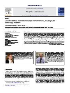
50 nm by controlling the corrosion time in the range of 5 min to 24 h. Intermediate porous structure was quenched by distilled water and residual acid within nanopore channels was removed by water rinsing. The fabricated NPG films were placed onto copper grids for microstructural characterization by a JSM 7000F scanning electron microscope 共SEM兲. The nanopore sizes of NPG were determined by statistically measuring the length scales of the nanopores and gold ligaments using a rotational fast Fourier transform method.17 The concentration of residual silver in the dealloyed NPG films detected by SEM energy dispersive x-ray analysis was smaller than 5 at. %. Ultraviolet-visible 共UV-Vis兲 absorption spectra in the range of 400–850 nm were collected by a JASCO V-650 spectrophotometer with a standard component of 60 mm integrating sphere. During the measurements, the NPG films were sandwiched by two glass slides and solvents were injected into their interstices. The chemical media with variable refractive indices were listed in the following: water 共n = 1.33兲, ethanol 共n = 1.36兲, 3:1 ethanol/toluene 共n = 1.39兲, 1:1 ethanol/toluene 共n = 1.429兲, 1:3 ethanol/toluene 共n = 1.462兲, and toluene 共n = 1.495兲. Scanning rate was 100 nm/ min and the bandwidth was selected as 0.1 nm. A baseline correction procedure was executed in the bare glass without a NPG film covering. Figure 1 shows the representative SEM micrographs of the NPG films with different pore sizes. All specimens exhibit an analogous microstructure in which the hollow channels and gold ligaments look identical in morphology and size. Cross-sectional morphology reveals that nanopores penetrate through the thin films in the lateral direction. The pore sizes can be tuned from 10 to 50 nm by controlling the corrosion time. For example, NPG with a length scale of 10 nm as shown in Fig. 1共a兲 is formed after 5 min dealloying at room temperature. The significant coarsening of the nanopores and ligaments has been observed with long etching time and, consequently, the pore size of ⬃50 nm can be achieved after 24 h etching 关Fig. 1共b兲兴. UV-Vis extinction spectra of the NPG films with pore sizes of 10, 15, 20, 25, 30, 40, and 50 nm soaked into the water are shown in Fig. 1共c兲. Regardless of nanopore sizes, two characteristic resonant bands can be detected in all the NPG samples. The band position at a low wavelength of
98, 093701-1
© 2011 American Institute of Physics
093701-2
Lang et al.
FIG. 1. 共Color online兲 SEM micrographs of NPG synthesized by means of free corrosion for 5 min 共a兲 and 24 h 共b兲. 共c兲 UV-Vis extinction spectra and 共d兲 the resonant peak position of 1 and 2 of NPG with the pore sizes of 10–50 nm in water. For comparison, the dashed line represents the size dependence resonant band of gold nanoparticles.
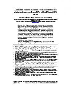
about 490 nm does not change when pore sizes reduce from 50 to 10 nm, whereas the band at a high wavelength shifts from 590 to 545 nm 关Fig. 1共d兲兴. The spectral features of NPG are apparently different from those of other nanostructured gold. For instance, gold nanoparticles are characterized by an isolate SPR band and gold nanorods exhibit one transverse plasmon band at ⬃520 nm.18,19 The low wavelength peak of NPG at 490 nm originates from the resonant absorption of gold films.20 The resonant location mainly relies on film thickness rather than nanopore and ligament sizes. Thus, it is nearly independent of nanopore sizes. In contrast, the high wavelength band, arising from LSPR, represents the significant redshift with the increase in pore sizes, which expectedly results from the effective electron oscillation lengths that are determined by the gold ligament sizes. Metal nanostructures have been developed as plasmonic sensors based on the SPR band shift with dielectric media.13–16 Due to the excellent reproduction of absorption spectra and intense SPR peaks, the NPG films can be utilized as reliable plasmonic sensors. Figures 2共a兲–2共c兲 show the extinction spectra of the NPG films immersed into a series of organic dielectric media. The SPR peak at 490 nm 共1兲 does not show detectable shift when the refractive indices of the media 共n兲 increase from 1.33 to 1.495. In contrast, the SPR band at ⬃540 nm 共2兲 represents evident redshift with the refractive indices. The shift magnitude of 2 has a linear relationship with the refractive index as shown in Fig. 2共d兲, and the sensitivity factor of NPG, defined by the peak shift per refractive index unit 共RIU兲 d2 / dn, can be determined from the slope of the regression line. For the 2 SPR band, the NPG films with larger pore sizes show more significant redshift with the refractive index, resulting in larger sensitivity factors of ⬃210 nm/ RIU and ⬃264 nm/ RIU for 30 nm and 50 nm porous gold, respectively 关see Fig. 2共d兲兴. Thus, the refractive index sensitivity of NPG is comparable to those of other nanostructured gold, such as gold nanorods,19 hollow sphere and cylindrical holes.21–23 It is known that plasmon coupling occurs when the interspace between neigh-
Appl. Phys. Lett. 98, 093701 共2011兲
FIG. 2. 共Color online兲 UV-Vis extinction spectra of porous gold films with 共a兲 10 nm; 共b兲 30 nm, and 共c兲 50 nm are recorded by immersing them into various dielectric environments. Refractive index of these solution increases from left to right: water 共n = 1.33兲, ethanol 共n = 1.36兲, 3:1 ethanol/toluene 共n = 1.39兲, 1:1 ethanol/toluene 共n = 1.429兲, 1:3 ethanol/toluene 共n = 1.462兲, and toluene 共n = 1.495兲. 共d兲 Dependence of resonance 共1, empty symbols兲 and LSPR 共2, solid symbols兲 peaks of NPG films on refractive index.
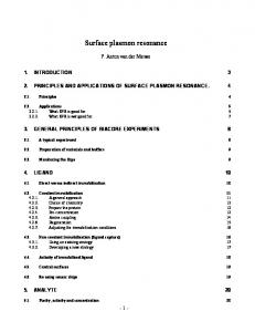
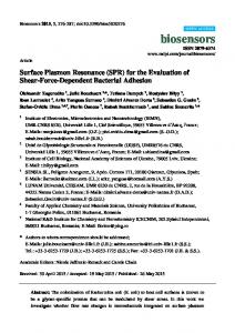
boring nanostructured metals is smaller than 10–20 nm, which leads to the reduction in plasmonic sensitivity. Apparently, the nanopore size dependence of the sensitivity factors of NPG, i.e., sensitivity factors decrease with nanopore sizes, is associated with the increased plasmon coupling between gold ligaments in NPG with a nanopore size smaller than 20 nm. Because of the recognizable dependence of plasmonic sensitivity on the nanopore sizes, the NPG films with different pore sizes give rise to distinct plasmon peak shift even in the same surrounding media 关Figs. 1共c兲 and 1共d兲兴. This can be employed for the application in biosensors for molecule identification by measuring the dependence of the plasmon peak shift with nanopore sizes. Figure 3共a兲 shows an example of the detection of human serum albumin 共HSA兲, in which the 2 peak of the UV/Vis spectra of HSA coated NPG significantly redshift with the increase in nanopore sizes. In contrast, only very limited peak shift can be observed in the extinction spectra of bare NPG films in air and water. Figure 3共b兲 illustrates the linear relationship of plasmon peak positions of NPG films as a function of pore size with the slopes of ⌬ / ⌬d ⬇ 1, 1.4, and 2.8 for the surrounding media of air, water, and HSA, respectively. From the obvious difference in the slopes, we can easily determine the molecules with different dielectric constants. Since the overlapping between
FIG. 3. 共Color online兲 共a兲 UV-Vis extinction spectra and 共b兲 pore size dependence of plasmon peak of bare NPG in air 共dashed lines兲, in water 共short dashed lines兲 and NPG coated with HSA monolayer 共solid lines兲.
093701-3
Appl. Phys. Lett. 98, 093701 共2011兲
Lang et al.
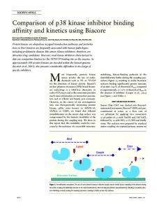
FIG. 4. 共Color online兲 共a兲 DDA simulated SPR spectra of NPG with various nanopore sizes. 共b兲 The relationship between the SPR band of NPG and dielectric index of a surrounding medium.
from 10 to 30 nm, again well consistent with the experimental observations 共Fig. 4兲. In summary, NPG films with 3D bicontinuous nanoporosity possess unique optical response with two characteristic SPR bands in absorption spectra. The resonant peak from LSPR excitation represents remarkable redshift with pore sizes and dielectric media, demonstrating that the NPG films can be utilized as plasmonic sensors. Moreover, the biocompatible NPG with a uniform nanostructure can be fabricated in the form of free-standing films for direct use in SPR devices without the requirement of a complex assembly process.
low and high wavelength bands, the patterns of NPG in air only show one broad band, which looks different from those of the NPG films in other surroundings with large dielectric indices, i.e., the UV-Vis spectra clearly show both low and high wavelength plasmon bands as a result of the redshift in the surface plasmon band 共2兲 while the low wavelength plasmon band 共1兲 remains constant. In view of the obvious changes in the slopes, ⌬ / ⌬d, with the different dielectric surrounding media, the UV-Vis extinction spectra of the NPG with different pore sizes can thus be utilized to detect adsorbed organic and biomolecules by measuring the nanopore size dependence of the plasmon band 共2兲 shift. The surface plasmonic properties of NPG were further investigated by near-field electromagnetic calculations using a discrete dipole approximation.24 A quasi-3D model abstracted from the real structure of NPG measured by electron tomography17 has been developed to describe a simplified local configuration of NPG with the variation in characteristic porous size and ligament length.25 The extinction spectra of this system have been calculated by considering external light incident normal to the top surface of the nanostructure. The peak positions of these spectra are corresponding to the SPR wavelength 2. Due to the nonperiodicity of the nanostructure in the simulation, we cannot obtain the SPR band 1 at ⬃490 nm that comes from the resonance of the gold films. Figure 4共a兲 shows the SPR spectra of simulated NPG with different nanopore sizes 共D兲, in which 2 shifts to red accompanying with the decrease in the intensity of localized electromagnetic fields when the nanopore sizes increase. The trend of peak shift with the characteristic nanopore size is well consistent with the experimental observations 共Fig. 2兲. In order to understand the dependence of 2 on dielectric environments, the extinction spectra are calculated by using a relative refractive index of a medium. The shift magnitude of 2 linearly depends on the refractive index as indicated in Fig. 4共b兲, agreeing with the experiment that the LSPR band, 2, is sensitive to dielectric environments. The refractive index sensitivity of NPG depends on the nanopore sizes and increases from 200 to 270 nm/RIU with the nanopore sizes
E. Ozbay, Science 311, 189 共2006兲. A. J. Forty, Nature 共London兲 282, 597 共1979兲. 3 J. Erlebacher, M. J. Aziz, A. Karma, N. Dimitrov, and K. Sieradzki, Nature 共London兲 410, 450 共2001兲. 4 F. Yu, S. Ahl, A. M. Caminade, J. P. Majoral, W. Knoll, and J. Erlebacher, Anal. Chem. 78, 7346 共2006兲. 5 J. Biener, G. Nyce, A. M. Hodge, M. M. Biener, A. V. Hamza, and S. A. Maier, Adv. Mater. 20, 1211 共2008兲. 6 L. H. Qian, X. Q. Yan, T. Fujita, A. Inoue, and M. W. Chen, Appl. Phys. Lett. 90, 153120 共2007兲. 7 M. C. Dixon, T. A. Daniel, M. Hieda, D. M. Smilgies, M. H. W. Chan, and D. L. Allara, Langmuir 23, 2414 共2007兲. 8 T. Fujita, H. Okada, K. Koyama, K. Watanabe, S. Maekawa, and M. W. Chen, Phys. Rev. Lett. 101, 166601 共2008兲. 9 M. E. Stewart, C. R. Anderton, L. B. Thompson, J. Maria, S. K. Gray, J. A. Rogers, and R. G. Nuzzo, Chem. Rev. 108, 494 共2008兲. 10 K. Raether, Surface Plasmons on Smooth and Rough Surfaces and Gratings 共Springer, Berlin, 1988兲. 11 C. F. Bohren and D. R. Huffman, Absorption and Scattering of Light by Small Particles 共Wiley, New York, 1998兲. 12 S. Link, M. B. Mohamed, and M. A. El-sayed, J. Phys. Chem. B 103, 3073 共1999兲. 13 X. Y. Lang, L. Zhang, P. F. Guan, T. Fujita, and M. W. Chen, Appl. Phys. Lett. 96, 073701 共2010兲. 14 M. D. Malinsky, K. L. Kelly, G. C. Schatz, and R. P. Van Duyne, J. Am. Chem. Soc. 123, 1471 共2001兲. 15 X. Xu and M. B. Cortie, Adv. Funct. Mater. 16, 2170 共2006兲. 16 J. Homola, Surface Plasmon Resonance Based Sensors 共Springer, Berlin, 2006兲. 17 T. Fujita, L. H. Qian, K. Inoke, J. Erlebacher, and M. W. Chen, Appl. Phys. Lett. 92, 251902 共2008兲. 18 T. Okamoto, I. Yamaguchi, and T. Kobayashi, Opt. Lett. 25, 372 共2000兲. 19 S. M. Marinakos, S. H. Chen, and A. Chilkoti, Anal. Chem. 79, 5278 共2007兲. 20 U. Kreibig and M. Vollmer, Optical Properties of Metal Clusters 共Springer, Berlin, 1995兲. 21 A. Dahlin, M. Zäch, T. Rindzevicius, M. Käll, D. S. Sutherland, and F. Höök, J. Am. Chem. Soc. 127, 5043 共2005兲. 22 D. Gao, W. Chen, A. Mulchandani, and J. S. Schultz, Appl. Phys. Lett. 90, 073901 共2007兲. 23 A. Lesuffleur, H. Im, N. C. Lindquist, and S. H. Oh, Appl. Phys. Lett. 90, 243110 共2007兲. 24 B. T. Braine and P. J. Flatau, User guide for the discrete dipole approximation code DDSCAT7.0, http://arxiv.org/abs/0809.0337 25 X. Y. Lang, P. F. Guan, L. Zhang, T. Fujita, and M. W. Chen, J. Phys. Chem. C 113, 10956 共2009兲. 1 2