Abstract: An acutely locked knee is a painful and debilitating orthopaedic ...
surgery is never recommended during pregnancy, arthroscopic knee surgery can
be.
Case Report
Locked Knee During Pregnancy Kyle Flik, M.D., Kyle Anderson, M.D., William Urmey, M.D., and Thomas Wickiewicz, M.D.
Abstract: An acutely locked knee is a painful and debilitating orthopaedic condition usually caused by a loose body or bucket-handle meniscal tear that requires surgery to remove or repair. We describe 2 cases of acute locking of the knee that occurred during pregnancy. Both patients underwent urgent arthroscopic treatment under spinal anesthesia without complication to the patient or fetus. Concerns regarding surgery during pregnancy revolve around the safety for the mother and fetus. Although local and regional anesthetics are safe with proper management, there is a slightly increased risk of spontaneous abortion when general anesthesia is used, especially in the first trimester. Although purely elective surgery is never recommended during pregnancy, arthroscopic knee surgery can be performed safely in emergency situations with proper planning by a coordinated team including the surgeon, anesthesiologist, and obstetrician. Key Words: Locked knee—Pregnancy—Anesthesia— Arthroscopy.
A
n acutely locked knee frequently represents a surgical urgency. However, pregnancy complicates the management of this and any orthopaedic condition, and is an appropriate factor in the decision to delay treatment. Although deferral of an elective surgical procedure in a pregnant woman until she has given birth is a common general recommendation,1,2 there is a void in the orthopaedic literature regarding the specific management of a locked knee in the gravid patient. We report 2 cases of locked knee presenting in different trimesters of pregnancy. Both were treated safely and effectively via prompt arthroscopic surgery under spinal anesthesia.
From the Departments of Sports Medicine and Anesthesia, The Hospital for Special Surgery, New York, New York (K.F., W.U., T.W.); and the William Clay Ford Center for Athletic Medicine, Detroit, Michigan (K.A.), U.S.A. Address correspondence and reprint requests to Kyle Flik, M.D., 310 East 71st St, Apt 4D, New York, NY 10021, U.S.A. E-mail:
[email protected] © 2004 by the Arthroscopy Association of North America 0749-8063/04/2002-3640$30.00/0 doi:10.1016/j.arthro.2003.12.001
CASE 1 A 37-year old woman in her seventh month of a high-risk pregnancy requiring cerclage of the cervix presented with a chief complaint of a locked left knee. She had stood up from the dinner table 2 weeks before when she “felt something snap” in her knee. She was unable to bear weight, and had a painful knee locked in slight flexion since this incident. Her history is significant for trauma 20 years earlier that required reconstructive surgery and aggressive rehabilitation for difficulties with range of motion. Nonetheless, she had been symptom free for the past decade. Physical examination revealed scars about her knee and an effusion. The knee was locked in 30° of flexion, and she had severe pain with any attempt at moving the joint. The ligamentous examination was limited but revealed no gross instability. Following consultation with an anesthesiologist and her obstetrician, the patient underwent arthroscopic removal of a large loose body in the intercondylar region. An isobaric spinal anesthetic was used as well as fetal Doppler monitoring. In addition, the patient’s obstetrician recommended that tocodynamometry be
Arthroscopy: The Journal of Arthroscopic and Related Surgery, Vol 20, No 2 (February), 2004: pp 191-195
191
192
K. FLIK ET AL. able to manipulate the knee to regain motion. This episode caused severe pain. She was seen by another physician and received an intra-articular injection of lidocaine, but was unable to tolerate the pain of an attempted manipulation. On examination, her knee was locked in a slightly flexed position with an effusion and medial joint-line tenderness. No endpoint was obtained on Lachman testing. Radiographs were not ordered. After discussion with her obstetrician, she was scheduled for arthroscopic treatment for a presumed locked buckethandle tear of her medial meniscus. On the following day, a partial medial meniscectomy under isobaric spinal anesthesia was performed for an irreparable medial meniscus bucket-handle tear. She was pain free with full motion of the knee at her first postoperative visit. She delivered a healthy boy at full term via spontaneous vaginal delivery.
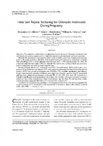
FIGURE 1. Arthroscopic view of loose body found in the intercondylar notch.
DISCUSSION performed. The patient was positioned supine in a slight left-lateral-decubitus position. No tourniquet was used. Arthroscopic evaluation via standard portals revealed severe degenerative osteoarthritis of the medial compartment, mild patellofemoral disease, and absence of the anterior cruciate ligament (ACL) and medial meniscus. A large 3 ⫻ 2 cm loose body, later confirmed by pathologic review to be an osteochondral fragment, was removed piecemeal from the intercondylar notch (Figs 1 and 2). She tolerated the uncomplicated procedure well and spent the immediate postoperative period on the obstetrical floor for monitoring before discharge. At her 2-week postoperative visit, her knee was back to baseline with full and pain-free motion. She delivered a healthy girl 8 weeks after surgery by caesarian section.
Locking implies an intra-articular derangement with a definite lesion within the knee in over 90% of cases.3 The most frequent cause is either a displaced buckethandle tear of the meniscus or a loose body, which lead to painful and limited range of motion of the joint, primarily in extension. The treatment of an acutely locked knee is timely diagnostic and therapeutic arthroscopy.3,4 The arthroscopic treatment for the 2 patients pre-
CASE 2 A healthy ACL-deficient 32 year-old woman in her 16th week of an uncomplicated pregnancy presented with a 24-hour history of a locked and painful right knee. She reported no unusual trauma, and was simply moving her leg when she felt a locking episode take place preventing full extension or flexion. She had a history of minor instability following an old ACL tear, as well as 2 surgical procedures to the right knee for removal of loose cartilage fragments. Although she reported prior locking episodes, she had always been
FIGURE 2.
The loose body being extracted from the notch.
LOCKED KNEE DURING PREGNANCY sented was straightforward. In case 1, the patient had an obvious large osteochondral loose body that was easily removed from the intercondylar notch region. Once this was performed, the knee regained its normal flexion and extension arc of motion. However, the second case presented a more difficult challenge because of the large size of the meniscal tear and the concern about future degenerative changes in the knee that would likely result from its removal. Nonetheless, the meniscus was not suitable for repair based on the poor quality of the tissue and lack of extension into or near the vascularized zone. Simple excision of the torn portion of meniscus provided timely relief without the need for postoperative immobilization. Pregnancy introduces several issues that must be addressed before any surgical procedure. Brodsky et al.2 and Steinberg and Santos1 have studied surgical anesthesia during pregnancy and fetal outcome, and advocate postponing purely elective surgery until the postpartum period and, if possible, deferring necessary surgery until after the first trimester. The urgency of a locked knee creates a unique clinical dilemma for the physician treating a pregnant woman with this painful and debilitating condition. In deciding on the appropriateness of surgical treatment, one must consider the potential risks to the mother and fetus as well as the disadvantages of postponing treatment. It is estimated that between 1.6% and 2.2% of all pregnant women require a surgical procedure unrelated to pregnancy at some time during gestation.1,2 A large study by Duncan et al.5 that included 2,565 women undergoing incidental surgery while pregnant, showed no increase in the rate of congenital anomalies, but a slight increase in rate of spontaneous abortion (7.1% v 6.5% in a matched control group). An earlier study also found an increased incidence of spontaneous abortion after procedures performed under general anesthesia during the first or second trimester.2 Although the teratogenicity of anesthetics has not been clearly elucidated, the critical period of vulnerability of major organ systems is known to be between the second and eighth weeks of gestation. The Collaborative Perinatal Project showed that the administration of local anesthetics, such as benzocaine, procaine, tetracaine, and lidocaine, during pregnancy did not result in an increased rate of fetal malformation.1 Thus, with the exception of cocaine, local anesthetics do not appear to be teratogenic when administered for clinical use. Spinal or epidural anesthesia is safe, but it is im-
193
portant to note that there is a decreasing drug requirement for spinal or epidural anesthesia with advancing gestation because epidural venous engorgement reduces the volume of cerebrospinal fluid and the epidural space.1 In the cases presented, a spinal anesthetic was chosen rather than a local knee block for the following reasons. First, a spinal requires a much smaller quantity of anesthetic, so that any inadvertent intravascular exposure would lead to a less significant dose to the fetus. Second, the greater amount of muscular relaxation afforded by a spinal is often beneficial, especially in cases of locked knee where the intra-articular pathology is unknown. If significant valgus stress to the knee is required during arthroscopy with pressure on the thigh against the lateral post, a local knee block may be insufficient. In addition, supplemental sedation, often required as an adjunct to local blocks, can be avoided with the use of a spinal anesthetic. Finally, if a local block is incomplete, the apprehension and discomfort that follow may lead to other physiologic responses including hypertension, which should be avoided during pregnancy. The left lateral decubitus position should be used during anesthesia in a patient in her second or third trimester to avoid compression of the inferior vena cava. Alterations in uteroplacental blood flow can also be avoided by maintaining adequate maternal mean arterial pressure. Maternal hypotension associated with sympathetic blockade from spinal or epidural anesthesia is a primary concern in that it can result in decreased uterine blood flow. Frequent blood pressure measurements should be obtained during any procedure. Finally, uterine activity should be monitored in the second and third trimesters to guard against spontaneous abortion. Tocodynamometry may be used with tocolytics available if required to arrest premature labor. A summary of the conclusions made in Steinberg’s review of surgical anesthesia during pregnancy is found in Table 1. It is possible that pregnancy itself may be a risk factor for knee injury due to physiologic alterations that occur, including increased body weight, altered proportions, and various hormonal changes. The patients presented in these case reports both had had chronic ACL injuries and therefore an inherent increased risk for instability and meniscal tears. Nonetheless, we surmise that pregnancy predisposed them to further knee injury by increasing both the load across and laxity within their knee joint. Abramson and Wilson6 first documented preg-
194
K. FLIK ET AL. TABLE 1. Surgical Anesthesia During Pregnancy
1. Postpone purely elective surgery until after delivery. 2. If surgery is necessary, delay until after the first trimester whenever possible, as long as the mother’s condition is not jeopardized. 3. Uterine displacement during transport, anesthesia, surgery, and recovery should be routine from the second trimester onward via lateral decubitus positioning. 4. Supplementary oxygen should be administered perioperatively whenever maternal anesthetic exposure may result in desaturation. 5. Avoid hypotension during regional anesthesia. 6. The commonly used local anesthetics are all safe. 7. Monitoring techniques such as fetal heart rate should be employed whenever feasible, as variability is a good indicator of fetal well-being.
nancy-related changes in joint laxity in 1934 regarding the well-known laxity in the pubic symphysis. Three subsequent studies7-9 support the notion that laxity of peripheral joints increases during pregnancy as a result of an increased level of the hormone relaxin. Eddie et al.10 and O’Byrne et al.11 reported that relaxin levels increased early in the first trimester and slowly decreased in the second and third trimesters. Dumas et al.12 support this in their study of knee laxity during pregnancy. They state that “laxity reaches a constant level before the fifth month of pregnancy, which suggests that laxity increases early in pregnancy.” Sciore et al.13 studied sex hormone receptors and found estrogen and progesterone receptor transcripts expressed in ligament tissue of male and female rabbits and humans. They reported that alterations in expression of the receptors occur in ligaments during pregnancy. A study by Schauberger et al.14 found definite increase in laxity in 5 of 7 peripheral joints over the course of pregnancy and the postpartum period. The exact role of relaxin and estrogen during pregnancy with regard to joint laxity and stability remains unclear. Nonetheless, it is seems reasonable to conclude that increased joint laxity combined with the weight gain during pregnancy may increase the risk for knee injuries and episodes of locked knee in the gravid patient. An investigation of 50 patients with locked knees by examination under anesthesia and diagnostic arthroscopy to investigate the causes of an acutely locked knee found that 42% of patients were involved in a sport when locking occurred and 42% reported a prior locking episode. A lesion of the meniscus was present in 68% and loose bodies were discovered in 6%.3 Other reported causes of locking include partial ACL tears,3,15 degenerative changes,3 superior dislocation of the patella,16,17 combined tear of the PCL and MCL,18 intra-articular tumor, and idiopathic locking.3 Allum and Jones reported an 8% incidence of locking without an identifiable cause.3
We recommend timely arthroscopic evaluation and treatment with the assistance of an experienced anesthesiologist if a pregnant woman presents with a locked knee. Radiographs are not crucial and should be avoided if possible in pregnant women. A coordinated, multidisciplinary approach involving the orthopaedic surgeon, anesthesiologist, and obstetrician is important for optimal outcome with lowest risk for both the patient and her fetus. Fetal monitoring should be employed when possible, and postoperative evaluation by an obstetrician is recommended. REFERENCES 1. Steinberg ES, Santos AC. Surgical anesthesia during pregnancy. Int Anesthesiol Clin 1990;28:58-66. 2. Brodsky JB, Cohen EN, Brown BW Jr, et al. Surgery during pregnancy and fetal outcome. Am J Obstet Gynecol 1980;138: 1165-1167. 3. Allum RL, Jones JR. The locked knee. Injury 1986;17:256258. 4. Critchley IJ, Bracey DJ. The acutely locked knee—Is a manipulation worthwhile? Injury 1985;16:281-283. 5. Duncan PG, Pope WD, Cohen MM, Greer N. Fetal risk of anesthesia and surgery during pregnancy. Anesthesiology 1986;64:790-794. 6. Abramson DRS, Wilson PD. Relaxation of the pelvic joints in pregnancy. Surg Gynecol Obstet 1934;58:595-613. 7. Ostgaard HC, Andersson GB, Schultz AB, Miller JA. Influence of some biomechanical factors on low-back pain in pregnancy. Spine 1993;18:61-65. 8. Calguneri M, Bird HA, Wright V. Changes in joint laxity occurring during pregnancy. Ann Rheum Dis 1982;41:126128. 9. Block RA, Hess LA, Timpano EV, Serlo C. Physiologic changes in the foot during pregnancy. J Am Podiatr Med Assoc 1985;75:297-299. 10. Eddie LW, Bell RJ, Lester A, et al. Radioimmunoassay of relaxin in pregnancy with an analogue of human relaxin. Lancet 1986;1:1344-1346. 11. O’Byrne EM, Carriere BT, Sorensen L, et al. Plasma immunoreactive relaxin levels in pregnant and nonpregnant women. J Clin Endocrinol Metab 1978;47:1106-1110. 12. Dumas GA, Reid JG, Wolfe LA, et al. Laxity of the knee cruciate ligaments during pregnancy. J Orthop Sports Phys Ther 1997;26:2-6. 13. Sciore P, Frank CB, Hart DA. Identification of sex hormone receptors in human and rabbit ligaments of the knee by reverse
LOCKED KNEE DURING PREGNANCY transcription-polymerase chain reaction: Evidence that receptors are present in tissue from both male and female subjects. J Orthop Res 1998;16:604-610. 14. Schauberger CW, Rooney BL, Goldsmith L, et al. Peripheral joint laxity increases in pregnancy but does not correlate with serum relaxin levels. Am J Obstet Gynecol 1996;174:667-671. 15. Chun CH, Lee BC, Yang JH. Extension block secondary to partial anterior cruciate ligament tear on the femoral attachment of the posterolateral bundle. Arthroscopy 2002;18:227-231.
195
16. Garner JP, Pike JM, George CD. Intra-articular dislocation of the patella: Two cases and literature review. J Trauma 1999; 47:780-783. 17. McWilliams TG, Binns MS. A locked knee in extension: A complication of a degenerate knee with patella alta. J Bone Joint Surg Br 2000;82:890. 18. MacDonald PB. Combined tear of the posterior cruciate and medial collateral ligaments resulting in a locked knee. Arthroscopy 1997;13:639-640.