STEM CELLS AND DEVELOPMENT Volume 22, Number 17, 2013 ! Mary Ann Liebert, Inc. DOI: 10.1089/scd.2012.0717
Low-Level Shear Stress Induces Human Mesenchymal Stem Cell Migration Through the SDF-1/CXCR4 Axis Via MAPK Signaling Pathways Lin Yuan,1,2 Naoya Sakamoto,3 Guanbin Song,1 and Masaaki Sato 2
Mesenchymal stem cells (MSCs) are able to home and migrate into damaged tissues and are thus, considered an optimal therapeutic strategy for clinical use. We previously demonstrated that higher shear stress ( > 2 Pa) hindered human MSC (hMSC) migration, whereas lower shear stress (0.2 Pa) induced cell migration through mitogen-activated protein kinase (MAPK) pathways. Here the mechanisms underlying shear stress-induced hMSC migration have been studied further. An MSC monolayer was mechanically wounded and subsequently exposed to low-level shear stress of 0.2 Pa. Image analysis was performed to quantify cell migration speeds under both flow and static conditions. hMSCs along both upstream- and downstream edges of the wound migrated at a similar speed to cover the wounded area under static conditions, whereas shear stress induced cells along the downstream edge of the wound to migrate significantly faster than those along the upstream edge. We also found that shear stress upregulated the secretion of stromal-derived factor-1 (SDF-1), which stimulated its receptor CXCR4 expression in hMSCs until the cells covered the wounded area. A CXCR4 antagonist repressed both cell migration and activation of c-Jun N-terminal kinase ( JNK) and p38 MAPK but did not affect extracellular signal-regulated kinase 1/2 (ERK1/2) phosphorylation. When MAPK activation in upstream- and downstream hMSCs was evaluated separately, ERK1/2 was activated earlier in downstream than in upstream cells. These results indicate that the SDF-1/CXCR4 axis mediates shear stress-induced hMSC migration through JNK and p38 MAPK pathways and that the difference in migration speeds between upstream- and downstream cells may be due to ERK1/2 activation.
Introduction
M
esenchymal stem cells (MSCs) exhibit the key properties of self-renewal and giving rise to cells of diverse lineages that can be isolated from many different adult tissues, including bone marrow, adipose tissue, liver, and muscle, as well as amniotic fluid and umbilical cord blood [1]. Concomitant with their convenient isolation and the lack of significant immunogenicity, MSCs have become the focus of emerging therapeutics to regenerate damaged tissues and treat inflammation resulting from sepsis, acute renal failure, cardiovascular disease, and myocardial infarction [2]. However, a significant barrier to the effective implementation of MSC therapy in clinical settings is the inability to target the homing or migration of these cells into tissues of interest with high efficiency. MSC homing is defined as the arrest of MSCs within a tissue vasculature followed by their transmigration across
the endothelium [3]. Our previous study demonstrated that shear stress, one of the important mechanical stimuli in the vasculature, can influence MSC migration through mitogenactivated protein kinase (MAPK) signaling pathways [4]. The precise mechanism by which a physical force is converted into a chemical signal in these cells is not clear. However, it appears to depend on physical structures, such as cytoskeletal elements and ion channels, or on specific sensory molecules, including growth factors, cytokines, and their receptors [5]. In the latter mechanism, also referred to as an autocrine signaling mechanism, autologous chemotaxis arises in a flow field where convection distributes autocrine factors creating a chemokine gradient, and then provides a chemotactic signal [6]. Previous studies confirmed that chemokines or growth factors act as migratory cues for MSC trafficking to an injured region [3] and that MSCs themselves express several chemokines and their receptors for regulation of cell migration [7–10]. Thus, we hypothesized that a
1 Key Laboratory of Biorheological Science and Technology, Ministry of Education, College of Bioengineering, Chongqing University, Chongqing, China. 2 Graduate School of Biomedical Engineering, Tohoku University, Sendai, Japan. 3 Department of Medical Engineering, Kawasaki University of Medical Welfare, Okayama, Japan.
2384
SHEAR STRESS-INDUCED MSC MIGRATION VIA SDF-1/CXCR4 AXIS possible mechanism involved in shear stress-induced MSC migration may be related to shear-regulated increase in factors as an autocrine mechanism, triggering chemoattractant activities, and leading to MAPK activation. The chemokine stromal cell-derived factor-1 (SDF-1), also known as CXCL12, is well known to play roles in organogenesis, hematopoiesis, and immune responses through its binding to the chemokine receptor CXCR4 [11,12]. A previous study showed that SDF-1 was expressed along the bronchoalveolar lavage of rat ventilated with an injurious tidal volume, and that is facilitates migration of epithelial cells to cover the denuded surface [13]. Flow cytometry and video monitoring analyses showed that the interactions between SDF-1 and CXCR4 mediated the trafficking of multiple myeloma cells homing in vivo [14]. Most importantly, Hattori et al. reported that the SDF-1/CXCR4 axis plays critical roles in both engraftment of hematopoietic stem cells into bone marrow and the mobilization of stem cells from bone marrow niches into the peripheral circulation, where these cells are then available for delivery to peripheral injury site [15]. However, the role of SDF-1/CXCR4 in shear stressinduced MSC migration has not yet been investigated. In the present study, we further clarify the mechanism of hMSC migration, which is enhanced by low-level shear stress, and determine whether SDF-1/CXCR4 plays roles in this process and the relationship between the SDF-1/CXCR4 axis and MAPK activations.
Materials and Methods Cell culture Bone marrow-derived human MSCs (hMSCs) were purchased from Lonza (Lonza). These cells were positive for CD29, CD44, CD105, and CD166, and negative for markers of the hematopoietic lineage, such as CD14, CD34, and CD45 by flow cytometric analysis. Moreover, these cells were capable of differentiation into mesodermal lineage cells, including osteoblasts, chondrocytes, and adipocytes under appropriate conditions. hMSCs were cultured in Dulbecco’s modified Eagle’s medium (Invitrogen) supplemented with 10% heat-inactivated fetal bovine serum ( JRH Bioscience), 100 U/mL penicillin, 100 mg/mL streptomycin, and 10 ng/ mL human bFGF (Austral Biologicals) at 37"C in a humidified incubator (Thermo Scientific) with 5% CO2. The culture medium was replenished two or three times a week, and subculture was performed by digestion with 0.05% trypsin/ 0.02% ethylenediaminetetraacetic acid (EDTA) when the cells were almost confluent. For the experiments, hMSCs at passages 4–8 were placed on glass-based dishes (j35; Asahi Techno Glass) coated with 0.1% gelatin.
Wound-healing assay Cell migration was measured using our previously described methods [4]. An hMSC monolayer was wounded using a 200-mm-thick plastic cell scraper (Corning) and observed under a microscope (IX81; Olympus) equipped with a 3CCD camera (ORCA-3CCD; Hamamatsu Photonics). Images of the wound under the static or flow condition were acquired every 10 min for 12 h using AquaCosmos software (Version 2.6). ImageJ software was used to determine cell migration speed expressed as migration distance/time (mm/min).
2385
Flow exposure experiment hMSCs were subjected to a shear stress of 0.2 Pa using a parallel flow chamber system as described previously [4]. In brief, the flow system was composed of a flow chamber, two collecting reservoirs, which were kept in a 37"C water bath, and a peristaltic flow pump (Masterflex) to circulate a perfusion culture medium. Because serum can influence cell migration to a great extent, the culture medium used for flow exposure experiments was serum-free but contained insulin transferring selenium-X supplement (Invitrogen). Control cells were maintained under static conditions (37"C in a humidified atmosphere 5% CO2) for the same length of time.
Sodium dodecyl sulfate-polyacrylamide gel electrophoresis and western blot analysis Cells at upstream- and downstream of a wound edge (Fig. 1A) were washed with ice-cold phosphate-buffered saline and separately scraped under a microscope immediately after exposure to laminar shear stress at scheduled time points. Proteins were extracted using cell lysis buffer [20 mM Tris– HCl (pH 7.2), 1 mM EDTA, 1 mM ethylene glycol tetraacetic acid, 1% NP-40, 0.5% sodium dodecyl sulfate (SDS) and 10 mM MgCl2] supplemented with phenylmethylsulfonyl fluoride and phosphatase inhibitor cocktails I and II (SigmaAldrich). Protein concentration was determined using a bicinchoninic acid protein assay (BCA method; Bio-Rad). After separation by 10% SDS–polyacrylamide gel electrophoresis, proteins were electroblotted onto polyvinylidene fluoride membranes (GE Healthcare). The membranes were blocked with Tris-buffered saline containing 0.1% Tween-20 (TBST) and 5% bovine serum albumin for 1 h at room temperature. Antibodies against ERK1/2, phospho-ERK1/2 (p-ERK1/2), JNK, phosphor-JNK (p-JNK), p38 MAPK, phosphor-p38 MAPK (p-p38 MAPK) (Cell Signaling Technology), and CXCR4 (Abcam) and a rabbit monoclonal antibody against b-actin (Cell Signaling Technology) were used according to the manufacturer’s protocols. The membrane was incubated overnight with these antibodies at 4"C with slight shaking. Next, the membranes were washed in TBST buffer and further incubated with a horseradish peroxidase-conjugated antibody (goat anti-rabbit IgG; SigmaAldrich) for 1 h at room temperature. To visualize the protein bands, an enhanced electrogenerated chemiluminescence system (Amersham) was used. A semi-quantitative evaluation of the band intensities was performed by densitometry. The levels of ERK1/2, p-ERK1/2, JNK, p-JNK, p38 MAPK, p-p38 MAPK, and CXCR4 proteins were determined by normalizing to those of the housekeeping protein b-actin.
Inhibitor experiment Inhibitors for CXCR4 (AMD3100), JNK (SP600125) (SigmaAldrich), p38 MAPK (SB203580), or ERK1/2 (U0126) (Cell Signaling Technology) were dissolved in dimethyl sulfoxide, aliquoted, and stored at - 20"C. These inhibitors were diluted immediately before use in the cell culture medium and applied to hMSCs at a final concentration of 20 mM 2 h prior to shear stress.
2386
YUAN ET AL.
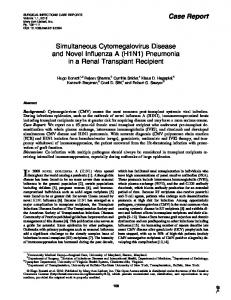
FIG. 1. (A) A side view schematic diagram that illustrates upstream- and downstream human mesenchymal stem cells (hMSCs) stimulated by shear stress. (B) Migration speed of hMSCs along both wound edges under static conditions (control) and when exposed to a shear stress of 0.2 Pa. Data are expressed as mean – SD (n = 6); *P < 0.05.
SDF-1 quantification using ELISA
SDF-1 secretion under shear stress
hMSCs were exposed to shear stress for 3, 6, 12, or 24 h with a subsequent static culture for 24 h. SDF-1 concentration in the cell culture supernatant was determined using a quantitative sandwich ELISA kit (R&D Systems, Inc.), according to the manufacturer’s instructions. The samples were evaluated using an ELISA plate reader at 450 nm with correction at 570 nm.
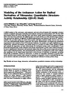
To investigate whether SDF-1 secretion increased with shear stress, we measured the amount of SDF-1 released by shear-stressed cells into the culture medium. As shown in Fig. 2, after applying a shear stress of 0.2 Pa for 3 h with subsequent static culture for 24 h, there was a significant increase in SDF-1 secretion (1.00 – 0.34 ng/mL), which was approximately 3.3-fold higher than that in the control group (P < 0.01). Although the amount of SDF-1 released decreased slightly as the duration of shear stress prolonged to 6 or 12 h, there was still a significant difference compared with the
Statistical analysis The results were analyzed using Student’s t test and analysis of variance (ANOVA). Bonferroni post hoc tests were used when the P-value (by ANOVA) indicated a statistically significant difference among groups. Data were expressed as mean – SD. P < 0.05 was considered statistically significant.
Results Effects of shear stress on hMSC migration speeds The migration speeds of hMSCs under static conditions or when exposed to a shear stress of 0.2 Pa are shown in Fig. 1B. In the absence of flow (control), no significant difference in the migration speed was observed between the cells along the upstream- and downstream edges of the wound (upstream: 0.17 – 0.06 mm/min; downstream: 0.14 – 0.08 mm/ min). In contrast, when stimulated by shear stress, cells along the downstream edge migrated significantly faster than those along the upstream edge (upstream: 0.20 – 0.1 mm/min; downstream: 0.39 – 0.1 mm/min; P < 0.05). Interestingly, the average migration speed of cells along the upstream edge in response to shear stress was not greater than that under static conditions. These results suggest that shear stress influences the migration patterns of hMSCs and cells along the downstream edge contribute more to wound closure under shear stress conditions than the cells along the upstream edge.
FIG. 2. Effect of shear stress on stromal-derived factor-1 (SDF-1) production. hMSCs were cultured under static conditions (control) or exposed to a shear stress (SS) of 0.2 Pa for 3, 6, 12, or 24 h with subsequent culture for 24 h. The amount of SDF-1 released was measured by ELISA. Data are expressed as mean – SD (n = 6); *P < 0.05, **P < 0.01 versus control.
SHEAR STRESS-INDUCED MSC MIGRATION VIA SDF-1/CXCR4 AXIS control group (6 h: 0.89 – 0.25 ng/mL and 12 h: 0.75 – 0.25 ng/ mL, respectively; P < 0.05).
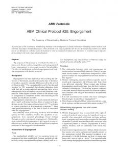
Shear stress enhances CXCR4 expression in hMSCs We examined CXCR4 expression using western blot analysis. Shear stress applied for 3 h increased CXCR4 expression in both upstream- and downstream cells, which showed 1.23 and 1.19-folds, respectively, higher compared with the control group (P < 0.05) (Fig. 3A, B). CXCR4 expression continued to increase significantly as the duration of shear stress prolonged and reached a peak at 6 h, which showed 1.27 (upstream) and 1.49 (downstream) folds compared with the control group (P < 0.05). However, CXCR4 expression decreased to the level of control 12 h after wounding, and no significant differences were observed between the upstream- and downstream cells at all detected time points.
CXCR4 regulates shear stress-induced hMSC migration and MAPK activation To further determine whether CXCR4 has a functional role in shear stress-induced hMSC migration and to clarify the relationship between CXCR4 expression and MAPK activation, we examined the effect of AMD3100, a specific inhibitor
FIG. 3. Detection of CXCR4 expressions by western blot analysis. hMSCs were cultured under static conditions or exposed to a SS of 0.2 Pa for 3, 6, 12, and 24 h. (A) Representative bands showing SS-induced CXCR4 expressions of in upstream (U-CXCR4) and downstream (D-CXCR4) cells. (B) Results of densitometric analysis of CXCR4 expression normalized to b-actin intensities. The dash line at 1.0 value depicts CXCR4 expression of the control group. Data are expressed as mean – SD (n = 6); *P < 0.05.
2387
of CXCR4 activity, on shear stress-induced hMSC migration. Both upstream- and downstream cells treated with this inhibitor decreased CXCR4 expression, which had been enhanced by low-level shear stress, to 0.91 (upstream) and 1.13 (downstream) folds compared with the static control group at 3 h, and continuously decreased to 0.78 (upstream) and 0.99 (downstream) folds at 6 h (Fig. 4A). Based on the usage of inhibitor-containing media for flow exposure experiments, we found that AMD3100 treatment partially delayed wound closure and the cells closed only 49% of the original wound at 6 h (Fig. 4B). Moreover, activation of JNK and p38 MAPK, which are activated by shear stress as proved in our previous study [4], were abolished by the CXCR4 inhibitor (Fig. 4C, D). However, ERK1/2 phosphorylation was not affected by AMD3100, which increased at 60 min followed by a slight decrease. The phosphorylation of ERK1/2 was still significantly different at 120 min compared with the control group (P < 0.05) (Fig. 4E). These results indicate that CXCR4 mediates shear stress-induced hMSC migration through the JNK and p38 MAPK signaling pathways but does not mediate ERK activation.
MAPK expression in upstreamand downstream hMSCs To understand the mechanism by which downstream cells migrated faster than upstream cells, we examined ERK1/2, JNK, and p38 MAPK phosphorylation after exposure to shear stress for 15, 30, 60, and 120 min in upstream- and downstream hMSCs separately. As shown in Fig. 5A and B, ERK1/2 phosphorylation occurred more rapidly in downstream cells than in upstream cells, which reached a plateau at 60 min and was 2.3-fold higher than the static control (P < 0.05). In particular, a significant difference in ERK1/2 activation between downstream- and upstream cells could be observed at 60 min (P < 0.05). Similar to ERK1/2 phosphorylation, JNK activation in upstream cells was not detected until 120 min (Fig. 5C, D). JNK phosphorylation in downstream cells showed a tendency to be higher compared with the upstream cells at 60 min, although this was not significantly different. Following 30 min of shear stress, p38 MAPK phosphorylation in upstream cells occurred simultaneously with the phosphorylation in downstream cells (compared to control, 2.5-fold higher in upstream versus 1.9fold higher in downstream; P < 0.05). However, p38 MAPK activity in upstream cells decreased rapidly and there are no statistically significant difference at 60 min and 120 min compared to the control (Fig. 5E, F). Further, hMSCs were pretreated with U0126 (an inhibitor of ERK1/2), SP600125 (an inhibitor of JNK), or SB203580 (an inhibitor of p38 MAPK). The average migration speeds at both wound edges were analyzed (Fig. 5G). After treatment with ERK1/2 or p38 MAPK inhibitor, the migration speed of downstream cells decreased dramatically to 0.2 – 0.07 mm/min (P < 0.05) and 0.05 – 0.03 mm/min (P < 0.01), respectively, compared with that of cells without inhibitor treatment. The migration speed of upstream cells was not affected by the ERK1/2 or p38 MAPK inhibitor. However, the migration speeds of both upstream- and downstream cells were significantly lower after treatment with the JNK inhibitor. Thus, these results further demonstrate that the contribution of MAPK differentially affects hMSC migration at each side of a wound area
2388 and that the kinetics of activation of the MAPK signal transduction pathway plays important roles in shear stressinduced hMSC migration.
Discussion Stem cell migration is not only a key component of normal homeostasis within an in vivo environment but is also crucial for wound healing and tissue regeneration processes [16]. Numerous studies have confirmed that systemically infused MSCs can migrate to injured, inflamed tissues, and exert therapeutic effects [17,18]. However, the mechanisms that regulate MSC trafficking after intravenous, intra-arterial, or
YUAN ET AL. local intratissue application remain unclear. Our previous study demonstrated that wound closure of hMSCs under flow is critically sensitive to shear stress levels; lower shear stress (0.2 Pa) induced hMSC migration through MAPK pathways, whereas greater shear stress ( > 2 Pa) hindered cell migration notably [4]. In the present study, we also found that unlike endothelial cells (ECs) [19] and trophoblast cells [20], which migrate faster in the direction of flow, hMSC migration was more significant along the downstream edge of the wound than along the upstream edge. Coincidentally, when stimulated by interstitial flow, breast cancer cells that migrated upstream moved faster, whereas those that migrated in the direction of flow moved at slower speeds but
FIG. 4. Effects of a CXCR4 inhibitor (AMD3100) on SS-induced hMSC migration and mitogen-activated protein kinase (MAPK) activation. (A) Representative bands for SS-induced CXCR4 expressions in upstream (U-CXCR4) and downstream (D-CXCR4) cells after treatment with 20 mM AMD3100. The dash line at 1.0 value depicts CXCR4 expression of the control group. (B) Representative micrographs of wound closure and time course of changes in the wound closure rate under a SS of 0.2 Pa in hMSCs treated with the CXCR4 inhibitor AMD3100. (C2E) Effects of SS on MAPK activation in hMSCs exposed to a SS of 0.2 Pa for 15, 30, 60, and 120 min in the presence of AMD3100. P: positive control (representative western blotting bands of strongest phosphorylation of JNK, p38 MAPK, or ERK1/2 detected in hMSCs were exposed to SS of 0.2 Pa without AMD3100). Representative bands showing the effects of the CXCR4 inhibitor on the expression of (C) p-JNK and total-JNK (tJNK), (D) p-p38 MAPK and total-p38 MAPK (t-p38 MAPK), (E) p-ERK1/2 and total-ERK1/2 (t-ERK1/2). Results of densitometric analysis of CXCR4, JNK, p38 MAPK, and ERK1/2 protein expression normalized to b-actin expression. Data are expressed as mean – SD (n = 4); *P < 0.05, **P < 0.01.
SHEAR STRESS-INDUCED MSC MIGRATION VIA SDF-1/CXCR4 AXIS
FIG. 4.
2389
(Continued).
with higher directedness [21]. The same phenomenon was also observed during the migration of other types of tumor cells. Polacheck et al. demonstrated that tumor cells moved in the opposite direction of flow depending on cell density, interstitial flow rate, receptor activity, and focal adhesion kinase (FAK) phosphorylation [6]. Interestingly, in our study, the migration speed of hMSCs along the upstream edge in response to shear stress was similar to that under static conditions. This was also reported by Gojova et al. who used an endothelial migration model [19]. The reason for this phenomenon remains unknown and warrants investigation. It is well documented that autologously secreted chemokines can form local pericellular diffusion gradients scattered by fluid convection, and cells subsequently chemotaxis up the flow-directed gradient [22,23]. Among several types of cytokine/receptor interactions, the SDF-1/CXCR4 axis plays an important role in the recruitment of MSCs into injured tissues [24,25]. Using a Boyden chamber assay, Yu et al. provided compelling evidence that CXCR4 overexpression effectively enhances MSC migration toward SDF-1 [26]. Son et al. also reported that CXCR4 expression not only facili-
tated the attraction of MSCs toward SDF-1 in a concentration-dependent manner but also increased the recruitment of cells to injured sites [27]. However, it is worthwhile to note that chemokine receptors and their ligands are highly promiscuous and able to bind multiple receptor/ligands. Several studies mentioned that besides SDF-1, macrophage migration-inhibitory factor (MIF) competed with cognate ligands for CXCR4, but this chemokine prefers to bind to CXCR2 and CD74 directly [28–30]. Moreover, O’Connor group demonstrated that partly through CD74, MIF inhibited MSC migration and might impair the ability of MSCs to home to injured tissues [31–33]. Thus, we do not think MIF is the ligand which binds to CXCR4 that mediates shear stress induced hMSC migration. On the other hand, recent studies by Saini and colleagues provided evidence to suggest that ubiquitin has an ability to activate CXCR4 [34–35]. However, since we hypothesized that a possible mechanism involved in shear stress-induced hMSC migration may be related to shear-regulated increase in factors as an autocrine mechanism, triggering chemoattractant activities, and Saini et al. demonstrated that ubiquitin possessed weaker
2390
YUAN ET AL.
FIG. 5. Effects of SS on the activation of MAPK proteins in hMSCs along the upstream- and downstream edges of the wound. hMSCs were exposed to a SS of 0.2 Pa for 15, 30, 60, and 120 min. (A, C, E) Representative bands showing SS-induced expression of (A) p-ERK1/2 and t-ERK1/2 in upstream (U-p-ERK1/2; U-t-ERK1/2) and downstream (D-p-ERK1/2; D-tERK1/2) cells, (C) p-JNK and t-JNK in upstream (U-p-JNK; U-t-JNK) and downstream (D-p-JNK; D-t-JNK) cells, and (E) pp38 MAPK and p38 MAPK in upstream (U-p-p38; U-p38) and downstream (D-p-p38; D-p38) cells. (B, D, F) Results of densitometric analysis of ERK1/2, JNK and p38 MAPK activation normalized to b-actin activation. (G) Migration speeds of MSCs along both wound edges under SS without inhibitors or treated with SP600125 (an inhibitor of JNK), SB203580 (an inhibitor of p38 MAPK), or U0126 (an inhibitor of ERK1/2). Data are expressed as mean – SD (n = 4); *P < 0.05, **P < 0.01.
chemotactic activity than SDF-1 in cell migration assay [35], we chose to study the effect of shear stress on the only cognate CXCR4 ligand SDF-1 secretion. Furthermore, CXCR4 has been shown to be ubiquitinated after bonded with ubiquitin, sorted to the lysosome, and degraded [36], but there was no significant difference in CXCR4 expressions between the experimental group and the control group after
hMSCs were exposed to shear stress for 12 or 24 h in our present study, which may indicate that SDF-1, other than ubiquitin, is the ligand bonded with CXCR4 and mediated shear stress induced hMSC migration. In our study, shear stress upregulated CXCR4 expression from 3 to 6 h both in upstream- and downstream cells, followed by a decrease to the control level at 12 h. Although we
SHEAR STRESS-INDUCED MSC MIGRATION VIA SDF-1/CXCR4 AXIS confirmed by immunofluorescence that expression of CXCR4 was induced not only in hMSCs at the edge of the wound but also in cells distant to the wound (data not shown). The reason may be related to wound closure that was completed at approximately 6-h recording period in our migration system. This suggested that hMSC migration and CXCR4 expression are interrelated and interactional. To demonstrate this, an hMSC monolayer was wounded using a wider scraper (approximately 1 mm) and then subjected to shear stress. We found that cells could not cover the wound area 24 h after wounding, but persistent and high CXCR4 expressions was observed both in upstream- and downstream cells compared with the control group (data not shown). Interestingly, we also observed that SDF-1 production by hMSCs decreased as the cells along both wound edges reached confluence. This indicates that a positive/negative control mechanism may exist to regulate cell biological processes. In this case, shear stress promoted hMSC migration through an upregulated SDF-1/CXCR4 axis, whereas after covering the wound area, the cell migration ability was reduced and further produced a ‘‘feedback signal’’ to reduce SDF-1 productions as well as CXCR4 expressions. Numerous studies have reported a direct role of SDF-1 in triggering CXCR4 activation [37,38]. Thus, we speculate that reduced CXCR4 expression is accounted for by decreased SDF-1 production in our migration system. We showed that shear stress-induced hMSC migration involved up-regulation of the expression of the SDF-1 receptor CXCR4, although no significant difference in CXCR4 expression was observed in upstream- and downstream cells. This indicated that migration along chemokine gradients was not the direct reason that caused downstream cells to migrate faster than upstream cells. After treatment with the ERK1/2, p38 MAPK, or JNK inhibitor, varying hMSC migration patterns were observed depending on the inhibitor used. In particular, the inhibition of either JNK or p38 MAPK almost completely blocked hMSC migration. This may indicate that JNK and p38 MAPK were involved in the same signaling cascade, with one being an upstream activator of the other. On the other hand, the JNK inhibitor retarded cell migration along both wound edges, and no difference in migration speed was observed between upstream- and downstream cells. The ERK1/2 and p38 MAPK inhibitors decreased only downstream cell migration and not upstream cell migration, which may explain that hMSC migration, was more significant along the downstream edge of a wound because of ERK1/2 or p38 MAPK activation. However, signal transduction pathways is a complex network involving a number of signaling molecules which may interact with each other to regulate cell biological process. The relationship among JNK, ERK1/2, and p38 MAPK are still unclear and need to be clarified in the further work. The precise mechanisms by which cells sense a shear stress signal and move against the direction of flow remain unclear. One possibility is that in addition to SDF-1, shear stress upregulates the expression of other growth factors [39,40] or chemokines [41], which mainly activate a downstream p38 MAPK or ERK1/2 signaling pathway. Alternatively, the cue may be provided by mechanical force, particularly shear stress-induced force acting on the leading edge of downstream cells that have no neighboring cells upstream. Recent work has demonstrated that the formation of focal adhesions
2391
(FAs) can be promoted by a flow-induced force [42,43] and the formation of new FAs is a key event in the regulation of cytoskeletal reorganization, cellular adhesion, survival, and migration [44,45]. Polacheck et al. proved that increased FAK activation is due to flow-induced stress gradients, which lead to FAs activation as well as guide tumor cell migration toward an upstream edge [6]. Li et al. also showed the directional migration of ECs in response to mechanical force and supposed that shear stress may cause FA aggregation along the direction of flow [43]. With regard to shear stress-induced hMSC migration, the role of FAK has not yet been studied. However, Lee et al. demonstrated that during hypoxia-stimulated MSC migration, FAK was involved in signal transduction pathways associated with the transient and early activation of MAPK [46]. Further studies are necessary to determine whether a shear stress-induced force may increase FAK activation and whether the activated FAK along with a signaling cascade plays a crucial role in downstream cells migrating upstream.
Conclusion This study showed that two types of signaling pathways may be involved in low-level shear stress-induced hMSC migration. One is the SDF-1/CXCR4 - JNK/p38 MAPK pathway and the other is an unknown growth factor/chemokine - ERK1/2 signaling pathway. These findings are briefly summarized in Fig. 6. We also demonstrated that shear stress induced cells along the downstream edge of a wound to migrate significantly faster than those along the upstream edge, which may involve p38 MAPK and ERK1/2 signaling molecules. However, much remains to be elucidated regarding the specific mechanisms involved in downstream cells migrating faster than upstream cells. Further studies on the effects of tension stress on downstream cell migration will undoubtedly provide additional insights
FIG. 6. Schematic representation of the signaling pathways affected by SS causing hMSC migration. On stimulation with a SS of 0.2 Pa, MSCs secrete SDF-1 and have increased CXCR4 expression, leading to the activation of two signaling pathways. One leads to JNK phosphorylation, which affects both upstream- and downstream cell migration, and the other leads to p38 MAPK activation, which may regulate downstream cell migration. In addition to SDF-1, secretion of other cytokines or growth factors by hMSCs activates ERK1/ 2 phosphorylation, which affects downstream migration of hMSCs.
2392 into the effects of the physical microenvironment on cell biological reactions and offer a rational basis for developing new stem cell-based therapeutic strategies in tissue engineering and tissue repair.
YUAN ET AL.
12.
Acknowledgments This study was supported in part by Grants-in-Aid for Scientific Research (Specially Promoted Research) from the Japan Society for the Promotion of Science (no. 20001007), National Natural Science Foundation of China (nos. 30770530, 11032012 and 11102240), and Fundamental Research Funds for the Central Universities (CDJXS11232243).
13.
14.
Author Disclosure Statement No competing financial interests exist.
15.
References 1. Salem HK and Thiemermann C. (2010). Mesenchymal stromal cells: current understanding and clinical status. Stem Cells 28:585–596. 2. Phinney DG and Prockop DJ. (2007). Concise review: mesenchymal stem/multipotent stromal cells: the state of transdifferentiation and modes of tissue repair-current views. Stem Cells 25:2896–2902. 3. Karp JM and Leng Teo GS. (2009). Mesenchymal stem cell homing: the devil is in the details. Cell Stem Cell 4:206–216. 4. Yuan L, Sakamoto N, Song GB and Sato M. (2012). Migration of human mesenchymal stem cells under low shear stress mediated by mitogen-activated protein kinase signaling. Stem Cells Dev 21:2520–2530. 5. Tan A, Sumpio BE, Lee S and Seifalian AM. (2010). The implications of human stem cell differentiation to endothelial cell via fluid shear stress in cardiovascular regenerative medicine: a review. Curr Pharm Des 16:3848–3861. 6. Polacheck WJ, Charest JL and Kamm RD. (2011). Interstitial flow influences direction of tumor cell migration through competing mechanisms. Proc Natl Acad Sci U S A 108:11115–11120. 7. Ponte AL, Marais E, Gallay N, Langonne´ A, Delorme B, He´rault O, Charbord P and Domenech J. (2007). The in vitro migration capacity of human bone marrow mesenchymal stem cells: comparison of chemokine and growth factor chemotactic activities. Stem Cells 25:1737–1745. 8. Ji JF, He BP, Dheen ST and Tay SS. (2004). Interactions of chemokine and chemokine receptor mediate the migration of mesenchymal stem cells to the impaired site in the brain after hypoglossal nerve injury. Stem Cells 22:415–427. 9. Chamberlain G, Wright K, Rot A, Ashton B and Middleton J. (2008). Murine mesenchymal stem cells exhibit a restricted repertoire of functional chemokine receptors: comparison with human. PLoS One 3:e2934. 10. Ringe J, Strassburg S, Neumann K, Endres M, Notter M, Burmester GR, Kaps C and Sittinger M. (2007). Towards in situ tissue repair: human mesenchymal stem cells express chemokine receptors CXCR1, CXCR2 and CCR2, and migrate upon stimulation with CXCL8 but not CCL2. J Cell Biochem 101:135–146. 11. Wojakowski W, Tendera M, Micha1owska A, Majka M, Kucia M, Mas´lankiewicz K, Wyderka R, Ocha1a A and Ratajczak MZ. (2004). Mobilization of CD34/CXCR4 + , CD34/ CD117 + , c-met + stem cells, and mononuclear cells expressing early cardiac, muscle, and endothelial markers into
16. 17.
18. 19.
20.
21.
22.
23.
24. 25.
26.
27.
peripheral blood in patients with acute myocardial infarction. Circulation 110:3213–3220. Hannelien V, Karel G, Jo VD and Sofie S. (2012). The role of CXC chemokines in the transition of chronic inflammation to esophageal and gastric cancer. Biochim Biophys Acta 1825:117–129. Ghosh MC, Makena PS, Gorantla V, Sinclair SE and Waters CM. (2012). CXCR4 regulates migration of lung alveolar epithelial cells through activation of Rac1 and matrix metalloproteinase-2. Am J Physiol Lung Cell Mol Physiol 302:L846–L856. Alsayed Y, Ngo H, Runnels J, Leleu X, Singha UK, Pitsillides CM, Spencer JA, Kimlinger T, Ghobrial JM, et al. (2007). Mechanisms of regulation of CXCR4/SDF-1 (CXCL12)-dependent migration and homing in multiple myeloma. Blood 109:2708–2717. Hattori K, Heissig B and Rafii S. (2003). The regulation of hematopoietic stem cell and progenitor mobilization by chemokine SDF-1. Leuk Lymphoma 44:575–582. Rando TA. (2006). Stem cells, ageing and the quest for immortality. Nature, 441:1080–1086. Chapel A, Bertho JM, Bensidhoum M, Fouillard L, Young RG, Frick J, Demarquay C, Cuvelier F, Mathieu E, et al. (2003). Mesenchymal stem cells home to injured tissues when co-infused with hematopoietic cells to treat a radiation-induced multi-organ failure syndrome. J Gene Med 5:1028–1038. Gurtner GC, Werner S, Barrandon Y and Longaker MT. (2008). Wound repair and regeneration. Nature 453:314–321. Gojova A and Barakat AI. (2005). Vascular endothelial wound closure under shear stress: role of membrane fluidity and flow-sensitive ion channels. J Appl Physiol 98:2355– 2362. Liu W, Fan Y, Deng X, Li N and Guan Z. (2008). Effect of flow-induced shear stress on migration of human trophoblast cells. Clin Biomech (Bristol, Avon) 23:S112–S117. Haessler U, Teo JC, Foretay D, Renaud P and Swartz MA. (2012). Migration dynamics of breast cancer cells in a tunable 3D interstitial flow chamber. Integr Biol (Camb) 4:401–409. Shields JD, Fleury ME, Yong C, Tomei AA, Randolph GJ and Swartz MA. (2007). Autologous chemotaxis as a mechanism of tumor cell homing to lymphatics via interstitial flow and autocrine CCR7 signaling. Cancer Cell 11:526–538. Randolph GJ, Angeli V and Swartz MA. (2005). Dendriticcell trafficking to lymph nodes through lymphatic vessels. Nat Rev Immunol 5:617–628. Lapidot T, Dar A and Kollet O. (2005). How do stem cells find their way home? Blood 106:1901–1910. Lapidot T. (2001). Mechanism of human stem cell migration and repopulation of NOD/SCID and B2mnull NOD/SCID mice. The role of SDF-1/CXCR4 interactions. Ann N Y Acad Sci 938:83–95. Yu X, Chen D, Zhang Y, Wu X, Huang Z, Zhou H, Zhang Y and Zhang Z. (2012). Overexpression of CXCR4 in mesenchymal stem cells promotes migration, neuroprotection and angiogenesis in a rat model of stroke. J Neurol Sci 316:141– 149 Son BR, Marquez-Curtis LA, Kucia M, Wysoczynski M, Turner AR, Ratajczak J, Ratajczak MZ and JanowskaWieczorek A. (2006). Migration of bone marrow and cord blood mesenchymal stem cells in vitro is regulated by stromalderived factor-1-CXCR4 and hepatocyte growth factor-c-met axes and involves matrix metalloproteinases. Stem Cells 24:1254–1264.
SHEAR STRESS-INDUCED MSC MIGRATION VIA SDF-1/CXCR4 AXIS 28. Bernhagen J, Krohn R, Lue H, Gregory JL, Zernecke A, Koenen RR, Dewor M, Georgiev I, Schober A, et al. (2007). MIF is a noncognate ligand of CXC chemokine receptors in inflammatory and atherogenic cell recruitment. Nat Med 13:587–596. 29. Schober A, Bernhagen J and Weber C. (2008). Chemokinelike functions of MIF in atherosclerosis. J Mol Med (Berl) 86:761–770. 30. Sanchez-Nin˜o MD, Sanz AB, Ruiz-Andres O, Poveda J, Izquierdo MC, Selgas R, Egido J and Ortiz A. (2013). MIF, CD74 and other partners in kidney disease: Tales of a promiscuous couple. Cytokine Growth Factor Rev 24:23–40. 31. Barrilleaux BL, Phinney DG, Fischer-Valuck BW, Russell KC, Wang G, Prockop DJ and O’Connor KC. (2009) Smallmolecule antagonist of macrophage migration inhibitory factor enhances migratory response of mesenchymal stem cells to bronchial epithelial cells. Tissue Eng Part A 15:2335–2346. 32. Fischer-Valuck BW, Barrilleaux BL, Phinney DG, Russell KC, Prockop DJ and O’Connor KC. (2010). Migratory response of mesenchymal stem cells to macrophage migration inhibitory factor and its antagonist as a function of colony-forming efficiency. Biotechnol Lett 32:19–27. 33. Barrilleaux BL, Fischer-Valuck BW, Gilliam JK, Phinney DG and O’Connor KC. (2010). Activation of CD74 inhibits migration of human mesenchymal stem cells. In Vitro Cell Dev Biol Anim 46:566–572. 34. Saini V, Marchese A and Majetschak M. (2010). CXC chemokine receptor 4 is a cell surface receptor for extracellular ubiquitin. J Biol Chem 285:15566–15576. 35. Saini V, Staren DM, Ziarek JJ, Nashaat ZN, Campbell EM, Volkman BF, Marchese A and Majetschak M. (2011). The CXC chemokine receptor 4 ligands ubiquitin and stromal cell-derived factor-1a function through distinct receptor interactions. J Biol Chem 286:33466–33477. 36. Marchese A and Benovic JL. (2001). Agonist-promoted ubiquitination of the G protein-coupled receptor CXCR4 mediates lysosomal sorting. J Biol Chem 276:45509–45512. 37. Chen G, Chen SM, Wang X, Ding XF, Ding J and Meng LH. (2012). Inhibition of chemokine (CXC motif) ligand 12/ chemokine (CXC motif) receptor 4 axis (CXCL12/CXCR4)mediated cell migration by targeting mammalian target of rapamycin (mTOR) pathway in human gastric carcinoma cells. J Biol Chem 287:12132–12141. 38. Schiraldi M, Raucci A, Mun˜oz LM, Livoti E, Celona B, Venereau E, Apuzzo T, De Marchis F, Pedotti M, et al. (2012). HMGB1 promotes recruitment of inflammatory cells to damaged tissues by forming a complex with CXCL12 and signaling via CXCR4. J Exp Med 209:551–563. 39. Xinaris C, Morigi M, Benedetti V, Imberti B, Fabricio AS, Squarcina E, Benigni A, Gagliardini E and Remuzzi G. (2013). A novel strategy to enhance Mesenchymal Stem Cell
40.
41.
42. 43.
44.
45. 46.
2393
migration capacity and promote tissue repair in an injury specific fashion. Cell Transplant 22:423–436. Watkins DJ, Zhou Y, Chen CL, Darbyshire A and Besner GE. (2012). Heparin-binding epidermal growth factor-like growth factor protects mesenchymal stem cells. J Surg Res 177:359–364. Smith H, Whittall C, Weksler B and Middleton J. (2012). Chemokines stimulate bidirectional migration of human mesenchymal stem cells across bone marrow endothelial cells. Stem Cells Dev 21:476–486. Lehoux S and Tedgui A. (2003). Cellular mechanics and gene expression in blood vessels. J Biomech 36:631–643. Li S, Butler P, Wang Y, Hu Y, Han DC, Usami S, Guan JL and Chien S. (2002). The role of the dynamics of focal adhesion kinase in the mechanotaxis of endothelial cells. Proc Natl Acad Sci U S A 99:3546–3551. Cox BD, Natarajan M, Stettner MR and Gladson CL. (2006). New concepts regarding focal adhesion kinase promotion of cell migration and proliferation. J Cell Biochem 99:35–52. Romer LH, Birukov KG and Garcia JG. (2006). Focal adhesions: paradigm for a signaling nexus. Circ Res 98:606–616. Lee SH, Lee YJ, Song CH, Ahn YK and Han HJ. (2010). Role of FAK phosphorylation in hypoxia-induced hMSCS migration: involvement of VEGF as well as MAPKS and eNOS pathways. Am J Physiol Cell Physiol 298:C847–C856.
Address correspondence to: Prof. Masaaki Sato Graduate School of Biomedical Engineering Tohoku University 6-6-01 Aramaki-Aoba, Aoba Sendai 980-8579 Japan E-mail:
[email protected] Dr. Guanbin Song Key Laboratory of Biorheological Science and Technology Ministry of Education College of Bioengineering Chongqing University Chongqing 400044 China E-mail:
[email protected] Received for publication December 26, 2012 Accepted after revision March 29, 2013 Prepublished on Liebert Instant Online April 2, 2013