1 Department of Neuroscience, Mayo Clinic, Jacksonville, Florida 32224, USA. 2 Division of Molecular Neurology, University Hospital of Erlangen, Erlangen, ...
LR R K 2 knockout mice display alterations in emotional and motor co-ordination behaviours Kelly M. Hinkle1, Mei Yue1, Bahareh Behrouz1, Justus C. Dächsel1, Sarah. J. Lincoln1, Erin E. Bowles1, Joel E. Beevers1, Brittany Dugger1 Beate Winner2, Iryna Prots2, Caroline B. Kent1, Kenya Nishioka1, Wen-Lang Lin1, Christopher Janus3, Dennis. W. Dickson1, Matthew J. Farrer1,4 and Heather. L. Melrose1
Department of Neuroscience, Mayo Clinic, Jacksonville, Florida 32224, USA. 2 Division of Molecular Neurology, University Hospital of Erlangen, Erlangen, Germany. 3 Center for Translational Research in Neurodegenerative Disease, Department of Neuroscience, University of Florida, Gainesville, Florida, USA. 4 Department of Medical Genetics, University of British Columbia, Vancouver, British Columbia V6T 2B5, Canada. 1
P040
Abstract Mutations in the LRRK2 gene are the most common cause of genetic Parkinson’s disease. Although the mechanisms behind the pathogenic effects of LRRK2 mutations are still not clear, data emerging from in vitro and in vivo models suggests roles in regulating neuronal polarity, neurotransmission, membrane and cytoskeletal dynamics and protein degradation. We have created mice lacking exon 41 that encodes the activation hinge of the kinase domain of LRRK2. We have performed a comprehensive analysis of this line up to 20 months of age, including evaluation of dopamine storage, release, uptake and synthesis and behavioral testing. Our results show that the dopaminergic system is essentially intact in LRRK2 knockout mice. In the open field, LRRK2 knockout mice display reduced exploratory behaviors, similar to anxiety-like phenotype, which could indicate that the limbic system is impacted by loss of LRRK2. Surprisingly, knockout mice outperformed their wild type littermates during rotarod testing, indicating cerebellar sensory changes and perhaps suggesting that lower levels of LRRK2 have some beneficial effects. Finally, we confirm that loss of LRRK2 causes degeneration in the kidney, which was accompanied by a compensatory increase in autophagy. These findings may be an important consideration for future anti-LRRK2 therapies.
Characterization of Exon 41 deleted LRRK 2 KO mice
Dopaminergic system is intact in LRRK 2 KO mice
Figure 2. Unbiased sterological estimates for TH-positive (A) neurons and (B) dendrites in the substantia nigra reveal no differences between WT and LRRK2 KO mice. Counting was performed using optical fractionator probes in TH stained sections from 18 month old WT and KO mice. (C-E) Dopamine axon terminal neurochemistry is normal in LRRK2 KO mice aged 18 months. HPLC analysis of total striatal content of dopamine (DA) and its metabolites 3,4-dihydroxyphenylacetic acid (DOPAC) and homovanillic acid (HVA). (F-G) Mean of individual animal turnover ratios of DA to metabolites DOPAC and HVA. (H) In vivo microdialysis reveals similar baseline and KCl-evoked levels of extracellular DA in 3-4 month old WT, HET and KO mice (I) Dopamine levels were plotted as a percentage of each individual animal’s mean B baseline levels to compare response over time. The % response did not differ between WT, HET and KO mice, although the peak response in HET and KO mice tended to be delayed. Dopamine levels were measured by HPLC from dialysates collected from the striatum. All data are presented as mean ± SEM.
Alterations in exploratory and motor co-ordination behaviour in LRRK 2 KO mice Figure 3.
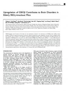
Figure 1. (A) Northern Blot hybridized with a probe to LRRK2 exon 24-27 showing absence of transcript in KO and diminished transcript in HET. A histone probe was used as a loading control. (B) Western blot with LRRK2 antibody 1182E, raised to amino acids 841-960 (B. Giasson) showing the absence of LRRK2 protein in KO and diminished signal in HET. GAPDH was used as a loading control. (C) Immunohistochemistry with MJFF2 c41-2 (Epitomics) antibody showing WT and KO brain sections at the level of the striatum. Specific signal is seen in the WT compared to KO. Rabbit IgG was used as an isotype control. Boxes depict enlarged images to the right. Scale Bar 50 microns.
Progressive degenerative kidney alterations and increased autophagy in LRRK 2 KO mice
In open field analyses 7-month KO mice (A) displayed significantly greater thigmotaxic (wall hugging) behaviour than WT at both age points, (B) spent significantly less time than WT exploring the inner area of the open field and (C) approached the novel object less frequently than WT mice; (D) movement traces in open field trials from WT and KO mice show thigmotaxic behaviour that was typical of LRRK2 KO mice. Open field was also performed on the same cohort at 16-months with similar results. For simplicity, only data at 7 months is shown, however longitudinal data was analyzed by repeated measures ANOVA and post-hoc analyses, followed by two a priori stated analyses using Student independent samples and matched-pairs sample tests respectively. (E) Motor co-ordination and balance were tested by (4-40rpm) accelerating rotarod analysis in an independent 7- month cohort of WT, HET and KO mice. KO mice significantly outperformed their WT littermates. Similar results were obtained in an older 16-month cohort. Data was analyzed by one-way ANOVA (p