Aerosol of a mixture of NAC and OVA for 30 min during each challenge. IV. GSEt together with methacholine during provocation of airway contractions,. 24 hours ...
6 Modulation of hyperresponsiveness by glutathione in a murine in vivo model of allergic asthma
J. Kloek1, I. Van Ark1, G. Folkerts1, F. De Clerck1,3, N. Bloksma1,2, F.P. Nijkamp1.
1
Department of Pharmacology and Pathophysiology, Utrecht institute for Pharmaceutical Sciences, Utrecht University, Utrecht, The Netherlands 2 Faculty of Biology, Utrecht University, Utrecht, The Netherlands 3 Department of Cardiovascular and Inflammation Pharmacology, Janssen Research Foundation, Beerse, Belgium
76 | Chapter 6
Abstract It is becoming increasingly clear that oxidative stress contributes to the pathogenesis of asthma. Therefore, this study addressed the question whether supplements of the endogenous antioxidant, glutathione (GSH), in a murine allergic asthma model would alleviate features of asthma. To this end, sensitized mice received aerosols of the GSH-donors, glutathione-ethyl ester (GSEt) or N-acetylcysteine, before or during respiratory allergen challenges, or during cholinergically provoked airway contractions one day after the last allergen challenge. Lung GSH levels were measured shortly after allergen challenge or after cholinergic provocation, and broncho-alveolar lavages (BAL) were performed to study the effect of GSH supplements on the influx of inflammatory cells into the airways. GSEt was found to decrease allergen-induced cholinergic airway hyperresponsiveness when given in combination with methacholine. However, when given before or during allergen challenge, both GSH-donors failed to decrease subsequent methacholine-induced airway contractility, BAL cell numbers, or increase lung GSH levels. In addition, allergen challenges of sensitized mice did not decrease lung GSH levels, suggesting that these challenges do not cause oxidative stress. Therefore, it is concluded that the failure of GSH-donors to decrease allergen-induced cholinergic hyperreactivity when given before or during allergen challenge may be explained by the lack of oxidative stress during the allergen challenge. In view of the oxidative stress upon allergen challenge observed earlier in the guinea pig and the importance of oxidative stress in the pathology of allergic asthma in humans, the guinea pig may be a more suitable species than the mouse to study oxidative stress in asthma.
Modulation of hyperresponsiveness by glutathione in mice |
77
Introduction Asthma is characterized by a chronic inflammatory condition of the lungs and reversible airflow obstruction. The inflammation is mild during remissions, but during exacerbations, inflammatory cells can produce large amounts of mediators, including reactive oxygen species (ROS). Exaggerated production of ROS may deplete anti-oxidant systems, leading to tissue damage and a disturbed biochemistry. This condition is referred to as oxidative stress and is increasingly appreciated in asthma (1, 2). It is still unclear how oxidative stress in asthma affects lung levels of glutathione (GSH), a prominent endogenous anti-oxidant. We recently demonstrated in a guinea pig model of asthma that allergen challenge induced GSH depletion of lung tissue during the early asthmatic response. In addition, supplementing GSH during an early asthmatic response in a lung perfusion model could prevent acute allergen-induced contractions. Besides allergen-induced acute airway contractions, airflow obstruction in asthmatics is manifest by exaggerated responses to contractile stimuli. This airway hyperreactivity (AHR) has a chronic character. Like allergeninduced acute airway contractions, AHR is generally considered to be a consequence of the inflammatory state of the lungs (3). Although the events contributing to its development are still ill understood, previous experiments in our lab have shown that isolated perfused lungs from GSH-depleted guinea pigs were hyperresponsive to histamine. In addition, perfusion of the lungs with GSH-ethyl ester (GSEt) prevented this hyperresponsiveness. Since these data suggest that GSH depletion may underlie AHR, the current study was done to address the question to what extent nebulization of GSH or GSH-precursors could counteract the development of airway inflammation and AHR in an in vivo murine model of asthma.
Methods Animals Animal care and use was in accordance with the guidelines of the local ethical committee for animal experiments. Specific pathogen-free male BALB/c mice (6 weeks) were obtained from the Utrecht University animal facilities department (Utrecht, The Netherlands) or from Charles River (Someren, Netherlands; study of protocol IV). The animals were housed in macrolon cages in a laminar flow cabinet and provided with food and water ad libitum.
78 | Chapter 6
Ovalbumin sensitization and challenge All animals were sensitized with an intraperitoneal injection (100 μl) of a mixture of 20 μg ovalbumin (grade V, Sigma, St Louis, MO; OVA) and 200 mg of the adjuvant, Al(OH)3, in 1 ml saline on days 0 and 7. On days 30, 33 and 36, the animals were given a challenge through inhalation of a nebulized OVA solution (1%, w/v) for 30 min. Control animals received saline instead of OVA. The aerosol (particle size 2.5 - 3.1 μm) was generated in a plexiglas exposure chamber with a Pari LC star nebulizer (Pari Respiratory equipment, Richmond, VA, USA) that was driven by compressed air (6 l/min). Intervention with thiol compounds Thiol compounds GSEt or N-acetyl cysteine (NAC) (Sigma, St. Louis, MO, USA) were nebulized in the above-mentioned nebulizer. Thiols were dissolved at 100 mM in saline; each solution was adjusted to pH 7. Controls received vehicle. Four treatment protocols were tested: I. II. III. IV.
GSEt aerosol for 5 min, 6 h before each OVA or saline challenge. GSEt aerosol for 5 min, 10 min before each OVA or saline challenge. Aerosol of a mixture of NAC and OVA for 30 min during each challenge. GSEt together with methacholine during provocation of airway contractions, 24 hours after the last OVA aerosol. In protocols I and II, each group consisted of three animals, while in protocols II and IV, eight animals per group were used. Measurement of airway responsiveness Twenty-four hours after the last allergen challenge, airway responsiveness was measured in conscious, unrestrained mice using barometric whole-body plethysmography (Buxco, EMKA Technologies, France). Enhanced pause (Penh) was taken as a measure of airway responsiveness, as described in detail previously (4). Mice were exposed to increasing concentrations of nebulized methacholine (acetyl-β-methylcholine chloride, Sigma, St. Louis, MO, USA; 1.6 - 13 mg/ml saline) in a plexiglas Buxco chamber. Each dose of methacholine was nebulized for 3 min and followed by a 3-min-period during which airway responses were recorded. Broncho-alveolar lavage Following airway responsiveness measurements, mice received a lethal dose of pentobarbitone sodium (Euthesate® 0.6 g/kg body weight, intraperitoneally). The
Modulation of hyperresponsiveness by glutathione in mice |
79
trachea was trimmed free of connective tissue, and a small incision was made to insert a cannula in the trachea. Via this cannula, the lavage was performed by filling the lungs with 1.0 ml sterile saline of 32°C. and withdrawing it after 10 seconds. The cell suspension was collected in a plastic tube on ice. This procedure was repeated three times to obtain a pooled cell suspensions from each animal. The cells were spun down by centrifugation at 400 g for 5 minutes at 4°C and resuspended in 150 μl ice-cold PBS. A sample of this was stained with Türk's solution and counted in a Bürker-Türk bright-line counting chamber to calculate the total number of cells. Another sample was used to prepare cytospin preparations. These were air dried, fixed, and stained with Diff-Quik (Merz + Dade A.G., Düdingen, Switzerland). The cells were differentiated morphologically into alveolar macrophages, eosinophils, lymphocytes and monocytes, and neutrophils by light microscopical observation. Glutathione measurements After the broncho-alveolar lavage, lungs were isolated and snap frozen in liquid nitrogen until analysis for GSH and oxidized glutathione (GSSG). To that end, lungs were crushed in liquid nitrogen using a mortar and pestle; the resulting powder was divided between two eppendorf tubes. To one tube, a mixture of 1 M HClO4 with 2 mM EDTA was added (1 ml per 250 mg powder) to assay total glutathione (GSH + GSSG), while the same mixture supplemented with 10 mM N-ethyl maleimide (NEM) was added to the other tube to assess levels of oxidized glutathione. After vigorous vortex mixing, the tubes were centrifuged at 5000 g for 10 min and total and oxidized glutathione concentrations in the supernatants were determined with a modified version (5) of the glutathione reductase-DTNB recycling assay according to Akerboom et al. (6). Values were expressed in nmol per g tissue (wet weight). Influence of OVA challenge on lung GSH/GSSG levels To investigate the effect of OVA challenge on lung GSH and GSSG levels, animals were sacrificed with pentobarbitone sodium (Euthesate® 0.6 g/kg body weight, intraperitoneally) immediately after the first or the third OVA aerosol, or 24 hours after the third challenge. Lungs were quickly isolated and snap-frozen in liquid nitrogen to be assayed for GSH and GSSG levels. Eight animals were used per time point. Statistics Dose response curves were statistically analyzed using a general linear model of repeated measures test, followed by the LSD post-hoc test for multiple comparisons.
80 | Chapter 6
In case the GSH levels and BAL cell counts were normally distributed, they were analyzed with a one-way ANOVA followed by the LSD post-hoc test for multiple comparisons. In case data were not normally distributed, they were analyzed with the Kruskal-Wallis test. Differences were considered statistically significant if P < 0.05.
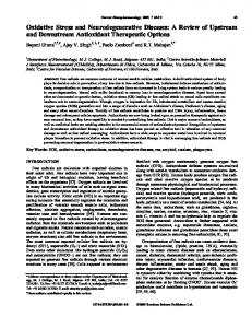
Results In animals that were treated with vehicle instead of GSEt 6 h before each challenge, saline challenge resulted in a normal dose-dependent increase of airway reactivity to methacholine (figure 1A and B).
4
4
A
3
Penh
Penh
3 2
2 1
1 0
B
0
1.6
3.0
6.3
methacholine (mg / ml)
13
0
0
1.6
3.0
6.3
13
methacholine (mg / ml)
Figure 1. Methacholine-induced airway contractions in OVA-sensitized mice that received vehicle (white bars) or GSEt (black bars) 6 h before each challenge with saline (panel A) or OVA (panel B). OVA challenges induced significant (P < 0.05) hyperresponsiveness as compared to saline challenges. GSEt-treatment had no influence on airway contractions of saline-challenged mice; neither did it alter OVAinduced hyperresponiveness. Bars indicate mean ± SEM
Compared to saline challenge, OVA challenge caused cholinergic hyperresponsiveness (P < 0.05). GSEt treatment did not affect cholinergic responsiveness as compared to the corresponding vehicle treatment. Lungs from the vehicle-pretreated, saline-challenged mice contained 300 and 90 nmol/g wet tissue weight GSH and GSSG, respectively (figure 2). These levels were not significantly changed by GSEt pretreatment or OVA challenge.
Modulation of hyperresponsiveness by glutathione in mice |
thiol (nmole / g)
750
500
250
0 pretreatment vehicle challenge
GSEt
saline
vehicle
GSEt
OVA
Figure 2. GSH (white bars) and GSSG (black bars) levels of lung tissue from OVAsensitized mice that received vehicle or GSEt 6 h before each challenge with saline or OVA. GSEt-treatment did not increase lung GSH of both saline- and OVA-challenged mice. Bars indicate mean ± SEM.
# cells (x 106)
5 4 3 2 1 0 pretreatment vehicle challenge
GSEt
saline
vehicle
GSEt
OVA
Figure 3. Numbers of inflammatory cells in broncho-alveolar lavage fluid from OVAsensitized mice that received vehicle or GSEt 6 h before each challenge with saline or OVA. OVA challenge significantly (P < 0.01) increased the number of inflammatory cells. Neither basal cell counts nor the OVA-induced increase of cell numbers were affected by GSEt-treatment. Dotted bars, macrophages; black bars, eosinophils; white bars, neutrophils; hatched bars, lymphocytes and monocytes. Bars indicate mean ± SEM.
81
82 | Chapter 6
Lung lavage fluid from the vehicle-pretreated, saline-challenged mice contained normal leukocyte numbers, largely consisting of macrophages (figure 3). OVA challenge significantly (P < 0.01) increased total cell numbers as particularly caused by an increased number of eosinophils and, to a lesser extent, lymphocytes and monocytes. GSEt pretreatment did not significantly change total and differential cell numbers as compared to the vehicle pretreatment.
4
4
A
3
Penh
Penh
3 2 1 0
B
2 1
0
1.6
3.0
6.3
methacholine (mg / ml)
13
0
0
1.6
3.0
6.3
13
methacholine (mg / ml)
Figure 4. Methacholine-induced airway contractions in OVA-sensitized mice that received vehicle (white bars) or GSEt (black bars) 5 min before each challenge with saline (panel A) or OVA (panel B). OVA challenges induced significant (P < 0.01) hyperresponsiveness as compared to saline challenges. GSEt-treatment did not alter basal airway responses in saline challenged animals or OVA-induced hyperresponiveness. Bars indicate mean ± SEM.
In animals that were treated with vehicle instead of GSEt 10 min before each challenge, saline challenge resulted in normal airway reactivity to methacholine (figure 4A and B) while OVA challenge caused cholinergic hyperresponsiveness (P < 0.01). GSEt treatment did not affect cholinergic responsiveness as compared to the corresponding vehicle treatment. GSH and GSSG levels of lungs from the vehicle-pretreated, saline-challenged mice were 390 and 16 nmol/g, respectively (figure 5), and were not significantly changed by GSEt pretreatment or OVA challenge. Lung lavage fluid from the vehicle-pretreated, saline-challenged mice contained normal leukocyte numbers, mainly macrophages (figure 6). OVA challenge significantly (P < 0.01) increased total cell numbers, eosinophils and lymphocytes and monocytes in particular. GSEt pretreatment did not significantly change total and differential cell numbers as compared to the vehicle-pretreated groups.
Modulation of hyperresponsiveness by glutathione in mice |
thiol (nmole / g)
500 400 300 200 100
0 pretreatment vehicle challenge
GSEt
vehicle
saline
GSEt
OVA
Figure 5. GSH (white bars) and GSSG (black bars) levels of lung tissue from OVAsensitized mice that received vehicle or GSEt 5 min before each challenge with saline or OVA. GSEt-treatment did not increase lung GSH of either saline- or OVAchallenged mice. Bars indicate mean ± SEM.
# cells (x 106)
4 3 2 1
0 pretreatment vehicle challenge
GSEt
saline
vehicle
GSEt
OVA
Figure 6. Numbers of inflammatory cells in broncho-alveolar lavage fluid from OVAsensitized mice that received vehicle or GSEt 5 min before each challenge with saline or OVA. OVA challenge significantly (P < 0.05) increased the number of inflammatory cells; this increase was not moderated by GSEt-treatment. Dotted bars, macrophages; black bars, eosinophils; white bars, neutrophils; hatched bars, lymphocytes and monocytes. Bars indicate mean ± SEM.
83
84 | Chapter 6 3
A
2
Penh
Penh
3
1
0
B
2
1
0
1.6
3.0
6.3
methacholine (mg / ml)
13
0
0
1.6
3.0
6.3
13
methacholine (mg / ml)
Figure 7. Methacholine-induced airway contractions in OVA-sensitized mice that received vehicle (white bars) or NAC (black bars) mixed with each saline (panel A) or OVA (panel B) challenge. OVA challenges induced significant (P < 0.01) hyperresponsiveness as compared to saline challenges. NAC did not alter airway contractions of saline-challenged mice, or OVA-induced hyperresponiveness significantly. Bars indicate mean ± SEM.
In animals challenged with saline aerosol without NAC, airway reactivity to methacholine was normal (figure 7A), while cholinergic hyperresponsiveness was observed after challenge with OVA aerosol (figure 7B; P < 0.05). Addition of NAC to the saline aerosol did not affect the airway response to methacholine. However, the addition of NAC to the OVA aerosol tended to decrease the hyperresponsiveness, but the effect was not statistically significant. Lungs from the saline-challenged mice contained 580 and 47 nmol/g GSH and GSSG, respectively (figure 8). These levels were not significantly changed by NAC or OVA challenge. Airway responses to methacholine and to methacholine mixed with GSEt did not differ in animals challenged with saline instead of OVA (figure 9A). In OVAchallenged mice, however, the hyperresponsiveness to methacholine was significantly attenuated by the addition of GSEt to the methacholine (P < 0.05). Lungs from mice challenged with methacholine one day after saline challenge contained 270 and 23 nmol/g GSH and GSSG, respectively (figure 10). These levels were not significantly changed by the addition of GSEt to the methacholine, or by the OVA challenge. OVA challenge significantly (P < 0.01) increased lung leukocyte numbers, particularly eosinophils and lymphocytes and monocytes, as compared to saline challenge. The cell numbers were not changed by challenge with methacholine or methacholine together with GSEt (figure 11).
Modulation of hyperresponsiveness by glutathione in mice |
thiol (nmole / g)
750
500
250
0 pretreatment vehicle challenge
GSEt
vehicle
saline
GSEt
OVA
Figure 8. GSH (white bars) and GSSG (black bars) levels of lung tissue from OVAsensitized mice that received vehicle or GSEt mixed with each saline or OVA challenge. NAC did not increase lung GSH of either saline- or OVA-challenged mice. Bars indicate mean ± SEM.
3
A
2
Penh
Penh
3
1
0
B
2
1
0
1.6
3
methacholine (mg / ml)
6.3
0
0
1.6
3
6.3
methacholine (mg / ml)
Figure 9. Methacholine-induced airway contractions in OVA-sensitized mice that received vehicle (white bars) or GSEt (black bars) mixed with methacholine, 24 h after the last challenge with saline (panel A) or OVA (panel B). OVA challenges induced significant (P < 0.01) hyperresponsiveness as compared to saline challenges. GSEt did not alter normal airway responses in saline-challenged mice, but it significantly (P < 0.05) decreased the OVA-induced hyperresponsiveness to methacholine. Bars indicate mean ± SEM.
85
86 | Chapter 6
thiol (nmole / g)
400 300 200 100
0 MCh challenge - GSEt allergen challenge
+ GSEt
- GSEt
saline
+ GSEt
OVA
Figure 10. GSH (white bars) and GSSG (black bars) levels of lung tissue from OVAsensitized mice that received vehicle or GSEt mixed with methacholine, 24 h after the last challenge with saline or OVA. GSEt-treatment did not increase lung GSH of either saline- or OVA-challenged mice. Bars indicate mean ± SEM.
# cells (x 106)
1.5
1.0
0.5
0.0 MCh challenge - GSEt allergen challenge
+ GSEt
saline
- GSEt
+ GSEt
OVA
Figure 11. Numbers of inflammatory cells in broncho-alveolar lavage fluid from OVA-sensitized mice that received vehicle or GSEt mixed with methacholine, 24 h after the last challenge with saline or OVA. OVA challenge significantly (P < 0.01) increased the number of inflammatory cells; this increase was not moderated by GSEt treatment. Dotted bars, macrophages; black bars, eosinophils; white bars, neutrophils; hatched bars, lymphocytes and monocytes. Bars indicate mean ± SEM.
Modulation of hyperresponsiveness by glutathione in mice |
500
A thiol (nmole / g)
thiol (nmole / g)
500 400 300 200 100 0
1
2
3
87
B
400 300 200 100 0
1
2
3
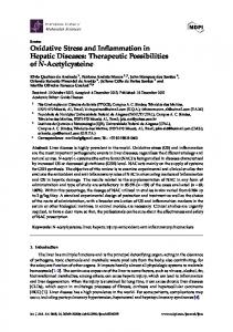
Figure 12. Influence of challenges with saline (panel A) or OVA (panel B) on tissue levels of GSH (white bars) and GSSG (black bars) in lungs of OVA-sensitized mice at different time points: 1, immediately after first challenge; 2, immediately after last challenge; 3, twenty-four hours after the last challenge. Neither GSH levels nor GSSG levels were significantly altered at any time point. Bars indicate mean ± SEM.
Influence of OVA challenge on lung GSH/GSSG levels GSH and GSSG levels were not significantly changed immediately after the first or third OVA challenge, or 24 h after the latter challenge, as compared to challenge with saline (figure 12A and B).
Discussion This chapter describes the effects of the GSH-donors, GSEt and NAC, in a murine model of allergic asthma on airway responsiveness, lung GSH and GSSG levels, and on the influx of inflammatory cells in the airways. Administration of the GSH-donors before or during allergen-challenge appeared to be an inadequate way to moderate airway hyperresponsiveness, although NAC given during OVA challenge tended to decrease airway hyperresponsiveness (figure 7). In these studies, the GSH-donors did not increase lung GSH and GSSG levels as measured one day after the last OVA challenge, immediately after the assessment of cholinergic airway responses (figures 2, 5, and 8). The unchanged GSH levels and BAL values one day after the last allergen challenge, and the observation that addition of GSEt to methacholine decreased cholinergic responsiveness (figure 9) probably explains the lack of effect of GSH donors on airway contractility when given before or during allergen challenge. The design of these treatment protocols was based on previous experiments in guinea pigs, showing a clear drop in lung GSH levels and an increase in GSSG levels during an OVA-induced early asthmatic
88 | Chapter 6
response. In OVA-sensitized mice, however, allergen challenge did not affect lung GSH levels (figure 12), suggesting that there is no need to replenish GSH during allergen challenge in mice. The combined absence of GSH-depletion and a symptomatic early asthmatic response upon allergen challenge in mice versus the presence of both features in guinea pigs (chapter 4), apparently reflects a species difference. This observation also supports the conclusion in chapter 4 that an early asthmatic response and a decrease of lung GSH are related. Although GSH supplementation decreased cholinergic responsiveness only when given during methacholine challenge, the protocols in this paper provide useful information regarding future studies on administration of GSH donors. The failure of GSH donors to enhance GSH levels in the lung can be explained in several ways. Firstly, the assessment of lung GSH levels, 24 h after the last treatment, was possibly too late to detect a, possibly transient, increase. Additionally, GSH supplementation may have led to a compensatory feedback inhibition of GSH synthesis, since GSH is a feedback inhibitor of its own synthesis (7). Secondly, the effect of GSEt nebulization may have been limited to the epithelium and the subepithelial tissues. Since GSH levels have been measured in whole lungs, an increase of GSH in (sub-) epithelial tissues may have been obscured. Finally, the GSHdonors may not have entered the tissue, but instead may have remained in the epithelial lining fluid. In the protocol in which GSEt was administered in combination with methacholine challenges, the broncho-alveolar lavage had probably washed away most of the GSEt. OVA challenges consistently increased BAL cell counts in the sensitized mice but, although such an inflammatory cell influx has been related to oxidative stress (8, 9), anti-oxidant treatment around the time of each OVA challenge did not change BAL cell numbers in our experiments. This may also be related to an absence of oxidative stress during the allergen challenge as indicated by the lack of GSH-depletion in the lungs (figure 12). In view of the evidence for an oxidative imbalance in a guinea pig model of allergic asthma (chapters 4 and 5), as well as in asthmatics (2, 10-13), the role of oxidative stress in the recruitment of inflammatory cells to the airways may be smaller in mice as compared to guinea pigs and humans. It is concluded that the decrease in airway contractility in mice when GSEt was given in combination with a contractile agonist is in line with earlier experiments in guinea pigs. However, the murine model lacks an early asthmatic response and concomitant GSH depletion in the lungs, while these features are both prominent in guinea pigs. In view of the presence acute asthmatic responses in humans and the
Modulation of hyperresponsiveness by glutathione in mice |
89
increasing evidence for a role of oxidative stress in asthmatics, guinea pig models may be more suitable than murine models to study the relation between oxidative stress and allergic asthma.
90 | Chapter 6
References
1.
2. 3.
4.
5.
6. 7.
8. 9. 10. 11.
12. 13.
Wood, L.G., D.A. Fitzgerald, P.G. Gibson, D.M. Cooper, and M.L. Garg. 2000. Lipid peroxidation as determined by plasma isoprostanes is related to disease severity in mild asthma. Lipids 35, no. 9:967. Dworski, R. 2000. Oxidant stress in asthma. Thorax 55 Suppl 2:S51. Vrugt, B., and R. Aalbers. 1993. Inflammation and bronchial hyperresponsiveness in allergic asthma and chronic obstructive pulmonary disease. Respir Med 87 Suppl B:3. Hamelmann, E., J. Schwarze, K. Takeda, A. Oshiba, G.L. Larsen, C.G. Irvin, and E.W. Gelfand. 1997. Noninvasive measurement of airway responsiveness in allergic mice using barometric plethysmography. Am J Respir Crit Care Med 156, no. 3 Pt 1:766. Vandeputte, C., I. Guizon, I. Genestie-Denis, B. Vannier, and G. Lorenzon. 1994. A microtiter plate assay for total glutathione and glutathione disulfide contents in cultured/isolated cells: performance study of a new miniaturized protocol. Cell Biol Toxicol 10, no. 5-6:415. Akerboom, T.P., and H. Sies. 1981. Assay of glutathione, glutathione disulfide, and glutathione mixed disulfides in biological samples. Methods Enzymol 77:373. Rahman, I., and W. MacNee. 1999. Lung glutathione and oxidative stress: implications in cigarette smoke-induced airway disease. Am J Physiol 277, no. 6 Pt 1:L1067. MacNee, W. 2000. Oxidants/Antioxidants and COPD. Chest 117, no. 5 Suppl 1:303S. Kelly, F.J. 1999. Gluthathione: in defence of the lung. Food Chem Toxicol 37, no. 9-10:963. Mohan, I.K., and U.N. Das. 1997. Oxidant stress, anti-oxidants, nitric oxide and essential fatty acids in bronchial asthma. Med. Sci. Res. 25, no. 5:307. Rahman, I., D. Morrison, K. Donaldson, and W. MacNee. 1996. Systemic oxidative stress in asthma, COPD, and smokers. Am J Respir Crit Care Med 154, no. 4 Pt 1:1055. Kelly, F.J., I. Mudway, A. Blomberg, A. Frew, and T. Sandstrom. 1999. Altered lung antioxidant status in patients with mild asthma. Lancet 354, no. 9177:482. Montuschi, P., M. Corradi, G. Ciabattoni, J. Nightingale, S.A. Kharitonov, and P.J. Barnes. 1999. Increased 8-isoprostane, a marker of oxidative stress, in exhaled condensate of asthma patients. Am J Respir Crit Care Med 160, no. 1:216.