Green, C. L., and A. C. Allison. 1978. Extensive prolonga- tion of rabbit kidney ... Rodt, and H. J. Kolb (ed.), Immunobiology of bone marrow transplantation.
INFECTION AND IMMUNITY, Sept. 1983, p. 1226-1233 0019-9567/83/091226-08$02.00/0 Copyright © 1983, American Society for Microbiology
Vol. 41, No. 3
Effect of Cyclosporine on the Response of Normal Human Lymphocytes to Cytomegalovirus In Vitro PAUL J. CONVERSE,* ALLAN D. HESS, PETER J. TUTSCHKA,
AND
GEORGE W. SANTOS
Bone Marrow Transplantation Program, The Johns Hopkins School of Medicine, Baltimore, Maryland 21205
Received 28 December 1982/Accepted 29 June 1983
Lymphocytes from healthy volunteers seropositive for cytomegalovirus (CMV) demonstrated strong lymphoproliferative responses to CMV-infected and glutaraldehyde-fixed foreskin fibroblasts (CMVFFX) when cocultured for 6 days. Addition of the immunosuppressive drug cyclosporine (CsA) to the cultures resulted in a 10-fold reduction (P < 0.001) in counts per minute of [3H]thymidine uptake. The proliferative response to noninfected fixed fibroblasts was also reduced 10-fold. Kinetic studies showed an inhibition of the lymphoproliferative response and not an alteration in the time course kinetics in CsA-treated cultures. Cytotoxicity to CMV-infected and unfixed fibroblasts by lymphocytes primed by CMVFFX in the presence of 0.5 ,ug of CsA per ml was significantly reduced (P < 0.01) as compared with untreated cultures but remained significantly above background level (P < 0.01). The cytotoxic response was still present but reduced at concentrations of .1.0 p,gIml. Cytotoxicity to noninfected fibroblasts by CMVFFx-primed lymphocytes could be eliminated by as little as 0.5 ,ug of CsA per ml. Stimulation of lymphocytes by CMVFFX but not by noninfected fixed fibroblasts resulted in the in vitro generation of suppressor cells. CsA at a final concentration of 1.0 or 2.5 p.g/ml did not significantly impair the induction of cells capable of suppressing the lymphoproliferative response of fresh autologous mononuclear cells to CMVFFX. The findings described above may have important clinical implications in that a degree of protective immunity to CMV-infected cells is maintained even in the presence of CsA.
Studies of human in vitro lymphocyte responses to cytomegalovirus (CMV) as measured by the incorporation of [3H]thymidine in proliferating lymphocytes or by lymphocytotoxicity to CMV-infected fibroblasts (19, 21-23) have generally shown a positive correlation of these tests for cell-mediated immunity with humoral immunity as determined by immunofluorescence or complement fixation antibody titers. Such immune responses are of particular interest because of CMV-induced complications in transplant and other immunosuppressed patients (7, 8, 18, 20) and because of a possible relationship of CMV and Kaposi's sarcoma recently reported in patients in the United States as well as the association recorded in young Africans (6, 10). Cyclosporine, previously known as cyclosporin A (CsA), a unique undecapeptide with immunosuppressive properties (1, 2), has been shown to facilitate organ transplants in various species (4, 11) and to prevent graft versus host disease after bone marrow transplantation in rats and humans (26, 27). The drug is believed to act selectively on T cells (1, 2, 12, 17, 29). Previous studies (12, 13) have demonstrated
diminished lymphoproliferation to alloantigens in primary and secondary cultures in the presence of CsA; the data have revealed a preferential blocking of the induction of cytotoxic but not suppressor cells in primary cultures. Since CsA is now widely used in clinical transplantation and since CMV is associated with complications after transplantation, it seemed worthwhile to examine the effects of this drug in vitro on the cell-mediated immune responses of healthy CMV-seropositive individuals. MATERIALS AND METHODS Subjects. Healthy volunteers were tested for serum antibody to CMV by complement fixation (W. H. Bums, The Johns Hopkins Hospital, Baltimore, Md.) and by lymphoproliferative assays (see below) for responsiveness to CMV. Those who responded to CMV-infected fibroblasts fixed with glutaraldehyde and irradiated (CMVFFX) but not to uninfected, fixed, and irradiated fibroblasts (NFFX) (hereafter referred to as responders, or A') were used in subsequent suppressor and cytotoxicity assays. All cells used for stimulation were irradiated with 1,500 rad from a "37Cs source. Mononuclear cell and lymphocyte isolation. Mononuclear cells were separated from the buffy coat of
1226
VOL. 41, 1983
CYCLOSPORINE AND RESPONSES TO CYTOMEGALOVIRUS
defibrinated blood by Ficoll-Hypaque density centrifugation by established techniques (12), washed three times in RPMI 1640 (GIBCO, Grand Island, N.Y.), and suspended at 2.0 x 10⁶ cells per ml in complete medium, consisting of RPMI 1640, 20% normal human CMV antibody-negative heat-inactivated serum, 1% glutamine (2 mM) supplement, 1% penicillin (100 U/ ml) and streptomycin (100 p.g/ml). Virus. CMV, Towne strain, was obtained from cultures maintained by R. LaFemina in The Johns Hopkins Medical Institutions laboratory of W. Gibson. Fresh cultures of foreskin fibroblasts were infected by the addition of 0.25 ml of a 10-4 dilution of infected fibroblast supernatant to monolayers at approximately 85% confluence in 25 ml of growth medium as previously described (16). Growth medium consisted of 10% fetal bovine serum (GIBCO) in Eagle minimal essential medium (GIBCO) with 1% gentamycin (40 ,ug/ml; Schering Pharmaceutical Corp., Kenilworth, N.J.). Medium was changed every 3 to 4 days. To assume expression of viral antigens on the surface of the majority of cells, infection was allowed to proceed until 100%o cytopathic effect was observed (10 to 14 days); within 7 days after the observation of complete cytopathic effect, infected cells were gently trypsinized, centrifuged, and kept at 4°C in growth medium for short-term storage or at -70°C in 10% dimethyl sulfoxide for long-term storage. A portion of the cells was suspended in phosphate-buffered saline without Ca2" or Mg2", pelleted, and fixed in 0.08% glutaraldehyde; fixed cells were similarly kept for short- and long-term storage. Uninfected cells were treated in the same manner. Fixed cells were not capable of infecting fibroblast monolayers nor, presumably, cultured lymphocytes. Cultures. Bulk cultures were set up in 25-cm2 flasks with 3 ml of lymphocytes at 2 x 10⁶/ml, 3 ml of CMVFFX or NFFX at 2 x 10⁴/ml, or 3 ml of irradiated lymphocytes at 2 x 10⁶/ml and 1.5 ml of complete medium with or without CsA. For some experiments cultures were established in flat-bottomed macrotiter (Cluster 24; Costar, Cambridge, Mass.) or microtiter (Microtest II; Falcon Plastics, Oxnard, Calif.) plates at the same ratio of fresh lymphocytes to fibroblasts or irradiated lymphocytes. CsA. CsA in powdered form, a generous gift from Sandoz Ltd., Biological and Medical Research Division, Basel, Switzerland, was dissolved in 95% ethanol and diluted to different concentrations (as indicated in the text) in complete medium. Measurement of lymphoproilferative responses. Proliferation of responder lymphocytes to CMVFFX, NFFX, allogeneic, or syngeneic lymphocytes (stimulating cells) was measured at various times after initiation of culture by the addition of 1 ,Ci of [3H]thymidine (New England Nuclear Corp., Boston, Mass.; specific activity, 6.7 Ci/mmol) to triplicate wells of microtiter plates containing 0.1 ml of responding lymphocytes, 0.1 ml of stimulating cells at concentrations as indicated above, and complete medium with or without CsA or to wells containing 0.1-ml samples of bulk cultures. After 18 h of additional incubation, the cultures were harvested onto glass fiber filter paper (grade 93 4 AH; Reeve Angel, Clifton, N.J.) with a multiple automated sample harvester (MASH II; Microbiological Associates, Bethesda, Md.). The filter paper was dried and processed for liquid scintillation
1227
counting. Results are expressed as the mean counts per minute [3H]thymidine incorporation ± standard error of the mean (SEM). Suppressor cell assays. Specific and nonspecific suppressive effects of lymphocytes primed in the presence or absence of CsA were assessed. Specific suppressive effects are arbitrarily defined as suppression of the response offresh autologous lymphocytes to CMVFFX by mononuclear cells primed by CMVFFX, whereas nonspecific suppressive effects are defined as the suppression of the response of fresh autologous lymphocytes to an antigen other than the antigen that stimulated the primed lymphocytes. Bulk cultures were harvested on day 7, incubated in complete medium to wash out CsA, and further washed two times in complete medium (12). The cells were resuspended at 2 x 10⁶/ml; 0.1-ml portions were set up in triplicate microtiter wells with 0.1 ml of 2 x 10⁵ fresh responding lymphocytes and 0.1 ml of 2 x 10³ fresh CMVFFX. The primed cells were also diluted in complete medium at ratios of 1:5 and 1:10, primed to fresh lymphocytes, and set up in triplicate wells. Control cultures consisted of irradiated autologously primed lymphocytes added to fresh lymphocytes and CMVFFX to control for cell density. The cultures were incubated for 1 week and pulsed with [3H]thymidine as described above. Percent suppression was calculated as follows: % suppression = 1 -
cpm of test culture x 100 cpm of control culture
Cell-mediated cytotoxicity. Cells cultured for 8 days with CMVFFX in the presence or absence of CsA were assayed for their ability to mediate lysis of CMVinfected or noninfected fibroblasts. The primed cells were washed as described above and set up at a ratio of 50:1 in round-bottomed microtiter plates (Titertek; Linbro Chemical Co., New Haven, Conn.) with nonfixed CMV-infected or noninfected target cells (105/ ml) labeled with 51Cr by standard techniques (12) in a final volume of 0.2 ml. After 4 to 5 h of incubation at 37°C, plates were centrifuged, and 100 ,utl of supernatant was harvested and counted for released 51Cr. Spontaneous and maximum release were measured by harvesting supernatants in wells to which 0.1 ml of medium or 0.1 ml of 1 N HCI, respectively, had been added to target cells. Percent specific 5'Cr release was calculated by the following formula:
% specific 5'Cr release = cpm test culture - cpm spontaneous x 100 cpm maximum - cpm spontaneous NK assays. Natural killer (NK) cell activity was measured against the K562 myeloid cell line maintained in RPMI 1640 with 10% fetal calf serum, gentamycin (40 ,ug/ml; Schering Pharmaceutical Corp.), penicillin (100 U/ml), and streptomycin (100 ,ug/ml). Effector cells were either lymphocytes primed by CMVFFX in the presence or absence of CsA for 8 days and harvested as described above for cytotoxicity assays or fresh mononuclear peripheral blood leukocytes (PBL) separated on a Ficoll-Hypaque gradient as
1228
CONVERSE ET AL.
INFECT. IMMUN.
TABLE 1. Lymphoproliferative responses to CMVFFX and control antigens as measured by tritiated thymidine uptake on day 6 of culturea
Respondersb (cpm)
Antigen
Nonrespondersb (cpm)
CMVFFX 38,899 ± 5,674 (11) 2,282 ± 436 (16) CMVFFX with CsA (1.0 ,ug/ml) 1,288 ± 429 (6) NDc CMVFFX with CsA (2.5 ,ug/ml) 810 ± 183 (5) ND NFFX 3,281 ± 941(9) 2,615 ± 491 (11) A,, 4,207 ± 1,131 (9) 3,111 ± 659 (10) B, 40,752 ± 4,101 (11) 57,930 ± 7,102 (16) a Data are expressed as mean counts per minute ± SEM. The CsA quantities are the final concentrations in culture. b Numbers in parentheses are numbers of responders or nonresponders. ' ND
=
Not done.
described above and washed twice in RPMI 1640. A portion of cells (10 x 106 to 15 x 106) was then suspended in 10 ml of RPMI 1640 with 20% autologous heat-inactivated serum and incubated at 37°C in a plastic petri dish for 1 h. The nonadherent cells were gently pipetted off, and the plate was washed two to three times with RPMI 1640. The cells were centrifuged, and the pellet was resuspended in RPMI 1640 with 10% fetal calf serum (FCS) and adjusted to a concentration of 5 x 10⁶/ml. Chromium-labeled K562 target cells (105/ml) were incubated in microtiter plates with the effector cells at effector to target cell ratios of 50:1, 25:1, 12.5:1, and, in some experiments, 6.25:1 for 4 h, after which supematants were harvested and counted as described above. Separation of T and non-T cells by E rosette formation. Cells cultured for 8 days were harvested, washed twice, counted, and resuspended at 107/ml. An equal volume of sheep erythrocyte-absorbed FCS was added to a portion of the cells. Neuraminidase-treated sheep erythrocytes at 108/ml were added in a volume equal to the FCS and the cultured cells. The mixture was centrifuged at 100 x g for 5 min at room temperature and then incubated for a further 15 min at room temperature. The pellet was resuspended by gentle rotation of the tube and then layered over FicollHypaque and centrifuged at 300 x g for 40 min. The nonrosetting cells found at the interface were harvested, washed, and resuspended to the original volume of cultured cells. Rosetted cells found in the pellet were recovered by NH4Cl lysis of the erythrocytes (25) and also resuspended to the original volume of the cultured cells. Statistical analysis. Results were analyzed for statistical significance 'by the Student t test.
RESULTS Proliferative responses. Twenty-seven individuals were tested for responses to CMVFFX and allogeneic lymphocytes (By). Control antigens were autologous lymphocytes (Ax) or NFFX or both. After 6 days in culture, CMV-seropositive individuals showed a threefold or greater increase in proliferation to CMVFFX compared to NFFX or Ax (Table 1). CMV seronegative individuals responded to infected fibroblasts at lev-
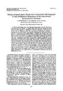
els comparable to their responses to NFFX. All individuals responded to Bx and none to A,. The effect of CsA on lymphoproliferation to CMVFFX was also measured. CsA was found to significantly (P < 0.001) reduce the proliferation of lymphocytes in response to CMVFFX. At final concentrations of 1.0 and 2.5 pg/ml (Table 1), the lymphoproliferative response was reduced to levels comparable to the levels of response to NFFX or A,. Kinetics of the response. In individuals with positive lymphoproliferative responses to CMVFFX, the kinetics of the response were similar to the responses to B,. Responses to virally infected fibroblasts in the presence of CsA as well as responses to NFFX peaked on day 5 or 6 but were significantly lower than the response to infected fibroblasts without drug (P < 0.001). Data from a representative experiment are shown in Fig. 1. Suppressor cell assays. To determine whether lymphocyte stimulation by CMV-infected fibroblasts led to the activation of suppressor cells, lymphocytes cultured for 7 days in the presence of CMVFFX, with or without CsA, were harvested and tested at different cell concentrations (2 x 10⁶/ml, and 4 x 10⁵ primed cells per ml with 2 x 10⁶ fresh responder cells for ratios of 1:1, 1:5, and 1:10, respectively) for their ability to suppress the response of a constant number offresh mononuclear cells (2 x 10⁶/ml) to the CMVinfected fibroblasts. At a 1:1 ratio of primed lymphocytes to fresh responder cells, the CMVFFx-primed lymphocytes suppressed the response of fresh mononuclear cells by 70% (Table 2). The suppressive effect was dependent on the ratio of primed cells added to fresh cells in the test system for cells derived from both CsA-treated and control bulk cultures. A more rapid decline in percent suppression was often observed in cells from CsAtreated cultures than from control cultures upon dilution (Fig. 2). Although there was little proliferation found in cultures incubated with CsA,
CYCLOSPORINE AND RESPONSES TO CYTOMEGALOVIRUS
VOL. 41, 1983 15
O
A+x
CMVFF.
Cl (A x CMVFFx )'x Atx CMVFFx
0
100
*--
((xCMVFFu CsA 2.5ug/ml)'xaAxCMVFFu
*--'(AA*xCMC VFFxCsA I.OAg/m
90
1229
)'
x
AOx CMVFFx
80
0
70
._
0 c
X 60
U,
E
aL
50
*n
40
30
I
20 0
l0 au
1:1
1:5
1:10
Ratio of Primed Lymphocytes to Fresh Responder Cells FIG. 2. A representative experiment showing mean percent suppression
of the response of fresh
autologous lymphocytes to CMVFF,, SEM mediated by lymphocytes primed by CMVFF. in the presence of CsA (A, 1.0 ,ug/ml; *, 2.5 tLg/ml) or absence of CsA (0). ±
Days FIG.
1.
Kinetics
of
the
lymphoproliferative
sponse to CMVFF,, (0), B,, (0), and presence of 1.0 p.g of CsA per ml
re-
CMVFF,, in the (A) in a repre-
sentative experiment.
after thorough washing these cells could substantially suppress the response of fresh cells to CMVFFX. To test the specificity of CMVFF,,-induced suppressor cells, their ability to suppress re-
sponses to alloantigens was also assessed. It was found that the CMVFF,,-induced suppressor cells were not able to suppress responses to alloantigens, whereas alloantigen-primed lymphocytes suppressed not only the response to the original alloantigen but also the response to CMVFFX (Table 2). The presence of CsA in the original lymphocyte cultures with alloantigen did not alter the suppressive effect. NFFX did
TABLE 2. Specific versus nonspecific suppression with and without CsAa Mean % suppression ± SEM at the following primed cell to fresh responder ratios:
Primed lymphocytes added to fresh cultures
responders
(A+ x CMVFFX)' (A+ x CMVFFX CsA 1.0 ,.g/ml)' (A+ x CMVFFX CsA 2.5 jig/ml)'
A+ A+ A+
CMVFFx CMVFFx CMVFFx
(A+ x CMVFFx)' (A+ x CMVFFX CsA 1.0 ,ug/ml)'
A+ A+
B, Bx
24 ± 2
(AXBx)' (A x Bx CsA 1.0 p.g/ml)' (A+ x Bx)'
A+ A+
Bx Bx
96±2
A+ A+
CMVFFx CMVFFx
48 ± 7 59 ± 8
(A+ x Bx CsA 1.0 ,ug/ml)'
Fresh
Stimulators
1:1
1:5
1:10
70 + 11 63 ± 4 71 ± 2
47 ± 13 49 ± 5 45 ± 8
34 ± 9 39 ± 6 36 ± 6
29 ± 12
83 ± 5
77±6 71 ± 5
(A+ x NFFx)' A+ 16 ± 9 CMVFFx (A+ x NFF,,)' Bx A+ 64 6 a Results are based on at least three separate experiments. A+, Seropositive individual responding to
CMVFFx.
1230
CONVERSE ET AL.
INFECT. IMMUN.
TABLE 3. Effect of CsA at the initiation of culture on lymphocytotoxicity to CMVF and NF on day 8 of culturea Primed lymphocytes (A' x CMVFFx)' Target
No CsA
CsA, 0.5
CsA, 1.0
,ug/ml 15 ± 4 3±1
,ug/ml 11 ± 1 3±1
CMVF 35 ± 3 NF 15 ± 1 a Data are expressed as mean percent specific 5 Cr release ± SEM at an effector to target cell ratio of 50:1 and based on at least six separate experiments in five individuals except as indicated. Mean spontaneous/ maximum 5"Cr release: CMVF = 26 + 6; NF = 27 + 6. A', Individual responding to CMVFFx.
70 -
c 60
(A x CMVFFx) - K562
X-X (A x CMVFFx)'-. CMVF
X 50"
40)
u
30
0._ v)
20
cn 0
iK
10 .
_ 50 25 12.5
FIG. 3. Results of four experiments showing mean percent specific lysis of CMVF (x) and K562 (A) cells by cells cultured for 8 days with CMVFFX (A' x CMVFFx)'.
not induce the activation of cells suppressing the response of fresh cells to CMV-infected fibroblasts. Cytotoxicity studies. The potential of CMVFFX to also stimulate the development of cytotoxic ing lysis of CMVF, experiments were performed cells and the ability of CsA to inhibit the induc- to compare the ability of day 8 cultured cells (A' tion of cytotoxicity were examined. Previous x CMVFFX) and fresh plastic nonadherent PBL studies with CsA (12, 29) had indicated that to lyse K562 cells, the conventional target for inhibition of the generation of cytotoxic lympho- assessing human NK cell activity. As shown in cytes occurred when fresh lymphocytes were Fig. 4, at four effector to target cell ratios tested cocultured with allogeneic lymphocytes and simultaneously, the curves for cultured cells and CsA for 8 to 10 days. In the current studies, fresh nonadherent PBL overlapped in three indilymphocytes were cocultured with CMVFFX in viduals tested. Mean lysis of K562 cells by the presence or absence of CsA. On day 8, the cultured cells in four experiments is shown in cells were harvested, washed, and set up at an Fig. 3. Day 8 cultured cells that had been treated effector to target cell ratio of 50:1 and in some with 0.5 or 1.0 ,ug of CsA per ml showed a experiments 25:1 and 12.5:1. The targets were statistically significant (P < 0.05) reduction of either unfixed CMV-infected fibroblasts (CMVF) or unfixed normal fibroblasts (NF) labeled with 51Cr. Primed cells from control cul80 SS tures effectively lysed CMV-infected targets 0-0 Fresh NA-PBL--K562 (Table 3). Lysis of noninfected fibroblasts by 470 + these primed cells was significantly greater than oe 65tAD 2 (AxCMVFFx)'-.K562 baseline levels, i,e., no lysis, but was significant60 ly less (P = 0.00001) than the lysis of infected a 50 0 target cells. The addition of CsA at the initiation of culture resulted in a significant reduction (P < 40 0.01) of cell-mediated cytotoxicity to both infected and noninfected target cells. There was no significant lysis of NF by cells cultured with 3-GS 0.5 or 1.0 ,ug of CsA per ml. However, at these levels of CsA, significant (P < 0.01) lysis of oe 10 CMVF was still observed. Mean spontaneous release of 51Cr was nearly identical for NF and CMVF. 50 25 12.5 6.25 Experiments were also carried out at different Effector: Target Ratio effector to target cell ratios to determine whether lysis of CMVF was dependent on the concenFIG. 4. Results of experiments in three individuals tration of primed cells in the assay. As shown in (SS, AD, and GS) demonstrating that fresh nonadherFig. 3, percent lysis diminished with decreasing ent (NA) PBL (0) and cells primed by CMVFFX in effector cell concentration. Similar results were vitro for 8 days (A' x CMVFFX)' (A) mediate equivafound in CsA-treated cultures (data not shown). lent levels of NK activity as measured against K562 In an attempt to characterize the cells mediat- target cells. -
-
-
VOL. 41, 1983
CYCLOSPORINE AND RESPONSES TO CYTOMEGALOVIRUS
K562 lysis compared with control cultures at an effector to target cell ratio of 50:1 only at the higher drug concentration. Mean percent 5tCr release mediated by CsA-treated cells at three effector to target cell ratios (50:1, 25:1, and 12.5:1) was 37 ± 6, 26 ± 7, and 20 + 10. To determine whether both T and non-T lymphocytes mediated killing of CMVF targets, experiments were carried out separating cells that formed rosettes (R+) with sheep erythrocytes from cells that did not form rosettes (R-). As shown in Table 4, in cultures not treated with CsA, killing of K562 cells was almost entirely carried out by R- or non-T cells. Both R- and R' cells mediated killing of CMVF, indicating a mixed effector cell population. However, reduced activity in the R+ population was presumably complicated by erythrocyte membrane binding to the T cells.' Similarly, mixed cell populations also appeared to be involved in lysis of CMVF mediated by cells that had been treated with 0.5 ,ug of CsA per ml. No lysis of K562 targets was observed in the R+ population of CsA-treated cells. DISCUSSION Previous studies have demonstrated that CsA diminishes lymphoproliferative responses to alloantigens and mitogens. The reports have also demonstrated that CsA blocks the induction of cytotoxic T cells in primary mixed lymphocyte reaction but allows for the expression of suppressor lymphocytes, leading to specific alloantigen tolerance (12, 13). The experiments reported here examined the effect of CsA on cell-mediated immune responses to a clinically important virus and investigated not only the proliferative response but also cytotoxic and suppressor cell activation. Mononuclear cells from responder individuals were capable of recognizing CMV antigens and proliferating as would be expected in a secondary immune response. Major histocompatibility complex antigens presumably present but at low density on the pooled fibroblasts (24) failed to provoke a lymphoproliferative response. The response was in fact comparable to the response to autologous lymphocytes. Therefore, proliferation above these baseline levels was entirely due to the presence of CMV antigens on the cells, a finding compatible with the results of M0ller-Larsen et al. and Schirm et al. (19, 21). It is not known when the responders were previously exposed to CMV, but the exposure was presumably not recent and was certainly not clinically apparent. Previous work in this laboratory has demonstrated that CsA blocks the production of interleukin 2 (IL-2)-like factors and the acquisition of responsiveness to such factors
1231
TABLE 4. Cell-mediated lysis of CMVF and K562 cells by day 8 primed cells with and without separation into R+ and R- fractionsa Target
Effector cell
population
CMVF
K562
(A' x CMVFFX)' control
44 ± 13 55 ± 2
unfractionated R+ R-
66± 2 4±1 12 ± 5 27 ± 9
(A+ x CMVFFX)' CsA 0.5 j±g/ml unfractionated R+
7± 2
30 ± 11
7 ± 2 0±0 55± 2 5±5 a Data are expressed as mean percent 5"Cr specific release ± SEM and are based on at least three separate experiments. The effector to target cell ratio was 50:1 before fractionation. After fractionation, cells were adjusted to the original volume. Mean percent spontaneous 51Cr release for CMVF = 27 ± 8 of maximum release. Mean percent spontaneous 51Cr release for K562 = 17 ± 4 of maximum release. A+, CMVseropositive individual. K562, Myeloid cell line, conventional target for human NK cells. R-
lymphocytes were not in an activated state, that is, they were not producing lymphocyte growth factors (e.g., T cell growth factor or IL-2). It also seems possible that they were not presenting "receptors" for these growth factors, a fact which remains unclear for in vivo sensitized lymphocytes. CsA presumably maintained the inactive state of the lymphocytes exposed to CMVFFX. The kinetic studies indicate an early inhibition of the responding lymphocytes rather than an alteration of the kinetics. CMV, at least in a live form, is postulated (5) to be immunosuppressive in itself. In the experiments reported here, CMV antigens present on fibroblasts were not capable of inducing a global suppression of the immune response. Lymphoproliferative responses to alloantigens were not inhibited by cells activated by CMVFFX. Only responses to the original stimulating CMV antigens were suppressed. Suppression of the response to CMV was also mediated by cells primed by CMVFFX in the presence of CsA, and the suppression was nearly as strong as in cultures lacking the drug. Whether CMVFFxprimed lymphocytes can also suppress the lymphoproliferative response to other microbial antigens as an explanation for CMV-induced global immunosuppression is being assessed. Responsive clones to microbial antigens are fewer (28) and might be more susceptible to suppression than are clones responsive to alloantigens. Experiments thus far support the (14, 15). Since CsA could block the induction of contention that suppression induced by a proliferative response, it seems likely that the CMVFFX to other microbial antigens (tetanus
1232
CONVERSE ET AL.
and herpes simplex) is weak but stronger than the suppression of responses to alloantigens (unpublished observations). Responder lymphocytes by definition had been previously exposed to CMV antigens. Lymphocytes cultured with CMVFFX and no drug proliferated and formed cytotoxic cells that were capable of lysing not only CMV-infected fibroblast target cells but, to a lesser extent, noninfected fibroblast targets as well. Cultured lymphocytes exposed to CMVFFX in the presence of CsA failed to proliferate, as indicated by an absence of [3H]thymidine uptake, but were still capable of lysing CMV-infected target cells. However, CsA-treated lymphocytes did not lyse noninfected fibroblast target cells. These data imply that recognition of fibroblast antigens, whether major histocompatibility complex or other antigens, or the induction of clones of lymphocytes responding to these antigens was blocked by CsA. The experiments fractionating the primed cell effectors by E-rosette formation indicate that a mixed population of T and non-T cells are mediating the lysis of CMV-infected fibroblasts in both CsA-treated and nontreated cultures. It is possible that in vivo primed T cells maintain their ability to kill CMV-infected fibroblasts after in vitro culture with CsA and CMV antigens. Furthermore, substantial levels of NK cell activity persist in CsA-treated cultures, as shown by the assay for cytotoxicity to K562 cells. The data may have important therapeutic implications in bone marrow transplantation, in which donor cells already primed by CMV or other microbes would have largely intact cytotoxic responses to infected recipient cells but would not develop an aggressive response to host minor alloantigens leading to graft versus host disease. The results suggest that CMV-specific cytotoxic and suppressor cells are activated by CMV-infected fibroblasts, but amplification of the cytotoxic arm of the response is particularly hampered by CsA. These results are in accord with those of others (14, 15, 17) who have found that CsA inhibits IL-2 production, a critical component for T-cell amplification and maturation, but the drug does not itself induce suppressor cells in the absence of antigen. On the other hand, our results would suggest that perhaps the amplification of suppressor cells is either independent of IL-2 or is mediated by another factor, such as the recently demonstrated T suppressor inducer factor (9). It will be important to establish which soluble mediators are effective in inducing a lymphocyte response to CMV that might help in controlling severe disease caused by this agent. Recent studies in a number of laboratories (3, 14, 15) have shown that the development of
INFECT. IMMUN.
cytotoxic T cells is dependent on the presence of lymphokines and monokines, such as IL-1 and IL-2, that are produced in response to mitogen or alloantigen stimulation. As stated above, reports from this laboratory (14, 15) and others (3, 17) have indicated that CsA limits the production of IL-2 and T cell growth factors as well as the induction of responsiveness to IL-2. A subsequent report will examine the production of growth factors found in supernatants of lymphocytes cultured with CMVFFX, the influence of CsA on both the production of growth factors and responsiveness to them, and the functional characteristics of cells exposed to crude and commercially prepared factors. ACKNOWLEDGMENTS This work was supported by Public Health Service grants CA 153% and iROl AM 25602 from the National Institutes of Health. A.D.H. is a scholar of the Leukemia Society of America. We gratefully acknowledge the expert secretarial assistance of Connie Rutter and the useful discussions with Albert Donnenberg. LITERATURE CITED 1. Borel, J. F. 1976. Comparative study of in vitro and in vivo drug effects on cell-mediated cytotoxicity. Immunology 31:631-641. 2. Borel, J. F., C. Feurer, C. Maynee, and H. Stahelin. 1977. Effects of the new anti-lymphocyte peptide cyclosporin A in animals. Immunology 32:1017-1025. 3. Bunjes, D., C. Hardt, M. Rollinghoff, and H. Wagner. 1981. Cyclosporin A mediates immunosuppression of primary cytotoxic T cell responses by impairing the release of interleukin 1 and interleukin 2. Eur. J. Immunol. 11:657-661. 4. Caine, R. Y., D. J. C. White, K. Rolies, D. P. Smith, and B. M. Herbertson. 1978. Prolonged survival of pig orthotopic heart grafts treated with cyclosporin A. Lancet i:11831185. 5. Carney, W. P., R. H. Rubin, R. A. Hoffman, W. P. Hansen, K. Healey, and M. S. Hirsch. 1981. Analysis of Tlymphocyte subsets in cytomegalovirus mononucleosis. J. Immunol. 126:2114-2116. 6. Centers for Disease Control. 1982. Update on acquired immune deficiency syndrome (AIDS)-United States. Morbid. Mortal. Weekly Rep. 31:507-508, 513-514. 7. Dowling, J. N., A. R. Saslow, J. A. Armstrong, and M. Ho. 1976. Cytomegalovirus infection in patients receiving immunosuppressive therapy for rheumatologic disorders. J. Infect. Dis. 133:399-408. 8. Elfenbein, G. J., and R. Saral. 1981. Infectious disease during immune recovery after bone marrow transplantation, p. 157-1%. In J. D. Allen (ed.), Infection and the compromised host, clinical correleations and therapeutic approaches, 2nd ed. The Williams & Wilkins Co., Baltimore. 9. Flood, P., K. Yamauchi, and R. K. Gershon. 1982. Analysis of the interactions between two lymphocyte molecules that are required for the expression of Ly-2 suppressor cell activity. J. Exp. Med. 156:361-371. 10. Giraldo, G., and E. Beth. 1980. The relationship of cytomegalovirus to certain human cancers, particularly to Kaposi's sarcoma, p. 57-73. In G. Giraldo and E. Beth (ed.), The role of viruses in human cancer. Elsevier/ North-Holland, Amsterdam. 11. Green, C. L., and A. C. Allison. 1978. Extensive prolongation of rabbit kidney allograft survival after short term cyclosporin A treatment. Lancet i:1182-1183.
- VOL. 41, 1983
CYCLOSPORINE AND RESPONSES TO CYTOMEGALOVIRUS
12. Hess, A. D., and P. J. Tutschka. 1980. Effect of cyclosporin A on human lymphocyte responses in vitro. I. CsA allows for the expression of alloantigen activated suppressor cells while preferentially inhibiting the action of cytolytic effector lymphocytes in MLR. J. Immunol. 124:2601-2608. 13. Hess, A. D., P. J. Tutschka, and G. W. Santos. 1981. Effect of cyclosporin A on human lymphocyte responses in vitro. II. Induction of specific alloantigen unresponsiveness mediated by a nylon wool adherent suppressor cell. J. Immunol. 126:961-968. 14. Hess, A. D., P. J. Tutschka, and G. W. Santos. 1982. Effect of cyclosporin A on human lymphocyte responses in vitro. III. CsA inhibits the production of T lymphocyte growth factors in secondary mixed lymphocyte responses but does not inhibit the response of primed lymphocytes to TCGF. J. Immunol. 128:355-359. 15. Hess, A. D., P. J. Tutschka, P. Zhang, and C. W. Santos. 1982. Effect of cyclosporin A on human lymphocyte responses in vitro. IV. Production of T cell stimulatory growth factors and development of responsiveness to these growth factors in CsA treated primary MLR cultures. J. Immunol. 128:360-367. 16. LaFemina, R. L., and G. S. Hayward. 1983. Replicative forms of human cytomegalovirus DNA with joined termini are found in permissively infected human cells but not in non-permissive Balb/c-3T3 mouse cells. J. Gen. Virol. 64:373-389. 17. Larsson, E.-L. 1980. Cyclosporin A and dexamethosone suppress T cell responses by selectively acting at distinct sites of the triggering process. J. Immunol. 124:28282833. 18. Linnemann, C. C., C. A. Kaufman, M. R. First, G. M. Schiff, and J. P. Phair. 1978. Cellular immune response to cytomegalovirus after renal transplantation. Infect. Immun. 22:176-180. 19. M0oler-Larsen, A., H. K. Andersen, I. Heron, and I. Sarov. 1975. In vitro stimulation of human lymphocytes by purified cytomegalovirus. Intervirology 6:249-257. 20. Quinnan, G. V., N. Kirmani, A. H. Rook, J. F. Manischewitz, L. Jackson, G. Moreschi, G. W. Santos, R.
21.
22.
23.
24. 25.
26.
27.
28.
29.
1233
Saral, and W. H. Burns. 1982. Cytotoxic T cells in cytomegalovirus infection. N. Engl. J. Med. 307:7-13. Schirm, J., H. W. Rosenhorst, and T. H. The. 1980. Comparison of in vitro lymphocyte proliferations induced by cytomegalovirus-infected human fibroblasts and cellfree cytomegalovirus. Infect. Immun. 30:621-627. Starr, S. E., B. Dalton, T. Garrabrant, K. Paucker, and S. A. Plotkin. 1980. Lymphocyte blastogenesis and interferon production in adult human leukocyte cultures stimulated with cytomegalovirus antigen. Infect. Immun. 30:17-22. Thong, Y. H., S. A. Hensen, M. M. Vincent, D. A. Fuccillo, W. A. Stiles, and J. A. Bellanti. 1976. Use of cryopreserved virus-infected target cells in a lymphocytotoxicity 51Cr release microassay for cell-mediated immunity to cytomegalovirus. Infect. Immun. 13:643-645. Thorsby, E., and S. Lie. 1968. Antigens on human fibroblasts demonstrated with HLS antisera and antihuman lymphocyte sera. Vox Sang. 15:44-53. Tsoi, M. S., J. Aprile, S. Dobbs, S. Goehle, and R. Storb. 1982. Enrichment (and depletion) of human suppressor cells with monoclonal antibodies and immunoglobulincoated plates. J. Immunol. Methods 53:293-305. Tutschka, P. J., W. E. Beschorner, and A. D. Hess. 1980. Use of cyclosporin A (CsA) in a rat model of allogenic marrow transplantation, p. 241-253. In S. Thierfelder, H. Rodt, and H. J. Kolb (ed.), Immunobiology of bone marrow transplantation. Springer-Verlag, New York. Tutschka, P. J., A. D. Hess, W. E. Beschorner, and G. W. Santos. 1982. Cyclosporin A in allogeneic bone marrow transplantation: preclinical and clinical studies, p. 519538. In D. J. G. White (ed.), Cyclosporin A: proceedings of an international conference. Elsevier Biomedical Press, New York. van Oers, M. H. J., J. Pinkster, and W. P. Zeilemaker. 1978. Quantification of antigen reactive cells among human T lymphocytes. Eur. J. Immunol. 8:477-484. Wang, B. S., E. H. Heacock, K. H. Cullins, I. F. Hutchinson, N. L. Tilney, and J. A. Mannick. 1981. Suppressive effects of cyclosporin A on the induction of alloreactivity in vitro and in vivo. J. Immunol. 127:89-93.