[Downloaded free from http://www.pediatricneurosciences.com on Wednesday, September 28, 2016, IP: 197.157.108.206]
Neuroimaging Magnetic resonance imaging in pantothenate kinase-2-associated neurodegeneration Paramdeep Singh, Kavita Saggar, Maneet Kaur, Davinder Singh Pannu Department of Radiodiagnosis, Dayanand Medical College and Hospital, Ludhiana, Punjab, India
Address for correspondence: Dr. Paramdeep Singh, Department of Radiodiagnosis, Dayanand Medical College and Hospital, Ludhiana, Punjab, India. E-mail:
[email protected]
ABSTRACT Pantothenate kinase-2-associated neurodegeneration (PKAN) is a rare autosomal recessive pediatric neurodegenerative disorder characterized by rigidity, dystonia, impaired postural reflexes, and progressive dementia. On T2-weighted magnetic resonance imaging images, marked low signal intensity is seen in the globus pallidus. This low signal intensity surrounds a central region of high signal intensity in the anteromedial globus pallidus, giving an eye-of-the-tiger appearance. Key words: Eye of the tiger, MRI, Pantothenate kinase-2-associated neurodegeneration
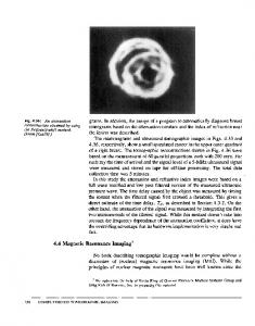
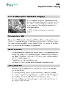
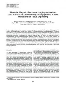
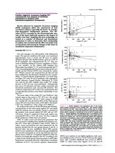
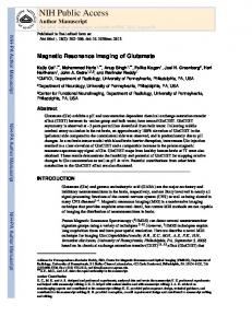
A 14-year-old boy presented with chief complaints of progressively increasing abnormal movements, gait disturbances, speech difficulty, and intellectual decline since the age of 10 years. He was born at full term to consanguineous parents. No history of perinatal hypoxic insult or delayed milestones was present. No relevant family history was present. Neurological examination revealed a generalized increase in limb tone, dsytonia, and hyperreflexia with extensor planter responses. Ophthalmic evaluation was normal. No evidence of Kayser–Fleischer rings or retinitis pigmentosa was seen. Serum copper, iron, and ceruloplasmin levels were within normal limits. Blood smear was positive for acanthocytes. Magnetic resonance imaging (MRI) of brain revealed bilateral anteriomedial hyperintensity surrounded by a region of hypointensity in the medial globus pallidus on T2- and T2*weighted images (eye-of-the-tiger sign). Similar hypointensity was also seen in dentate nuclei, red nuclei, and substantia nigra [Figures 1–6].
Figure 1: Axial T2-weighted image showing hypointensity (thick arrow) with a central region of hyperintensity (thin arrow) in the medial globus pallidus (eye-of-the-tiger sign)
Access this article online Quick Response Code:
Website: www.pediatricneurosciences.com
DOI: 10.4103/1817-1745.97618
Figure 2: Axial T2-weighted image showing hypointense signal localized in the dentate nuclei of the cerebellum (arrow)
2012 / Jan-Apr / Volume 7 / Journal of Pediatric Neurosciences / 27
[Downloaded free from http://www.pediatricneurosciences.com on Wednesday, September 28, 2016, IP: 197.157.108.206] Singh, et al.: MRI in PKAN
Figure 3: Axial T2-weighted image showing hypointense signal in the substantia nigra and red nuclei (arrow)
Figure 4: T2* weighted image showing hypointensity with a central region of hyperintensity in the medial globus pallidus (eye-of-the-tiger sign)
Figure 5: T2* weighted image showing hypointense signal localised in the dentate nuclei of the cerebellum
Figure 6: T2* weighted image showing hypointense signal in the substantia nigra and red nuclei
Neurodegeneration with brain iron accumulation (NBIA) is a clinically and genetically heterogenous group of disorders characterized by brain iron deposition and associated with neuronal death. The causes of NBIA comprise pantothenate kinase-associated neurodegeneration (PKAN), neuroferritinopathy, infantile neuroaxonal dystrophy, and aceruloplasminemia. [1] PKAN is a rare autosomal recessive neurodegenerative disorder. The diagnosis is usually made in first decade of life or in early adolescence. The clinical phenotype that is associated with mutations in the pantothenate kinase 2 gene, PANK2, is termed pantothenate kinase-associated neurodegeneration (PKAN) and is usually classified into classical or atypical forms. Classic PKAN is characterized by early age of onset, usually before 6 years, and rapid progression. The main clinical (extrapyramidal) features are dystonia, speech disturbance (dysarthria), and rigidity, with corticospinal tract involvement resulting in spasticity, hyperreflexia, and extensor planter responses. Patients with atypical PKAN have a later onset of symptoms and progression is slower. The average age of onset is 13–14 years. Iron levels in CSF and blood are within normal limits. [2]
Neuroferritinopathy and aceruloplasminemia present in adult or late life.[1] Iron deposition abnormalities are also seen in non-inherited disorders such as in beta–thalassemia major, HIV and Wilson disease, where T2* hypointensities of the globus pallidus, caudate nuclei, and putamen have been described.
28 / Journal of Pediatric Neurosciences / Volume 7 / Jan-Apr / 2012
The substantia nigra, dentate nuclei, and globus pallidus are iron-rich structures in the normal brain. The main MRI features associated with NBIA is due to high iron in the basal ganglia, appreciated as hypointense lesions in the globus pallidus and substantia nigra pars reticulata on T2-weighted images. A central region of hyperintensity with surrounding hypointensity of the globus pallidus on T2-weighted images (eye-of-the-tiger sign) is nearly pathognomonic for PKAN [Figure 1]. The hyperintense central region indicates edema or necrosis, while the surrounding hypointense region indicates high iron.[2] Pathological analysis of brain tissue from patients with PKAN shows triad of iron deposition, axonal spheroids, and gliosis in the globus pallidus. [3] The central hyperintensity seen in PKAN is absent in
[Downloaded free from http://www.pediatricneurosciences.com on Wednesday, September 28, 2016, IP: 197.157.108.206] Singh, et al.: MRI in PKAN
non-PKAN forms of NBIA; the MRI demonstrates only hypointense signal in the globus pallidus on T2-weighted images. [4] An eye-of-the-tiger sign, considered almost pathognomonic of PKAN, has been observed in cases of neuroferritinopathy. Eye-of-the-tiger sign can be seen in non-NBIA conditions such as corticobasal degeneration and progressive supranuclear palsy, and these should be considered in the differential diagnosis. This sign has also been reported in other extrapyramidal parkinsonian disorders, including cortical–basal ganglionic degeneration, Steele–Richardson–Olszewski syndrome, and early-onset levodopa-responsive Parkinsonism.[1,5] Thus, an eye-of-the-tiger sign must not be interpreted in isolation; scans should be looked for involvement of other basal ganglia nuclei and cerebral cortex.
References 1.
McNeill A, Birchall D, Hayflick SJ, Gregory A, Schenk JF, Zimmerman EA, et al. T2* and FSE MRI distinguishes four subtypes of neurodegeneration with brain iron accumulation. Neurology 2008;70:1614-9. 2. Gregory A, Hayflick SJ. Neurodegeneration with brain iron accumulation. Folia Neuropathol 2005;43:286-96. 3. Halliday W. The nosology of Hallervorden-Spatz disease. J Neurol Sci 1995;134:84-91. 4. Hayflick SJ, Hartman M, Coryell J, Gitschier J, Rowley H. Brain MRI in neurodegeneration with brain iron accumulation with and without PANK2 mutations. AJNR Am J Neuroradiol 2006;27:1230-3. 5. Guillerman RP. The eye-of-the-tiger sign. Radiology 2000;217:895-6. Cite this article as: Singh P, Saggar K, Kaur M, Pannu DS. Magnetic resonance imaging in pantothenate kinase-2-associated neurodegeneration. J Pediatr Neurosci 2012;7:27-9.
Source of Support: Nil. Conflict of Interest: None declared.
2012 / Jan-Apr / Volume 7 / Journal of Pediatric Neurosciences / 29