Surg Endosc (2012) 26:738–746 DOI 10.1007/s00464-011-1945-1
and Other Interventional Techniques
Management options for symptomatic stenosis after laparoscopic vertical sleeve gastrectomy in the morbidly obese Amit Parikh • Joshua B. Alley • Richard M. Peterson Michael C. Harnisch • Jason M. Pfluke • Donovan M. Tapper • Stephen J. Fenton
•
Received: 26 April 2011 / Accepted: 31 August 2011 / Published online: 2 November 2011 ! Springer Science+Business Media, LLC 2011
Abstract Background This study aimed to determine the incidence, etiology, and management options for symptomatic stenosis (SS) after laparoscopic sleeve gastrectomy (LSG). Methods A retrospective study reviewed morbidly obese patients who underwent LSG between October 2008 and December 2010 to identify patients treated for SS. Results In this study, 230 patients (83% female) with a mean age of 49.5 years and a mean body mass index (BMI) of 43 kg/m2 underwent LSG. In 3.5% of these patients (100% female; mean age, 42 years; mean BMI, 42.6 kg/ m2), SS developed. The LSG procedure was performed using a 36-Fr. bougie and tissue-reinforced staplers. Four patients had segmental staple-line imbrication, and seven patients underwent contrast study, with 71.4% demonstrating a fixed narrowing. Endoscopy confirmed shortsegment stenoses: seven located at mid-body and one located near the gastroesophageal junction. Endoscopic management was 100% successful. The mean number of dilations was 1.6, and the median balloon size was 15 mm. The mean time from surgery to initial endoscopic intervention was 48.8 days, and the mean time from the first dilation to toleration of a solid diet was 49.6 days. Two Presented at the SAGES 2011 Annual Meeting, March 30–April 2, 2011, San Antonio, TX. A. Parikh ! J. B. Alley ! R. M. Peterson ! J. M. Pfluke ! S. J. Fenton (&) Department of Surgery, University of Texas Health Sciences Center at San Antonio, Mail Code 7842, 7703 Floyd Curl Drive, San Antonio, TX 78229-3900, USA e-mail:
[email protected] J. B. Alley ! M. C. Harnisch ! J. M. Pfluke ! D. M. Tapper ! S. J. Fenton San Antonio Military Medical Center, San Antonio, TX, USA
123
patients were referred to our institution after undergoing LSG at another facility. The mean time to the transfer was 28.5 days. The two patients had a mean age of 35 years and a mean BMI of 42.3 kg/m2. Both patients experienced immediate SS after perioperative complications comprising one staple-line hematoma and one leak. Contrast studies demonstrated minimal passage of contrast through a longsegment stenosis. Both patients underwent multiple endoscopic dilation procedures and endoluminal stenting, ultimately requiring laparoscopic conversion to Roux-en-Y gastric bypass. The mean time from the initial surgery to the surgical revision was 77 days, and the mean time after the first intervention to tolerance of a solid diet was 82 days. Conclusion Symptomatic short-segment stenoses after LSG may be treated successfully with endoscopic balloon dilation. Long-segment stenoses that do not respond to endoscopic techniques may ultimately require conversion to Roux-en-Y gastric bypass. Keywords
Bariatric ! Complications ! Obesity
The vertical sleeve gastrectomy has been in use since the late 1980s. Hess and Hess [1] and Marceau et al. [2] were the first to report their experience with this new gastrectomy as part of the biliopancreatic diversion with a duodenal switch (BPD-DS) operation. They modified the Scopinaro BPD with the addition of the sleeve gastrectomy in an attempt to reduce marginal ulceration, provide gastric restriction, and maintain the regulation of gastric emptying. The first published report of a laparoscopic BPD-DS with a vertical sleeve gastrectomy occurred almost a decade later [3]. In 2003, Regan et al. [4] reported their experience with the two-stage gastric bypass for super–super-obese patients.
Surg Endosc (2012) 26:738–746
After weight loss from the sleeve gastrectomy, patients routinely underwent another operation such as the Roux-enY gastric bypass or duodenal switch. These authors concluded that this was a reasonable approach for these high-risk patients [5, 6]. Over the past 5 years, laparoscopic sleeve gastrectomy (LSG) is used increasingly as a primary and sole weightloss operation not only for the super–super obese but also for the morbidly obese. Several centers have reported a percentage of excess weight loss (%EWL) approaching that of the Roux-en-Y gastric bypass at both short- and mid-term follow-up assessment [7–13]. Due to the long staple line and altered intragastric pressures, LSG is associated with several complications. The most dreaded complication is a staple-line leak, reported to occur in about 1% of cases [14]. Some studies have reported a much higher leak rate, up to 3.9% in primary LSG procedures [7, 15, 16]. Several reports have discussed the challenges of managing a leak after LSG and techniques to minimize its occurrence [17, 18]. Sleeve stenosis also can occur due to the intentional narrow tubularization of the stomach. It currently is reported to occur in 0.26% to 4% of LSG operations [13, 15, 16, 19–22]. This may underestimate the true incidence of stenosis in current practice because early published series of LSG tended to use larger bougies with the intention of two-stage weight loss. Additionally, little literature exists regarding patient characteristics, operative techniques, and other variables that may contribute to the development of a sleeve stenosis, and few reports have described the subsequent management of these patients. [18, 19, 23]. This study aimed to review the incidence of symptomatic sleeve stenosis among patients at our institution who underwent LSG as the primary weight loss procedure.
Methods Institutional review board (IRB) approval was requested for a retrospective cohort analysis of patients who underwent LSG complicated by symptomatic sleeve stenosis from October 2008 to December 2010. The indications for entry into our bariatric surgery program conform to the 1991 National Institutes of Health (NIH) consensus statement, which specifies a body mass index (BMI) greater than 40 kg/m2 or a BMI of 35–39.9 kg/m2 together with significant obesity-related comorbidities [24]. At entry to the program, each patient undergoes a 6-month supervised diet, participation in our bariatric support group and informational seminars, and a standard preoperative bariatric workup. Additional preoperative testing is conducted based on individual patient comorbidities.
739
At completion of preoperative testing, each patient is presented with his or her surgical options, and an informed consent discussion is conducted summarizing the best available evidence for each procedure. The patients in our bariatric program choose to undergo LSG based on their preference and physician guidance. In this study, the surgery and perioperative management were performed as previously described by our institution [25]. A retrospective analysis of our IRB-approved prospective bariatric surgical database was performed to identify patients who underwent LSG during the study period. All patients who underwent LSG were reviewed, including those who underwent conversion from previous adjustable gastric band placement. At the time of the analysis, follow-up data were available for 227 (98.7%) of 230 patients at 1 month, 205 (89.1%) of 230 patients at 3 months, 171 (91.9%) of 186 patients at 6 months, 86 (67.2%) of 128 patients at 12 months, and 18 (62.1%) of 29 patients at 24 months. The mean follow-up period for the series was 8.7 months (range, 1–24 months). Weight loss and evidence of clinically significant sleeve stenosis were documented. Standard BMI definitions were used. The percentage of excess weight loss (%EWL) was defined as weight lost/preoperative weight – ideal body weight, with ideal body weight (IBW) defined by the Devine formula. For men, IBW is 50 kg ? 2.3 kg for each inch of height exceeding 5 ft, and for women, IBW is 45.5 kg ? 2.3 kg per inch of height exceeding 5 ft. Percentage of excess BMI loss (%EBL) was defined as BMI points lost/preoperative BMI—25. The medical record of patients identified as having clinically significant sleeve stenosis were reviewed for initial operative details, imaging results, details of endoscopic intervention, details of additional intervention, time to diagnosis, time to resolution of symptoms, and stricture characteristics. Basic statistics were used to summarize patient demographics. Continuous data were compared using the unpaired Student t-test. Categorical data were analyzed with Fisher’s exact test. All hypotheses tested were two-sided with an overall level of significance (a) set at 0.05. Individual hypothesis tests with p values less than 0.05 were considered statistically significant. The analysis tools were SAS 9.1.3 (SAS Institute Inc., Cary, NC, USA) and Microsoft Excel 2007 (Microsoft, Redmond, WA, USA).
Results During the study period, 230 patients underwent LSG. The baseline data are presented in Table 1. The mean age of the 230 patients (83% female) was 49.4 ± 10.6 years, and their mean preoperative BMI was 43.0 ± 6.4 kg/m2. For 11 of these patients (4.8%), laparoscopic conversion from a
123
740
Surg Endosc (2012) 26:738–746
adjustable gastric band to LSG was performed. None of these patients experienced a clinically symptomatic stenosis. Additionally, seven patients (3%) with a BMI of 30 to 34.9 kg/m2 (mean, 33.0 ± 1.3 kg/m2) were managed under protocol as part of an ongoing prospective controlled clinical trial (NCT00965302, http://www.clinicaltrials.gov). None of these patients experienced a clinically significant stenosis. Reoperation was required by 13 patients (5.7%), and 12 of these reoperations (92.3%) were performed laparoscopically (5 for staple-line bleeding, 3 due to suspicion of leak [all negative], 3 for staple-line leak, 1 for choledocholithiasis, and 1 negative laparoscopic reexploration for suspected incarcerated umbilical hernia). The overall complication rates are noted in Table 2. The diagnosis for eight patients (3.5%) was clinically symptomatic stenosis requiring intervention. All eight patients were women with a mean age of 42.0 ± 7.9 years (range, 28–54 years) and a mean preoperative BMI of 42.6 ± 6.3 kg/m2. All underwent LSG over a 36-Fr bougie using tissue-reinforced staplers. Four of the patients (50%) had a portion of the staple line (the superior 4 cm of the fundus staple line in 3 patients and a mid-body staple-line intersection point in 1 patient) imbricated with a running suture. As shown in Table 3, the patients with stenosis were significantly younger than those in the no stenosis group (42 vs. 49.7 years; p = 0.042). The patients with stenosis
Table 1 Baseline cohort
were found more often to be women (100% vs. 82.4%) of a minority race (100% vs. 25.2%) who had undergone segmental imbrication of the staple line (50% vs. 45.9%), although these differences were not statistically significant. At suspicion of stenosis, seven of the eight patients underwent an upper gastrointestinal (UGI) contrast study, with five patients (71.4%) demonstrating a focal narrowing of the sleeve. Endoscopy confirmed a short-segment stenosis in the mid-body of the sleeve in seven patients and a narrowing just distal to the gastroesophageal (GE) junction in the remaining patient. The mean time from surgery to the initial endoscopic intervention was 48.8 ± 42.0 days (range, 27–151 days). Each patient underwent endoscopic pneumatic balloon dilation. The mean number of dilations required was 1.6 ± 0.52 (range, 1–2), and the median balloon size was 15 mm (range, 15–18 mm). The mean time after the first dilation to tolerance of a solid diet was 49.6 ± 17.3 days (range, 27–72 days), and the mean time to resolution from the initial surgery was 98.4 ± 38.3 days (range, 55–181 days). The 6- and 12-month %EWLs were comparable between the two groups, as shown in Table 4. None of these patients required operative revision to correct the symptomatic stenosis. Table 3 Group comparisons Stenosis
No stenosis
p Value
Mean age (years)
42.0
49.7
0.042b
Mean preoperative BMI (kg/ m2)
42.6
43.0
0.859
n
n = 230 (%)
Female (%)
100.0
82.4
0.357
Female
83.0
Minority (%)
100.0
25.2
0.112
Diabetic
34.8
Imbricationa (%)
50.0
45.9
1.000
4.8
Nonstenosis complications (%)
12.5
18.9
1.000
6-Month postoperative %EWL ± SD
49.7 ± 12.1
45.5 ± 14.7
0.258
12-Month postoperative %EWL ± SD
60.1 ± 11.1
50.3 ± 19.7
0.131
Conversion Age (years) 2
Preoperative BMI (kg/m )
Mean ± SD
Range
49.4 ± 10.6
19–69
43.0 ± 6.4
30.6–66.1
SD standard deviation, BMI body mass index
BMI body mass index, SD standard deviation, %EWL percentage of excess weight loss
Table 2 Complication rates n Overall
%
a
Segmental oversewing of the staple line
b
p \ 0.05
51
22.2
Leak
3
1.3
Bleeding
7
3.0
Stenosis
8
3.5
Pulmonary embolus Wound
1 7
0.4 3.0
No. of balloon dilations
Negative exploration
4
1.7
Balloon size (mm)
Urinary
5
2.2
Time from first dilation to solid diet (days)
49.6 ± 17.3
27–72
Nausea that resolved
9
3.9
Time from LSG to solid diet (days)
98.4 ± 38.3
55–184
Other
7
3.0
123
Table 4 Management outcomes
Time to first dilation after surgery (days)
Mean ± SD
Range
48.8 ± 42.0
27–151
1.6 ± 0.5
1–2
16.0 ± 1.7
14–18
SD standard deviation, LSG laparoscopic sleeve gastrectomy
Surg Endosc (2012) 26:738–746
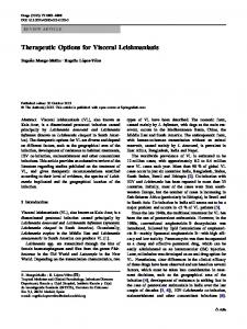
Two additional patients were referred to our institution for management of clinically significant sleeve stenosis after undergoing LSG at another facility. The first patient had undergone LSG 36 days before transfer to our hospital. The procedure was performed using the endoscope as a bougie and with tissue reinforcement. It was complicated by a large staple-line hematoma seen on computed tomography (CT) scan obtained on postoperative day (POD) 2. A contrast study performed on POD 1 did not demonstrate passage of contrast past the GE junction, and the patient reported inability to swallow saliva. No operative intervention was performed. This patient was discharged and readmitted two times for dysphagia and dehydration, with similar findings on repeated contrast studies. At the time of transfer, no parenteral nutrition had been administered, and the patient had lost 62 pounds (28 kg), having been administered only intravenous (IV) fluids. At admission to our facility, the patient underwent a UGI contrast study, which demonstrated a very thin stream of contrast passing just distal to the GE junction. Endoscopy was performed, which confirmed a stenosis just distal to the GE junction (Fig. 1). With a gentle push and rightward twist, the scope easily traversed the stricture. Dilation was attempted using an 18-mm balloon. The stenotic area easily dilated without any evidence of mucosal change. An 18 9 100-mm Polyflex covered esophageal stent (Boston Scientific, Natick, MA, USA) was inserted under fluoroscopic guidance. Fluoroscopy confirmed it to be in good position traversing the area of the stenosis. Almost immediately after deployment, the stent migrated up into the esophagus and required removal. The patient was discharged home with total parenteral nutrition for 4 weeks until normalization of albumin and prealbumin levels.
741
Laparoscopic exploration was performed 89 days after the LSG. Intraoperative findings demonstrated a twisting of the gastric sleeve just distal to the GE junction due to a large resolving hematoma on the neo-greater curvature of the mid-body of the sleeve. Attempts made to correct the twist failed. Therefore, conversion to a Roux-en-Y gastric bypass (LRYGBP) was performed, creating a pouch just proximal to the twist. The patient was discharged on POD 3 receiving a liquid diet, which was advanced per routine. Tolerance of a solid diet occurred 118 days after the LSG. The second patient had undergone LSG 21 days before transfer. The operative reports noted that intraoperatively, a staple ‘‘misfire’’ had occurred opposite the incisura, and the area had been oversewn with absorbable suture. The contrast study performed on POD 1 had demonstrated evidence of a leak, so the patient had been taken to the operating room, where exploratory laparotomy had been performed. The suspected leak had been confirmed and the area repaired using permanent suture. The patient had improved clinically but could not tolerate liquids. At this point, the patient was transferred to our facility for further management. A UGI contrast study demonstrated a stricture just proximal to the incisura. Endoscopy was performed, and a 4.5- to 6-cm stricture was traversed using a pediatric endoscope. The stricture also was noted to be narrow, approximately 4 mm in diameter. Two serial endoscopic dilations were attempted over several days using 9- to 12-mm balloons. A week later, a 18 9 70-mm Alimaxx-ES (Merit Medical Systems Inc, South Jordan, UT, USA) fully covered esophageal stent was placed. Liquids were tolerated by the patient, who was discharged from the hospital. Repeat esophagogastroduodenoscopy with stent removal
Fig. 1 Upper gastrointestinal (GI) contrast study and endoscopy. A A small stream of contrast is noted to pass just distal to the gastroesophageal junction. B Endoscopy demonstrating the functional stenosis
123
742
Surg Endosc (2012) 26:738–746
was performed 27 days later due to epigastric pain and discomfort. The patient still was able to tolerate a thin liquid diet, but subsequent attempts to advance the diet failed. The patient was hesitant to undergo a revisional surgery and continued on a high-protein liquid diet at home. Approximately 1 week later, the patient requested surgical management. A laparoscopic exploration was performed 65 days after the LSG. Extensive scar tissue was identified. The area of stenosis was located, and conversion to a LRYGBP was performed just proximal to this area. The patient’s hospital course was complicated by a hospital-acquired urinary tract infection treated with antibiotics, and the patient was discharged home on POD 6 receiving a liquid diet. The diet was routinely advanced, and the time from LSG to tolerance of a solid diet was 93 days. Table 5 summarizes the management of the transferred patients.
Discussion As federal employees, we practice in a unique environment unhampered by the biases of the paid and the payer. All three bariatric surgery procedures (laparoscopic adjustable gastric banding [LAGB], LSG, and LRYGBP) are offered to patients entering our program. The patients are educated about each procedure, including the risks, complications, and outcomes for each. Based on patient characteristics, the operating surgeon may recommend one procedure over the other, but ultimately, the choice of procedure rests with the patient. Since the introduction of LSG to our program in 2008, it has become the most common bariatric procedure we perform. We believe this may be due to personal interactions among our patients within a tight-knit military community as well as their participation in the bariatric support group. A recent report from the Bariatrics Outcomes Longitudinal Database (BOLD) demonstrated that between June 2007 and May 2009, LSG was the third most common bariatric procedure performed in the United States, Table 5 Management outcomes for transferred patients
Time to first dilation after surgery (days) No. of balloon dilations Balloon size (mm) Time from first dilation to stent (days) Time from LSG to operative revision (days) Time from LSG to solid diet (days)
Mean ± SD
Range
23.5 ± 4.9
20–27
2.5 ± 0.7
2–3
16.5 ± 2.1 20.0 ± 14.1
15–18 10–30
77.0 ± 17.0
65–89
105.5 ± 17.7
93–118
SD standard deviation, LSG laparoscopic sleeve gastrectomy
123
although it still represented a very small percentage (2.29%) of the overall cases managed [26]. Over the past 5 years, findings have shown LSG to be a safe and effective primary weight loss procedure [10, 13, 16, 27–29]. Short- and mid-term outcomes appear promising, with some even approaching those seen with LRYGBP. Increasingly, the sleeve also is used as a revisional procedure for patients who have failed weight loss after LAGB or vertical band gastroplasty (VBG) [30, 31]. Although LSG is less technically demanding than LRYGBP, pitfalls exist that can greatly complicate the patient’s postoperative course. Such complications are well documented, with staple-line leak, hemorrhage, return to the operating room, and stricture among those most often discussed [13, 15, 17, 18]. As LSG gains popularity among surgeons, patients, and insurers, it can be anticipated that such complications will become more prevalent. We found our symptomatic sleeve stenosis rate of 3.5% to be higher than previously reported in other studies. The significance of this report lies in its identification of factors contributing to sleeve stenosis in an attempt to prevent them. As more surgeons begin performing this procedure on a more routine basis, this will become ever more important. In a recent review of 36 studies evaluating LSG as a primary and staged procedure, Brethauer et al. [16] demonstrated that the rate of postoperative strictures requiring endoscopic or operative intervention was 0.6% in studies with more than 100 patients. With such results, the technique must be considered as the cause. These episodes of stenosis did not occur early in our experience. The first symptomatic stenosis identified and treated was in our 91st patient, well over a year after adoption of LSG into our practice. Our technique changed 7 months before the first stenosis episode and has remained constant since that time. It entails initiation of the sleeve approximately 3 to 4 cm orad from the pylorus, use of a 36-Fr bougie, tissue-reinforced staples, and selective segmental imbrication of the staple line. The change correlates mainly with a decrease in bougie size from 40 to 36 Fr and initiation of single-use loading unit stapler use with integrated bioabsorable reinforcement (Duet TRS; Covidien, Norwalk, CT, USA). When required, the selective segmental imbrication was performed without the aid of a bougie, after which an intraoperative endoscopy demonstrated passage of the endoscope into the antrum with visualization of the pylorus in all eight cases and no evidence of stenosis. Notably, only 2 of the 230 patients who underwent LSG had no staple line reinforcement, and our change in the type of stapleline reinforcement used occurred 7 months and 54 patients before the first symptomatic stenosis in this series. All eight patients had staple-line reinforcement, and four of them required selective segmental imbrication due to
Surg Endosc (2012) 26:738–746
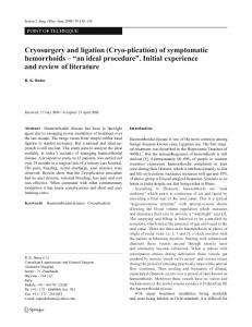
concerns of the staple line. One of these patients had staple-line reinforcement except at the very top of the sleeve (a single unreinforced 45-mm load). This area was imbricated and is believed to be the site at which the stenosis occurred. Two additional patients had the superior 3 cm of reinforced staple line imbricated but subsequently experienced a stenosis in the mid-body. In the final patient, a reinforced staple line was segmentally imbricated in the mid-body across a staple-line junction. The later diagnosis for this patient was a mid-body stenosis at the presumed area of imbrication. It is possible that the act of imbrication itself can lead to the development of a symptomatic stenosis. Therefore, care should be taken to limit the amount of tissue being imbricated, especially with reinforced staple lines, because this may significantly narrow the lumen and contribute to the development of symptomatic stenosis. An increase in the leak rate is reported to occur with decreasing size of the bougie [17]. Several studies have reported the size of the sleeve based on the size of the bougie used and its correlation with weight loss [5, 7, 16, 20, 32]. This literature is contradictory and ultimately suggests that the overall size of the sleeve does not correlate with better weight loss. Additionally, the literature remains contradictory regarding the correlation of stenosis rates with bougie size used. For example, Cottom et al. [20] reported using 46- to 50-Fr bougies with a stenosis rate of 3.9%, whereas Lalor et al. [18] reported using either a 44- or 52-Fr bougie with a stenosis rate of only 0.7%. This suggests another technical cause independent of bougie size contributing to the stenosis rate. Notably, Cottom et al. [20] did state that changing their overall technique from imbricating the staple line to covering it with fibrin glue caused their stenosis rate to disappear. This type of stenosis most likely occurred due to overnarrowing of the sleeve at the incisura. Care must be taken to leave plenty of tissue anteriorly in this area, especially when the sleeve starts closer to the pylorus. Narrowing here can occur as the clinician begins to ‘‘cut the corner’’ even with a larger bougie in place due to over-retraction of the greater curvature during stapling. The process of retracting the greater curvature where tension is progressively applied can cause stretch on the stomach during division. Once the bougie is removed, the stomach will recoil, resulting in a narrowing. A twisted or spiral sleeve is another cause of symptomatic stenosis. This twist has been previously described [21, 33]. Progressive rotation of the staple line in an anterior to posterior plane can lead to a narrowing despite a fairly normal luminal diameter. This curve can make passage of enteric contents difficult, resulting in a functional stenosis. This often is demonstrated by easy passage of the endoscope or balloon dilator through the narrowed area. Much like a clown twisting a straight balloon, an anterior
743
twist at the incisura can result in a functional stenosis (Fig. 2). An endoscope can pass through by pushing and twisting in the same direction, and a balloon dilator can be used to open the stenosis. However, the stenosis returns at withdrawal of the endoscope or deflation of the balloon dilator. A functional sleeve stenosis also can result from external sources such as a hematoma that causes the sleeve to scar in a kinked manner. Such complications should be promptly treated [32]. All eight patients in this study who experienced symptomatic stenosis after LSG were treated endoscopically. Only age differed between the two groups, although the study may have been underpowered to detect significant differences. It should be noted that 75% of these patients underwent a UGI contrast study on POD 1, with only one demonstrating mild delay in the passage of contrast. An abnormal appearance of the postoperative UGI contrast study is common and can be difficult to interpret. It therefore is not a good predictor of symptomatic stenosis [34, 35]. Although LSG is being used more frequently, many radiologists may be relatively inexperienced with this anatomy and may not know how to interpret correctly what they are seeing. It is therefore important that the operating surgeon be present during these studies to help direct their interpretation. It has been our practice to use this study selectively to exclude a leak, after which we advance the patient’s diet based on his or her clinical progression. The patients in this series presented about 7 weeks after surgery, although one presented nearly 5 months after
Fig. 2 Representation of the spiral sleeve. Much like the straight balloon a clown uses to make objects, the functional stenosis is caused by twisting of the sleeve, although the luminal diameter remains of a reasonable caliber
123
744
surgery. A UGI contrast study was obtained and demonstrated a focal narrowing of the sleeve in 71.4% of the patients. This is a reasonable initial test because it is relatively inexpensive and noninvasive. All the patients underwent endoscopy, which confirmed the stenosis, then allowed simultaneous treatment with balloon dilation. Two patients were found to have a functional stenosis due to a twist or kink of the stomach at the incisura. Although the diameter of the lumen may appear to be relatively normal in size, the twist of the sleeve must offer sufficient resistance to counter the passage of gastric contents. Symptoms consistent with this type of stenosis were encountered in our series as the patients were advanced to a more solid diet despite what appeared to be a normal UGI contrast study. On the average, almost two dilations to a diameter of 15 mm were required for symptom resolution and resumption of a solid diet. Between each dilation, the patients demonstrated improvement in oral tolerance (e.g., advancement to a pureed diet). These patients were most likely amenable to balloon dilation due to the anatomically short length of their stenoses. When compared with the nonstenosis patients, these patients had a greater %EWL at both 6 and 12 months. This was not found to be significant and is understandable given the prolonged period of oral intolerance. Two patients were transferred to our facility due to dysphagia and oral intolerance immediately after surgery. Findings showed that both conditions occurred after perioperative complications: a large staple-line hematoma in the one case and a staple-line leak in the other case. At endoscopy, both patients were found to have a long area of stenosis. Our initial concern was their nutritional status. Total parenteral nutrition was used in both cases, and as the patients were able, a high-protein liquid oral diet was initiated. Nasoenteric feeds proved to be difficult due to the sense of ‘‘drowning’’ both patients experienced at placement of a feeding tube. A percutaneous jejunal feeding tube was not placed to minimize distortion of the anatomy should revision be required. Initial nonsurgical management was attempted, including serial endoscopic dilation and placement of fully covered intestinal stents. Both patients experienced minimal relief from each dilation. The one patient was found to have a high functional stenosis just distal to the GE junction, most likely due to kinking from a large staple-line hematoma. Both the scope and balloon dilator passed easily through the stenosis, but the patient was unable to tolerate oral secretions. At attempted placement of the covered stent, it was promptly displaced superiorly into the esophagus. This was thought to result from the twisting of the sleeve itself. The other patient had successful stent placement. Although it did allow continued liquid oral intake, as
123
Surg Endosc (2012) 26:738–746
previously described, the stent caused a great deal of discomfort and pain requiring prolonged narcotic analgesia [34]. Despite keeping the stent for 27 days, no improvement in symptoms was observed. Both patients underwent a difficult laparoscopic revisional procedure, with successful conversion to an RYGBP and eventual progression to a solid diet. Although longitudinal seromyotomy in long-sleeve strictures has been described previously, it was not thought to be appropriate in these settings [23]. We propose the following management algorithm (Fig. 3) for patients who have undergone LSG with persistent nausea, vomiting, or dysphagia. First, an UGI contrast study should be obtained. If this study demonstrates an abnormal finding or if the symptoms persist over time, an esophagogastroduodenoscopy should be performed with anticipation of performing a dilation. Repeat dilation can be performed as long as the patient demonstrates improvement in oral tolerance. Placement of a stent also can be considered, although a stent often is poorly tolerated by the patient due to pain and discomfort [36]. Failure of progression to a normal diet warrants consideration of operative revision to an RYGBP.
Conclusion Caution should be taken in performing LSG to avoid the creation of sleeve stenosis. Clinically significant
Fig. 3 Management algorithm. UGI, upper gastrointestinal contrast study; EGD, esophagogastroduodenoscopy
Surg Endosc (2012) 26:738–746
short-segment stenoses may be treated successfully with endoscopic balloon dilation. Long-segment stenoses are less likely to respond to endoscopic techniques and may ultimately require conversion to Roux-en-Y gastric bypass. Disclosures Amit Parikh, Joshua B. Alley, Richard M. Peterson, Michael C. Harnisch, Jason M. Pfluke, Donovan M. Tapper, and Stephen J. Fenton have no conflicts of interest or financial ties to disclose. The opinions expressed are solely those of the authors and do not represent the views of the United States Air Force, the Department of Defense, or the United States Government or their endorsement.
References 1. Hess DS, Hess DW (1998) Biliopancreatic diversion with a duodenal switch. Obes Surg 8:267–282 2. Marceau P, Biron S, Bourque RA, Potvin M, Hould FS, Simard S (1993) Biliopancreatic diversion with a new type of gastrectomy. Obes Surg 3:29–35 3. Ren CJ, Patterson E, Gagner M (2000) Early results of laparoscopic biliopancreatic diversion with duodenal switch: a case series of 40 consecutive patients. Obes Surg 10:514–523; discussion 524 4. Regan JP, Inabnet WB, Gagner M, Pomp A (2003) Early experience with two-stage laparoscopic Roux-en-Y gastric bypass as an alternative in the super–super-obese patient. Obes Surg 13:861–864 5. Milone L, Strong V, Gagner M (2005) Laparoscopic sleeve gastrectomy is superior to endoscopic intragastric balloon as a first stage procedure for super-obese patients (BMI C 50). Obes Surg 15:612–617 6. Silecchia G, Boru C, Pecchia A, Rizzello M, Casella G, Leonetti F, Basso N (2006) Effectiveness of laparoscopic sleeve gastrectomy (first stage of biliopancreatic diversion with duodenal switch) on comorbidities in super-obese high-risk patients. Obes Surg 16:1138–1144 7. Himpens J, Dobbeleir J, Peeters G (2010) Long-term results of laparoscopic sleeve gastrectomy for obesity. Ann Surg 252: 319–324 8. Givon-Madhala O, Spector R, Wasserberg N, Beglaibter N, Lustigman H, Stein M, Arar N, Rubin M (2007) Technical aspects of laparoscopic sleeve gastrectomy in 25 morbidly obese patients. Obes Surg 17:722–727 9. Gluck B, Movitz B, Jansma S, Gluck J, Laskowski K (2010) Laparoscopic sleeve gastrectomy is a safe and effective bariatric procedure for the lower BMI (35.0–43.0 kg/m2) population. Obes Surg. doi:10.1007/s11695-010-0332-7:03 10. D’Hondt M, Vanneste S, Pottel H, Devriendt D, Van Rooy F, Vansteenkiste F (2011) Laparoscopic sleeve gastrectomy as a single-stage procedure for the treatment of morbid obesity and the resulting quality of life, resolution of comorbidities, food tolerance, and 6-year weight loss. Surg Endosc 28(8):2498–2504 11. Rubin M, Yehoshua RT, Stein M, Lederfein D, Fichman S, Bernstine H, Eidelman LA (2008) Laparoscopic sleeve gastrectomy with minimal morbidity: early results in 120 morbidly obese patients. Obes Surg 18:1567–1570 12. Tagaya N, Kasama K, Kikkawa R, Kanahira E, Umezawa A, Oshiro T, Negishi Y, Kurokawa Y, Nakazato T, Kubota K (2009) Experience with laparoscopic sleeve gastrectomy for morbid versus super morbid obesity. Obes Surg 19:1371–1376
745 13. Tucker ON, Szomstein S, Rosenthal RJ (2008) Indications for sleeve gastrectomy as a primary procedure for weight loss in the morbidly obese. J Gastrointest Surg 12:662–667 14. Gagner M, Deitel M, Kalberer TL, Erickson AL, Crosby RD (2009) The second international consensus summit for sleeve gastrectomy, March 19–21, 2009. Surg Obes Rel Dis 5:476–485 15. Frezza E, Reddy S, Gee LL, Wachtel MS (2009) Complications after sleeve gastrectomy for morbid obesity. Obes Surg 19:684–687 16. Brethauer S, Hammel J, Schauer P (2009) Systematic review of sleeve gastrectomy as staging and primary bariatric procedure. Surg Obes Rel Dis 5:469–475 17. Gagner M (2010) Leaks after sleeve gastrectomy are associated with smaller bougies: prevention and treatment strategies. Surg Laparosc Endosc Percutan Tech 20:166–169 18. Lalor PF, Tucker ON, Szomstein S, Rosenthal R (2008) Complications after laparoscopic sleeve gastrectomy. Surgery Obes Rel Dis 4:33–38 19. Zundel N, Hernandez JD, Galvao Neto M, Campos J (2010) Strictures after laparoscopic sleeve gastrectomy. Surg Laparosc Endosc Percutan Tech 20:154–158 20. Cottam D, Qureshi FG, Mattar SG, Sharma S, Holover S, Bonanomi G, Ramanathan R, Schauer P (2006) Laparoscopic sleeve gastrectomy as an initial weight-loss procedure for high-risk patients with morbid obesity. Surg Endosc 20:859–863 21. Lacy A, Ibarzabal A, Obarzabal A, Pando E, Adelsdorfer C, Delitala A, Corcelles R, Delgado S, Vidal J (2010) Revisional surgery after sleeve gastrectomy. Surg Laparosc Endosc Percutan Tech 20:351–356 22. Dapri G, Cadie`re GB, Himpens J (2009) Reinforcing the staple line during laparoscopic sleeve gastrectomy: prospective randomized clinical study comparing three different techniques. Obes Surg. doi:10.1007/s11695-009-0047-9 23. Dapri G, Cadie`re GB, Himpens J (2009) Laparoscopic seromyotomy for long stenosis after sleeve gastrectomy with or without duodenal switch. Obes Surg 19:495–499 24. National Institutes of Health (1992) Gastrointestinal surgery for severe obesity: National Institutes of Health consensus development conference statement. Am J Clin Nutr 55:615S–619S 25. Alley J, Fenton S, Harnisch MC, Angeletti MN, Peterson R (2010) Integrated bioabsorbable tissue reinforcement in laparoscopic sleeve gastrectomy. Obes Surg 21(8):1311–1315 26. Demaria EJ, Pate V, Warthen M, Winegar DA (2010) Baseline data from American Society for Metabolic and Bariatric Surgery: designated bariatric surgery centers of excellence using the bariatric outcomes longitudinal database. Surg Obes Rel Dis 6:347–355 27. Bohdjalian A, Langer FB, Shakeri-Leidenmu¨hler S, Gfrerer L, Ludvik B, Zacherl J, Prager G (2010) Sleeve gastrectomy as sole and definitive bariatric procedure: 5-year results for weight loss and ghrelin. Obes Surg 20:535–540 28. Menenakos E, Stamou MK, Albanopoulos K, Papailiou J, Theodorou D, Leandros E (2009) Laparoscopic sleeve gastrectomy performed with intent to treat morbid obesity: a prospective single-center study of 261 patients with a median follow-up of 1 year. Obes Surg 20(3):276–282 29. Nocca D, Krawczykowsky D, Bomans B, Noe¨l P, Picot MC, Blanc PM, De Seguin De Hons C, Millat B, Gagner M, Monnier L, Fabre JM (2008) A prospective multicenter study of 163 sleeve gastrectomies: results at 1 and 2 years. Obes Surg 18:560–565 30. Goitein D, Feigin A, Segal-Lieberman G, Goitein O, Papa MZ, Zippel D (2011) Laparoscopic sleeve gastrectomy as a revisional option after gastric band failure. Surg Endosc 23(7):1559–1563 31. Foletto M, Prevedello L, Bernante P, Luca B, Vettor R, FranciniPesenti F, Scarda A, Brocadello F, Motter M, Famengo S, Nitti D
123
746 (2010) Sleeve gastrectomy as revisional procedure for failed gastric banding or gastroplasty. Surg Obes Rel Dis 6:146–151 32. Parikh M, Gagner M, Heacock L, Strain G, Dakin G, Pomp A (2008) Laparoscopic sleeve gastrectomy: does bougie size affect mean %EWL? Short-term outcomes. Surg Obes Rel Dis 4:528–533 33. Uglioni B, Wo¨lnerhanssen B, Peters T, Christoffel-Courtin C, Kern B, Peterli R (2009) Midterm results of primary vs secondary laparoscopic sleeve gastrectomy (LSG) as an isolated operation. Obes Surg 19:401–406
123
Surg Endosc (2012) 26:738–746 34. Werquin C, Caudron J, Mezghani J, Leblanc-Louvry I, Scotte´ M, Dacher JN, Savoye-Collet C (2008) Early imaging features after sleeve gastrectomy. J Radiol 89:1721–1728 35. Goitein D, Goitein O, Feigin A, Zippel D, Papa M (2009) Sleeve gastrectomy: radiologic patterns after surgery. Surg Endosc 23:1559–1563 36. Eubanks S, Edwards CA, Fearing NM, Ramaswamy A, De La Torre RA, Thaler KJ, Miedema BW, Scott JS (2008) Use of endoscopic stents to treat anastomotic complications after bariatric surgery. J Am Coll Surg 206:935–938; discussion 938–939