APPLIED AND ENVIRONMENTAL MICROBIOLOGY, Oct. 1997, p. 3858–3865 0099-2240/97/$04.0010 Copyright © 1997, American Society for Microbiology
Vol. 63, No. 10
Detection and Characterization of Fungal Infections of Ammophila arenaria (Marram Grass) Roots by Denaturing Gradient Gel Electrophoresis of Specifically Amplified 18S rDNA GEORGE A. KOWALCHUK,* SASKIA GERARDS,
AND
JAN W. WOLDENDORP
Department of Plant-Microorganism Interactions, Center for Terrestrial Ecology, Netherlands Institute of Ecology, 6666 ZG Heteren, The Netherlands Received 8 April 1997/Accepted 3 August 1997
Marram grass (Ammophila arenaria L.), a sand-stabilizing plant species in coastal dune areas, is affected by a specific pathosystem thought to include both plant-pathogenic fungi and nematodes. To study the fungal component of this pathosystem, we developed a method for the cultivation-independent detection and characterization of fungi infecting plant roots based on denaturing gradient gel electrophoresis (DGGE) of specifically amplified DNA fragments coding for 18S rRNA (rDNA). A nested PCR strategy was employed to amplify a 569-bp region of the 18S rRNA gene, with the addition of a 36-bp GC clamp, from fungal isolates, from roots of test plants infected in the laboratory, and from field samples of marram grass roots from both healthy and degenerating stands from coastal dunes in The Netherlands. PCR products from fungal isolates were subjected to DGGE to examine the variation seen both between different fungal taxa and within a single species. DGGE of the 18S rDNA fragments could resolve species differences from fungi used in this study yet was unable to discriminate between strains of a single species. The 18S rRNA genes from 20 isolates of fungal species previously recovered from A. arenaria roots were cloned and partially sequenced to aid in the interpretation of DGGE data. DGGE patterns recovered from laboratory plants showed that this technique could reliably identify known plant-infecting fungi. Amplification products from field A. arenaria roots also were analyzed by DGGE, and the major bands were excised, reamplified, sequenced, and subjected to phylogenetic analysis. Some recovered 18S rDNA sequences allowed for phylogenetic placement to the genus level, whereas other sequences were not closely related to known fungal 18S rDNA sequences. The molecular data presented here reveal fungal diversity not detected in previous culture-based surveys. Furthermore, molecular data have been most useful in agricultural systems in which pathogens have already been well characterized, and the main goal has been to develop a fast, efficient screening for the presence of a particular disease (for a review, see reference 18). The use of highly variable regions of DNA, such as the ribosomal DNA (rDNA) internal transcribed spacer regions 1 and 2 (ITS1 and ITS2, respectively) as the targets for molecular analyses has allowed for such finetuned discernment between pathogenic and nonpathogenic fungi by use of restriction fragment length polymorphism (9, 14, 21) or specifically targeted oligonucleotide primers (34, 60). Several molecular analyses have successfully employed the use of the random amplified polymorphic DNA (6, 55) technique, probes derived from random amplified polymorphic DNA bands (10), or restriction fragment length polymorphism of total genomic DNA (24, 46) to detect and distinguish between closely related fungi, but again these methods are often targeted at discriminating known species or strains from each other. The above-described techniques are inadequate for the analysis of fungal communities from field sites where several different fungi may be simultaneously present and where their identities are unknown. Such broad analysis requires the use of a target region which is comparable across diverse taxa yet still contains enough relevant phylogenetic information, such as the 18S rRNA gene (4, 22, 57–59). The monophyletic nature of the true fungi (the Ascomycetes, Basidiomycetes, Zygomycetes, and Chytridiomycetes [3]) has allowed for the synthesis of fungal specific PCR primers (56). This report describes a
Fungal pathogens cause serious damage to agricultural crops and to some plants in their natural setting (20, 50). Although key fungal pathogens have been identified and can be specifically detected for many important agricultural diseases, most natural fungal pathosystems have been far-less-well characterized. Many fungi, especially those inhabiting roots, lack sufficient distinguishing morphological characteristics, making identification difficult and time-consuming, especially in species-rich systems. Identification of potential pathogens often requires that fungal strains first be cultured, prior to classification. These procedures make it difficult to detect strains which either grow slowly under the specified laboratory conditions or have an obligatory commensal/parasitic relationship with the plant or other organisms. Such biases may be particularly important in field situations where the dominant pathogens are unknown. The advent of molecular techniques, many based on the PCR (29, 37), has opened the door to more-sensitive and rapid methods of fungal detection (13, 41). A wide variety of molecular methods have already been employed to detect known crop pathogens (36, 53), yet there is little knowledge of the role of potential pathogens in natural ecosystems, in which several fungal species, as well as other organisms, may act in synergy.
* Corresponding author. Mailing address: Department of Plant-Microorganism Interactions, Netherlands Institute of Ecology, Center for Terrestrial Ecology, Boterhoeksestraat 22, Postbox 40, 6666 ZG Heteren, The Netherlands. Phone: 31 (0) 2647 91314. Fax: 31 (0) 2647 23227. E-mail:
[email protected]. 3858
VOL. 63, 1997
DGGE DETECTION OF FUNGI IN PLANT ROOTS
nested PCR strategy used to amplify a 569-bp 18S rDNA fragment, with an additional 59 36-bp GC-rich sequence introduced by the PCR, from fungal material contained within plant roots. This GC clamp (40) stabilizes the melting behavior of the DNA fragment, making it suitable for analysis by denaturing gradient gel electrophoresis (DGGE). This technique is capable of separating closely related sequences by their differential mobilities in a gradient of denaturants (33). DGGE has recently been used to study the ecology of bacterial communities from several different environments and has helped to identify important community members after PCR with general eubacterial 16S rDNA primers (31), primers specifically targeted to the 16S rRNA gene of monophyletic bacterial groups (26), or primers directed at other relevant structural genes (54). Community fingerprints can subsequently be interpreted by comparison with reference patterns of known phylogenetic origin (12, 26), by hybridization with oligonucleotide probes targeted for specific internal sites (43, 45), or by sequence analysis of excised bands (12, 26, 30). The last possibility also requires comparison with a suitable dataset to allow for valid phylogenetic analysis. Marram grass (Ammophila arenaria L.) is the most important plant species responsible for the stabilization of coastal foredunes in Europe, western North America, and Australia (19, 25). This plant species dominates and thrives in outer dunes, where fresh wind-blown sand accumulates. However, when sand accumulation diminishes, marram stands often become feeble and begin to degenerate. The reasons for this decline are thought to be plant-pathogenic fungi and nematodes (49, 50), which comprise a host-specific pathosystem contributing to the succession of coastal dune plant species (48). Identification of the pathogens responsible for the degeneration of A. arenaria is therefore an essential step in understanding these coastal dune ecosystems and allowing for their proper management. To this end, De Rooij Van Der Goes et al. (7) surveyed Dutch coastal foredunes to identify potentially important fungi and nematodes associated with A. arenaria. Laboratory pot experiments also showed that a mixture of common fungal species decreased the growth of A. arenaria and that the addition of nematodes exacerbated this effect (8). Although this survey identified some potentially important pathogenic fungi and produced a working strain collection of dune isolates, these investigators (7, 8) acknowledge limitations and potential biases of such culture-based analyses. It is hypothesized that specific fungi play an important role in the degeneration of A. arenaria and the primary succession of plant species in coastal sand dune systems (8, 48). To avoid the difficulties and biases of culture methods for surveying fungi, we sought to develop a PCR-based system for the analysis of fungal communities. Such a system must be able to amplify fungal DNA from plant material and to separate sequences for the total fungal community into the sample’s constituent parts. To this end, we developed a nested PCR system which was coupled with DGGE and sequence analysis of excised DGGE bands. Thus, the goal of this study was to test and describe this molecular strategy with pure fungal cultures and plants infected in the laboratory and to show how it can be applied to the analysis of field roots taken from healthy or degenerating A. arenaria stands. MATERIALS AND METHODS Fungal strains and culture conditions. Cultures (Table 1) were kept and grown according to the conditions recommended by the Centraalbureau voor Schimmelcultures (Baarn, The Netherlands), and “CBS” indicates strains which were obtained from this organization. Other fungal isolates referred to in this study are from the collection of De Rooij Van Der Goes et al. (7). Fungal
3859
TABLE 1. Fungal strains used in this studya Fungal strain
Partial 18S rDNA sequence determined
Acremonium sp. strain C158 ................................................... Plectosphaerella cucumerina CBS215.84 ................................ Plectosphaerella sp. strain C99 ................................................ Colletrichum lindsmuthianum SG2 ......................................... Apiospora sp. strain H37 ......................................................... Cladosporium cladosporioides CBS131.29 ............................. Cladosporium cladosporioides DR114.................................... Pleospora sp. strain C74 .......................................................... Microdochium bolleyi CBS137.64 ........................................... Microdochium sp. strain C95 .................................................. Verticillium albo-atrum CBS394.91......................................... Verticillium dahliae IPO1......................................................... Ulocladium atrum CBS193.67 ................................................. Ulocladium atrum SK46........................................................... Phoma exigua CBS833.84 ........................................................ Phoma exigua SK11.................................................................. Phoma leveillei SG1.................................................................. Fusarium oxysporum CBS619.87............................................. Fusarium oxysporum CWB1 .................................................... Fusarium oxysporum H309 ...................................................... Fusarium culmorum CBS122.73 ............................................. Fusarium culmorum IPO39-01................................................ Fusarium culmorum C27 .........................................................
2 1 1 1 2 1 1 1 1 1 1 2 1 1 1 1 1 1 1 1 1 1 1
a 1, the partial 18S rDNA sequence was determined during the course of this study and use in the phylogenetic analysis to construct the tree in Fig. 4.
cultures for DNA isolations were grown on potato dextrose agar (PDA) plates at 20°C in the dark for 1 to 3 weeks. Infection of control plants. A. arenaria and Triticum aestivum L. (wheat) seeds were surface sterilized (40 min, 5% [wt/vol] hypochlorite; 20 min, 1% [wt/vol] hypochlorite) and germinated on 1.5% (wt/vol) water agar plates. One week after germination, they were transferred to 1-liter flasks containing 400 ml of 1.2% (wt/vol) water agar (pH 6.0) with Hoagland solution (at a ratio of 1/4) and subjected to an artificial day/night schedule of 12 h of light at 18°C and 12 h of dark at 10°C. Inocula from fungi used to infect plants consisted of Phoma exigua (CBS833.84), Fusarium oxysporum (CBS619.87 or CWB1), and Fusarium culmorum (CBS122.73 or IPO39-01) cultures that were grown to confluence on PDA plates. Colony plugs (4 by 4 mm) were placed at the base of each A. arenaria seedling to infect the test plants. Upon harvest, root material which was not used for DNA isolation was incubated on PDA plates for 2 weeks at 20°C in the dark to confirm the presence of the fungus in question. White asparagus (Asparagus officinalis L.) roots infected with F. oxysporum CWB1 (2) were obtained from W. J. Blok (Department of Phytopathology, Agricultural University of Wageningen, Wageningen, The Netherlands). Field sample sites and material. Three sites were sampled from the Dutch coastal dunes near Oostvoorne (51°549N, 4°049E), near the Harlingvlietdam (51°509N, 4°059E), and near Ouddorp (51°499N, 3°579E). All sites have similar vegetation successions (48, 51). A. arenaria is healthy on the beachward dune face where it is exposed to wind-blown sand yet degenerates at more inland locations. Plant roots from the most-recent-year’s growth were gathered randomly from both healthy and degenerating A. arenaria stands at each location in July 1995. Samples were transported at 4°C to the laboratory and kept at 280°C until use. Roots were sonicated two times for 5 min each, first in 10 mM sodium pyrophosphate and then in distilled water (8), to remove root surface fungi. Root pieces from the different locations and types of A. arenaria stands were analyzed separately. A total of 44 1-cm root pieces were used for the PCR and DGGE analysis: six from each type (healthy and degenerating) of stand from Harlingvlietdam and eight root pieces from each type of stand from the other two locations. DNA isolation. DNA was isolated from fungal cultures by scraping 0.25 g of hyphal material from PDA plates and adding it to a 2-ml screw-capped polypropylene tube containing 0.5 g of glass beads (0.5-mm diameter; BioSpec Products, Techno Lab, Alkmaar, The Netherlands), 0.5 ml of 120 mM K2HPO4 buffer (pH 8.0), and 0.5 ml of water-saturated phenol (pH 8.0; Aquaphenol; Ampligene, Illkirch, France). Tubes were shaken three times for 30 s at 5,000 rpm in a minibeadbeater (BioSpec Products) and kept on ice between periods of shaking. After centrifugation (4 min, 5,000 3 g), 0.4 ml of the aqueous phase was removed for phenol-chloroform and chloroform extraction (39). DNA was precipitated for 2 h at 220°C with 2 volumes of ice-cold 96% (vol/vol) ethanol–0.1 volume of 3 M sodium acetate (pH 5.2). After centrifugation for 15 min (14,000 3 g at 4°C), the pellet was washed once with ice-cold 70% (vol/vol) ethanol, allowed to air
3860
KOWALCHUK ET AL.
dry, resuspended in 100 ml of sterile deionized water, and stored at 220°C until further use. DNA was isolated from root material by modified use of the Puregene DNA isolation kit (Gentra Systems, Research Triangle Park, N.C.). Root material (80 mg) was added to 600 ml of lysis solution–200 mg of 0.5-mm-diameter glass beads (for the destruction of fungal material)–300 mg of 1.0-mm-diameter zirconium beads (BioSpec Products) (for the destruction of plant material). Samples were beaten as described above and subsequently incubated for 1 h at 65°C. Samples were cooled to 37°C and treated with 3 ml of a 10 mM RNase solution for 15 min. After RNase treatment, the tubes were cooled on ice, and 200 ml of the Puregene protein precipitation solution (Gentra Systems) was added. Samples were mixed and centrifuged at 14,000 3 g for 3 min. The supernatant was transferred to a microcentrifuge tube, and DNA was precipitated with 600 ml of ice-cold isopropanol. The tubes were inverted 50 times and centrifuged at 14,000 3 g for 4 min at 4°C. The pellet was resuspended in 400 ml of TE (10 mM Tris, 1 mM EDTA [pH 8.0]) for phenol-chloroform extraction, reprecipitation, and washing (39). Pellets were allowed to air dry before resuspension in 50 ml of water. Nested PCR strategy and conditions. DNA isolated from root material was subjected to PCR with primers NS1 and NS8 (56) and the following thermocycling program: 2 min at 94°C (1 cycle); 30 s at 94°C, 45 s at 50°C, and 90 s at 72°C (30 cycles); and 5 min at 72°C (1 cycle). All reactions were carried out in a volume of 50 ml containing Thermus brokianus DNA polymerase (Dynazyme; Finnzymes, Ipoo, Finland) and 30 pmol of each primer, according to the reaction conditions recommended by the manufacturer. Reaction mixtures were overlaid with 50 ml of mineral oil (molecular biology grade; Sigma) and run in an OmniGene thermal cycler (Hybaid; Teddinton, Middlesex, United Kingdom). Each reaction mixture (30 ml) was analyzed by agarose gel electrophoresis (1.0% [wt/vol] agarose, 0.53 TBE [13 TBE is 90 mM Tris-borate {pH 8.3} and 2 mM EDTA]) with ethidium bromide staining. The band of the expected size, approximately 1.7 kb, was excised from the gel, and DNA was extracted with the aid of the Qiaex II Gel Extraction Kit (Qiagen, Inc., Chatsworth, Calif.). DNA was eluted in a final volume of 50 ml, 0.5 ml of which was used as the template in a second 50-ml PCR mixture with the forward primer NS1-GC (CGCCCG CCGCGCGCGGCGGGCGGGGCGGGGGCACGGGCCAGTAGTCATAT GCTTGTC [the GC clamp sequence is underlined]) and the reverse primer NS2110 (GAATTACCGCGGCTGCTGGC), which overlaps the site of the NS2 primer used by White et al. (56). Reaction conditions were as described above, with the following thermocycling program: 2 min at 94°C (1 cycle); 30 s at 94°C, 60 s at 55°C, and 75 s at 72°C (30 cycles); and 5 min at 72°C (1 cycle). PCR products were again analyzed by agarose gel electrophoresis (1.5% agarose). DGGE. DGGE of PCR products generated by the NS1-GC/NS2110 primer pair was performed according to the protocol of Muyzer et al. (32) with the use of the D-Gene system (Bio-Rad Laboratories, Hercules, Calif.). Polyacrylamide (8% [wt/vol]) gradient gels (1.5 mm thick, 0.53 Tris-acetate-EDTA [TAE] buffer; 37:1 ratio of acrylamide–bis-acrylamide; 25 to 45% denaturant; 20 by 20 cm) were poured with the aid of a gradient maker (C. B. S. Scientific Co., Del Mar, Calif.) and an Econo-pump (Bio-Rad Laboratories) at a speed of 5 ml min21 (40-ml gradient volume). The 100% denaturing acrylamide contained 7 M urea with 40% formamide (33). Gels were poured from bottom to top by pricking a hole through the sealing rubber gasket. To get well-polymerized slots, a 10-ml top gel containing no denaturants was added before polymerization was complete. Gels were run for 16 h at 85 V in 0.53 TAE buffer at a constant temperature of 60°C. Gels were stained in MilliQ (Millipore B. V., Etten-Leur, The Netherlands) water containing 0.5 mg of ethidium bromide liter21 and destained twice in MilliQ water prior to UV transillumination. Gel images were stored by using the Imager system (Ampligene). Recovery of DNA after DGGE. The middle portion of each DGGE band was excised for subsequent DNA isolation. The approximately 60 mg of acrylamide gel material per band was put into a 2-ml screw-capped polypropylene tube with 0.1 g of glass beads (0.1-mm diameter; BioSpec Products) and 0.1 ml of TE buffer and shaken in a minibeadbeater as described previously (12). After centrifugation at 3,000 3 g for 2 min, the gel fragments and buffer were removed with a pipette, transferred to a 1.5-ml microcentrifuge tube, and incubated overnight at 37°C. Buffer (1 ml) containing extracted DNA was used as the template for amplification of 50-ml reaction mixtures with the NS1 (no GC clamp) and NS2110 primers (56) under the conditions described above for the second PCR step. PCR products were purified by using Wizard PCR Preps (Promega, Madison, Wis.), with elution in a final volume of 30 ml of H2O; 5 ml of the eluted solution was subjected to DGGE analysis to approximate the DNA concentration, to confirm fragment purity, and to test for heteroduplexes (data not shown). DNA recovery was not successful for some very faint bands, as no PCR products could be detected after attempted reamplification (see Fig. 3B). Cloning, sequencing, and phylogenetic analysis. The purified PCR product (150 ng) from the 1.7-kb 18S rDNA fragments recovered from fungal cultures was ligated into the plasmid vector pGEM-T in a reaction volume of 10 ml (Promega Corp.). The ligation mix (1 ml) was used to transform E. coli (Epicurian Coli XL1-Blue MRF9 supercompetent cells; Stratagene, La Jolla, Calif.) by the protocol provided by the manufacturer. The presence of inserts with the expected sizes was confirmed by PCR with the flanking vector primers SP6 and T7 (Promega) and a small portion of untreated white colony as the template. The PCR protocol was 30 rounds at 94°C (30 s), 55°C (60 s), and 72°C (70 s), with 2 U of T. brokianus polymerase and the buffer supplied by the manufacturer in a
APPL. ENVIRON. MICROBIOL.
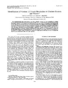
FIG. 1. DGGE of 18S rDNA fragments from 13 fungal species. Chromosomal DNA was isolated from each of 13 different fungal species for amplification of an 18S rDNA fragment by using the NS1-GC/NS2110 primer pair (56), and 5 ml of PCR product was subjected to DGGE analysis. Lanes: 1, Acremonium sp. strain C158; 2, strain CBS215.84; 3, strain SG2; 4, Apiospora sp. strain H37; 5, strain CBS131.29; 6, Pleospora sp. strain C74; 7, strain CBS137.64; 8, strain CBS394.91; 9, strain IPO1; 10, strain CBS193.67; 11, strain CBS833.84; 12, strain CBS619.87; 13, strain CBS122.73.
volume of 25 ml. Colonies containing reannealed vector generated a product with a size of approximately 160 bp, as predicted from the manufacturer’s map, and recombinants containing partial inserts showed products with sizes of ,1.8 kb. Recombinant colonies with inserts with the correct size were grown overnight at 37°C with shaking at 200 rpm in 3 ml of Luria-Bertani broth (Difco, Detroit, Mich.) supplemented with 50 mg of ampicillin ml21. For plasmid isolations, the Wizard mini-prep kit (Promega) was used. DNA sequences were generated by using the vector priming sites SP6 and T7 and the NS1, NS2, and NS4 primers (56). Sequencing reactions were carried out with an ABI PRISM Dye Terminator Cycle Sequence Ready Reaction Kit (Perkin Elmer, Foster City, Calif.) according to the manufacturer’s instructions. The products were analyzed by using an Applied Biosystems (San Jose, Calif.) automatic sequencer (model 373 with Stretch adapter) at the Department of Biotechnology, Wageningen, the Netherlands, and sequences were assembled by using the Sequence Navigator program (version 1.0, release 3.0.1; Applied Biosystems). PCR products recovered from isolated DGGE bands which had been purified as described above were subjected to sequence analysis with the same protocols as those for plasmid DNA but with only the NS1 and NS2 primers. The nucleotide sequences corresponding to positions 50 to 474 of the Saccharomyces cerevisiae 18S rRNA gene (28) were determined for 20 clones derived from fungal isolates and from a total of 52 DGGE bands. The sequence corresponding to this region of the A. arenaria 18S rRNA gene was also determined. Homology searches with the EMBL database were conducted by using the FastaA program (35). All sequences were aligned against a representative selection of fungal sequences extracted from the ribosomal nucleotide databases (27, 47). Sequences were manipulated by using the SeqApp program, version 1.9a169 (17). The alignment used for sequence analysis included 403 sites which could be unambiguously aligned for all recovered clones and published sequence data. All sequence analyses were implemented in PHYLIP 5.57 (11). For distance matrix analyses, the Jukes and Cantor (23) correction was used, tree topology was built by the neighbor-joining method (38), and boot-strapping analysis was conducted for 100 replicates by using the program SeqBoot (11). Nucleotide sequence accession numbers. All culture-derived sequences and unique band-derived sequences identified in this study have been deposited in the EMBL database under accession numbers Z94123 to Z94142 and Z94143 to Z94152, respectively. The sequence of the A. arenaria 18S rRNA gene from position 50 to 474 has been deposited in the EMBL database under accession number Z94153.
RESULTS DGGE of PCR products from pure fungal cultures. PCR products recovered from fungal isolates with the NS1-GC/ NS2110 primer pair were subjected to DGGE analysis to determine the ability of this region of the 18S rRNA gene to discriminate between different fungal species. DNA fragments from all fungal isolates tested migrated in the range of 30 to 36% denaturant concentration (Fig. 1). Although most fungal bands could be easily distinguished from each other, some showed very similar migration behaviors. Band position in the DGGE alone was not a good predictor of phylogenetic posi-
VOL. 63, 1997
DGGE DETECTION OF FUNGI IN PLANT ROOTS
3861
FIG. 2. Detection of known fungal plant infections by PCR and DGGE. PCR products recovered from characterized fungal isolates and plants infected with them are shown. Lanes: 1, sterile A. arenaria; 2, A. arenaria infected with Phoma exigua CBS833.84; 3, Phoma exigua CBS833.84; 4, A. arenaria infected with F. oxysporum CBS619.87; 5, F. oxysporum CBS619.87; 6, A. arenaria infected with F. culmorum CBS122.73; 7, F. culmorum CBS122.73; 8, sterile Triticum aestivum; 9, Triticum aestivum infected with F. culmorum IPO39-01; 10, F. culmorum IPO3901; 11, sterile Asparagus officinalis; 12, Asparagus officinalis infected with F. oxysporum CWB1; 13, F. oxysporum CWB1.
tion. This point is illustrated by the comparison of bands from Verticillium albo-atrum (Fig. 1, lane 8) and Cladosporium cladosporioides (lane 5), which, despite being distantly related, gave very similar DGGE results (around 33.5% denaturant). Verticillium dahliae (lane 9) is more closely related to V. alboatrum yet produces a more distant band (32.5% denaturant) (see Fig. 4) (57). To further characterize the discrimination achieved by DGGE of the NS1-GC/NS2110 PCR product, multiple isolates from several species were analyzed. No intraspecific variation was detected among nine F. culmorum, four F. oxysporum, four Phoma exigua, four Ulocladium atrum, three Cladosporium cladosporioides, three Microdochium bolleyi, and two Chaetomium funicola isolates tested, although these species could be distinguished from each other by DGGE after PCR with a mixed template containing all seven species (results not shown). Nested PCR for recovery of fungal 18S rDNA from infected plant controls. To test the recovery of fungal material from plant roots, nested PCR and DGGE were performed on DNA extracted from plant roots which had been inoculated with known fungal strains (Fig. 2). DNA samples obtained directly from infected roots were first subjected to PCR with the fungus specific NS1/NS8 primers (56). This product was subsequently used as the template in a second PCR with the NS1-GC/ NS2110 primer combination to produce fragments suitable for DGGE analysis. In control experiments, fungal DNA could be detected to a 1:1,000 ratio with competing plant DNA, by using the reaction conditions described (results not shown). In all cases, the fungus that was used to create the infection could be detected by DGGE, by using the band produced by the pure fungal isolate for comparison (Fig. 2). Residual plant material was still recovered in some cases, and its DGGE band could be aligned with that produced by the fragment generated by direct amplification with the NS1-GC/NS2110 primer pair from sterile plant material (for example, Fig. 2, compare lanes 1 and 4). Plant-derived fragments all migrated further than fungus-derived fragments, with the DGGE patterns of the monocots A. arenaria and Triticum aestivum being very similar (38% denaturant) and that of the dicot Asparagus officinalis being clearly distinguishable (37% denaturant). DGGE of field material from healthy and degenerating A. arenaria roots. The nested PCR strategy outlined above was used to recover fungal DNA fragments from field A. arenaria root samples. DGGE analysis revealed relatively simple band-
FIG. 3. (A) DGGE analysis of field root samples. The DGGE patterns of A. arenaria (lane 1), 8 of the 44 root samples examined (lanes 2 to 9), and nine fungal isolates (lanes 10 to 18) are shown. Root samples are from the Harlingvlietdam dune locations from both degenerating (lanes 2 to 4) and vital (lanes 5 to 9) A. arenaria stands. Lanes: 10, F. oxysporum CBS619.87; 11, F. culmorum CBS122.73; 12, U. atrum CBS193.67; 13, Microdochium bolleyi CBS137.64; 14, Phoma exigua CBS833.84; 15, Cladosporium cladosporioides CBS131.29; 16, Apiospora sp. strain H37; 17, Plectospharearella cucumerina CBS215.84; 18, V. dahliae IPO1. (B) Schematic drawing of DGGE pattern. The labels of bands which were excised for sequence analysis correspond with those used in Table 2 and Fig. 4. Faint bands for which DNA recovery was unsuccessful and bands from fungal isolate references have not been assigned labels. The band sequences EB-1, EB-2, EB-3, EB-4, and EB-11 were recovered from samples analyzed on this gel, whereas other band sequences (see Fig. 4 and Table 2) were recovered from the additional 36 root samples examined.
ing patterns, with only one to four distinguishable bands per root sample. In total, 44 root pieces with various degrees of fungal infection from the different sample locations were analyzed in this fashion. Eight representative root-derived DGGE patterns are shown in Fig. 3A, along with the DGGE patterns from A. arenaria and nine fungal species previously isolated from A. arenaria roots from the same locations (7). A DGGE band corresponding to the position of A. arenaria (Fig. 3A, lane 1) was detectable in some samples, especially when the roots showed little or no sign of infection. Although some of the bands detected in field samples could be aligned with those of fungal isolates, others appeared to contain sequences not represented in the collection of fungal isolates used as a source of comparison. Sequence analysis of 18S rDNA fragments recovered from DGGE gels and pure fungal isolates. The partial 18S rDNA sequences for 20 representative fungal strains from species isolated from A. arenaria roots (7) were determined to complement existing comparative sequence information. These sequences were included in the phylogenetic analysis of 18S rDNA sequences recovered after PCR and DGGE from A. arenaria roots (Fig. 4). Comparison of DGGE band-derived sequences revealed the identities of some bands obtained from different root samples, suggesting the presence of the same fungal species in multiple field specimens (Fig. 3B; Table 2). Some band sequences also
3862
KOWALCHUK ET AL.
APPL. ENVIRON. MICROBIOL.
FIG. 4. Neighbor-joining tree from partial 18S rDNA sequences recovered by DGGE. The neighbor-joining tree was constructed as described in the text. Sequences determined in this study from fungal isolates are indicated by asterisks, and those which were recovered by DGGE of environmental samples begin with EB (environmental band). Sequences EB-5 through EB-10 were obtained from root samples not shown in Fig. 3. Bootstrap values (.50) are given only for groups relevant to the discussion.
VOL. 63, 1997
DGGE DETECTION OF FUNGI IN PLANT ROOTS
3863
TABLE 2. Distribution of 18S rDNA sequences No. of root samples from which sequence was recovered at site indicated Sequence designationa
EB-1 EB-2 EB-3 EB-4 EB-5 EB-6 EB-7 EB-8 EB-9 EB-10
Organism with greatest sequence identity (% identity)b
Chytridium conferevae (89.6) Leptosphaeria doliolum (99.6) Microdochium bolleyi CBS137.64* (99.0) Phoma leveillei SG1* (100) Phoma exigua CBS833.84* (99.4) Xerocomus chrysenteron (94.7) Spizellomyces acuminatus (90.3) Mycosphaerella mycopappi (95.3) Aureobasidium pullulans (95.8) Microdochium sp. C95* (98.8)
Total
Oostvoorne
Harlingvlietdam
Ouddorp Total
Healthy
Deg.
Healthy
Deg.
Healthy
Deg.
1 0 0 1 0 0 2 0 0 2
1 0 0 0 0 0 0 3 0 5
1 1 3 2 0 1 0 0 0 0
2 2 3 0 0 0 0 0 3 0
2 2 1 1 2 3 0 0 0 0
1 0 4 0 0 0 0 3 0 0
8 5 11 4 2 4 2 6 3 7
6
9
8
10
11
8
52
a
The unique sequences of DNA products which were recovered from excised bands after DGGE analysis of environment root samples are shown with respect to the 18S rDNA sequence showing greatest identity. Note that only sequences EB-1, EB-2, EB-3, EB-4, and EB-11 were recovered from samples for which the results are shown in Fig. 3A, and other sequences come from root samples for which analysis results are not shown. EB11 was the band produced by DGGE analysis of sterile A. arenaria root samples. b Asterisks indicate the fungal species for which the partial 18S rDNA sequences were determined during the course of this study.
showed high degrees of identity with known fungal 18S rDNA sequences, consistent with phylogenetic placement of the identified sequences to a genus level (Fig. 4; Table 2). Genera which could clearly be detected included Microdochium, Phoma, and Septoria/Leptosphaeria. The first two genera have previously been isolated from these dune systems (7). Previous studies showed Phoma exigua to be a common species in these dune areas, but more recent inventories have revealed a large number of Phoma leveillei isolates (15). The 18S rDNA sequence of an isolate of this latter species was identical to band EB-4, which was isolated from multiple root samples (Fig. 4; Table 2). Other bands showed only modest levels of similarity with known fungal sequences, not allowing precise phylogenetic placement, and yet other bands (EB-1 and EB-7) yielded sequences which, although consistent with fungal origin, showed no clear affiliation with known fungal groups (Fig. 4; Table 2). The results presented here represent only a preliminary demonstration of the described techniques, and the small numbers of samples preclude statistical analysis. However, it is clear from Table 2 that the identification of some sequences was dependent on not only location but also the type of stand sampled. The EB-1 sequence was detected in all sample sets, whereas EB-4, EB-5, and EB-6 were represented only in roots from vigorous stands. EB-8 and EB-9 were the only sequences which were specifically detected in roots from degenerating stands. All recovered sequences, except those of the host plant, showed affiliation with fungi, demonstrating the ability of this nested PCR strategy to target fungal 18S rDNA. The DNA sequences derived from DGGE bands suspected of being from plant origin were identical to that determined for A. arenaria, showing greatest similarity (99.5%) with the small subunit (SSU) rDNA database entries for rice (accession number X00755) and wheat (accession number X02202). DISCUSSION 18S rDNA-based approach to the study of fungal communities. SSU rRNA genes, and the information they contain, have been recognized as powerful tools for both identifying organisms as well as inferring their phylogenetic relationships (1, 58,
59). This target has homologous structure and function for all known organisms, offering the possibility for phylogenetic comparison between diverse taxa (47). It also contains variable regions which can be used to identify and target specific organisms or groups of organisms. PCR has facilitated the recovery of SSU rRNA genes from natural environments, and recent advances in microbial ecology have extended the known diversity of bacteria and archaea (16), have contributed to a greater understanding of the evolutionary relationships between organisms (58, 59), and have allowed detailed description of microbial communities with respect to important environmental factors and introduced perturbations (12, 26, 43, 45). 18S rRNA genes have also been used to gain insight into the evolutionary relationships of the fungi (56, 57), but this report describes the first use of a PCR-based strategy targeting this gene to characterize total fungal communities. An additional advantage of an SSU rRNA-based approach is the existence of a rapidly growing nucleotide sequence database for this gene which can be used for reference in the analysis of unknown sequences recovered from isolated strains or directly from the environment (27). Unfortunately, the number of fungal taxa for which sequence information is available is still quite limited, and we found it necessary to determine the partial 18S rDNA sequences for a number of fungal species common to the investigated dune systems. Of course, the variation contained within the 18S rRNA gene will not be ideal for all levels of analysis. This is illustrated by the fact that our PCR-DGGE strategy could not be used to distinguish between different strains of a single species, although it must be stressed that this lack of intraspecific variation may not be true for all taxa. Distinguishing between such closely related isolates would require the use of a more variable target, such as the ribosomal ITS regions. ITS regions have been used successfully for this purpose (7, 18, 34), but their lack of structural homology across broader taxa preclude their use in higher level phylogenetic analyses. Nested PCR and DGGE strategy. The 18S rDNA region flanked by the NS1 and NS2110 primers was chosen as the target for this analysis because this fragment contains enough sequence information to allow phylogenetic inference and is of suitable size for DGGE analysis (45). The use of this rDNA region to analyze root-infecting fungi is complicated by the fact
3864
KOWALCHUK ET AL.
that the NS1/NS2110 primer pair may amplify plant material (56). To circumvent this problem, a nested PCR was performed, with the first step designed to target the fungi and the second designed to produce products suitable for DGGE. One potential drawback of using a PCR-based approach is the introduction of biases due to the preferential amplification of certain templates (44). The use of two rounds of PCR could augment such biases further. However, control experiments using mixtures of fungal DNAs as the template for PCR gave no reason to believe that detection of certain fungi failed because of poor amplification efficiency in the competitive PCR, although the possibility remains that some fungi may be more resistant than others to the cell lysis methods used. Furthermore, a wide range of isolates was recovered, with examples from both Ascomycetes and Basidiomycetes, suggesting that the techniques used do not systematically exclude major fungal groups. Despite these facts, a single PCR step would be preferred, and fungal PCR primer development should continue. As with any electrophoresis-based technique, the DGGE analysis performed suffers from the limitation that although different band mobilities show non-identity, similar band mobilities do not prove identity. As demonstrated above and elsewhere (26), DGGE banding pattern alone cannot be used to identify and phylogenetically place recovered PCR fragments. Furthermore, due to the relatively broad specificity of the given PCR strategy, especially in the absence, or with very low concentrations, of fungal 18S rDNA, some DGGE bands might represent DNA fragments not originating from fungal 18S rDNA templates. DGGE band excision and sequence determination were necessary for full interpretation of DGGE patterns (12, 26, 45). Phylogenetic analysis of recovered 18S rDNA sequences. It was our goal to identify key fungi infecting the roots of A. arenaria, yet in contrast to the number of bacterial 16S rDNA sequences in databases (27), the number of fungal 18S rDNA sequences available for comparison with the sequences recovered from environmental samples is limited. The partial sequencing of 18S rRNA genes from fungal species previously isolated from the analyzed dune locations helped to identify some recovered 18S rDNA sequences, but several sequences could not be identified with the available sequence information. Due to the incompleteness of the fungal 18S rDNA database, we could not determine if apparently novel sequences identified by the DGGE bands originated from novel fungal species which have to date eluded laboratory culturing or from known fungi whose 18S rRNA genes have not yet been sequenced. Thus, it remains to be seen if such molecular approaches will provide evidence for novel fungal lineages, as has been the case with the molecular analysis of Bacteria and Archaea (16). The usefulness of sequences recovered by such PCR-based strategies will increase as fungal 18S rDNA sequence information continues to accrue, especially for the ecologically important arbuscular mycorrhizal fungi. Continued efforts in sequencing both cultured fungal strains and 18S rDNA recovered from the field should also allow for the engineering of specific oligonucleotide primers and probes for the in situ detection of key fungal species. The molecular approach adopted here for the description of the community of fungi infecting marram roots should also prove applicable to the study of fungi in other important ecosystems. Fungi infecting health and degenerating A. arenaria roots. The results presented here are only in partial agreement with the results of previous culture-based studies (7) that described an abundance of the pathogenic fungi Fusarium culmorum,
APPL. ENVIRON. MICROBIOL.
Phoma exigua, and Microdochium bolleyi and the saprophytic fungi Penicillium spp., Trichoderma harzianum, and F. oxysporum in A. arenaria roots. The level of diversity per root as determined by the number of DGGE bands per root was consistent with that observed in attempts to culture root-infecting fungi on agar plates (7, 15), but the sequences of many recovered samples were not similar to those of culture collection strains. Although the genera Phoma and Microdochium were readily detected, many other genera which were thought to be potentially important inhabitants of A. arenaria roots were not detected. Notably, we detected no sequences related to F. oxysporum or F. culmorum. Clearly, methods designed to target these known species would not have been able to detect the majority of the recoverable fungal 18S rDNA sequences from these samples. We also failed to recover sequences which showed a close affinity with the few known sequences of arbuscular mycorrhizal fungi, although representatives of this fungal group have been observed with a microscope in roots of marram grass from other locations (5). It has not yet been possible to recreate a fully effective A. arenaria pathosystem in laboratory pot experiments (8). The molecular data presented here, however, illustrate that nonculturable fungal strains or strains that are difficult to culture may also be important in certain pathosystems, and laboratory attempts to reassemble such pathosystems must take this into account. It may be the case that sequences found only within degenerating stands come from fungal species which are important components in the pathosystem affecting A. arenaria, but sequence data provide no information on the ecological role of the fungi detected. It is also possible that certain fungal species exert positive effects on the plant due to protection from pathogens or increased nutrient uptake. The use of molecular techniques does not reduce the value of culture isolation and characterization, as the function of detected fungi can be determined only by pathogenicity studies of fungal isolates. Although the data presented in the demonstration of this PCRDGGE strategy are only preliminary in nature, the tools are now in place for the in-depth molecular analysis of the fungal community contributing to A. arenaria degeneration. ACKNOWLEDGMENTS We thank J. R. Stephen and J. P. Clapp for critical comments and help in the analysis of sequence data and W. J. Blok and A. Van Thermohuizen from the Department of Phytopathology of the Wageningen Agricultural University, Wageningen, The Netherlands, and C. H. A. Snijders of the CPRO-DLO, Wageningen, The Netherlands, for providing the strains F. oxysporum CWB1, Verticillium dahliae IPO1, and F. culmorum IPO39-01, respectively. G.A.K. was supported by a molecular ecology stimulation grant from The Netherlands Science Foundation for Fundamental Research. REFERENCES 1. Amann, R. I., W. Ludwig, and K.-H. Schleifer. 1995. Phylogenetic identification and in situ detection of individual microbial cells without cultivation. Microbiol. Rev. 59:143–169. 2. Blok, W. J., and G. J. Bollen. 1995. Fungi on roots and stem bases of asparagus in The Netherlands: species and pathogenicity. Eur. J. Plant Pathol. 101:15–24. 3. Bowman, B. H., J. W. Taylor, and T. J. White. 1992. Molecular evolution of the fungi: human pathogens. Mol. Biol. Evol. 9:893–904. 4. Bruns, T. D., T. J. White, and J. W. Taylor. 1991. Fungal molecular systematics. Annu. Rev. Ecol. Syst. 22:525–564. 5. Clapp, J. P. Personal communication. 6. Crowhurst, R. N., B. T. Hawthorne, E. H. A. Rikkerkink, and M. D. Templeton. 1991. Differentiation of Fusarium solani f. sp. cucurbitae races 1 and 2 by random amplification of polymorphic DNA. Curr. Genet. 20:391–396. 7. De Rooij Van Der Goes, P. C. E. M., W. H. Van Der Putten, and C. Van Dijk. 1995. Analysis of nematodes and soil-borne fungi from Ammophila arenaria (marram grass) in Dutch coastal foredunes by multivariate techniques. Eur. J. Plant Pathol. 101:149–162.
VOL. 63, 1997 8. De Rooij Van Der Goes, P. C. E. M. 1995. The role of plant-parasitic nematodes and soil-borne fungi in the decline of Ammophila arenaria (L.) link. New Phytol. 129:661–669. 9. Erland, S., B. Henrion, F. Martin, L. A. Glover, and I. J. Alexander. 1994. Identification of the ectomycorrhizal basidiomycete Trylospora fibrillosa Donk by RFLP analysis of the PCR-amplified ITS and IGS regions of ribosomal DNA. New Phytol. 126:525–532. 10. Fani, R., G. Damiani, C. Di Serio, E. Gallori, A. Grifoni, and M. Bazzicalupo. 1993. Use of random amplified polymorphic DNA (RAPD) for generating specific DNA probes for microorganisms. Mol. Ecol. 2:243–250. 11. Felsenstein, J. 1993. PHYLIP: phylogeny inference package. University of Washington, Seattle, Wash. 12. Ferris, M. J., G. Muyzer, and D. M. Ward. 1996. Denaturing gradient gel electrophoresis profiles of 16S rRNA-defined populations inhabiting a hot spring microbial mat community. Appl. Environ. Microbiol. 62:340–346. 13. Gardes, M., G. M. Mueller, J. A. Fortin, and B. R. Kropp. 1991. Mitochondrial DNA polymorphisms in Laccaria bicolor, L. laccata, L. proxima and L. amethystina. Mycol. Res. 95:206–216. 14. Gardes, M., and T. D. Bruns. 1996. ITS-RFLP matching for identification of fungi. Methods Mol. Biol. 50:177–186. 15. Gerards, S. Unpublished data. 16. Giovannoni, S. J., T. B. Britschgi, C. L. Moyer, and K. G. Field. 1990. Genetic diversity in Sargasso Sea bacterioplankton. Nature 345:60–63. 17. Gilbert, D. G. 1993. SeqApp sequence alignment editor. Indiana University, Bloomington, Ind. (Available from author by ftp [ftp.bio.indiana.edu].) 18. Henson, J. M., and R. French. 1993. The polymerase chain reaction and plant disease diagnosis. Annu. Rev. Phytopathol. 31:81–109. 19. Huiskes, A. H. L. 1979. Biological flora of the British isles: Ammophila arenaria (L.) Link (Psamma arenaria (L.) Roem. et Shult.: Calamagrostis arenaria (L.) Roth). J. Ecol. 67:363–382. 20. Jarosz, A. M., and A. L. Davelos. 1995. Effects of disease in wild plant populations and the evolution of pathogen aggressiveness. New Phytol. 129: 371–387. 21. Johnston, C. G., and S. D. Aust. 1994. Detection of Phanerochaete chrysosporium in soil by PCR and restriction enzyme analysis. Appl. Environ. Microbiol. 60:2350–2354. 22. Jørgensen, R. A., and P. D. Cluster. 1989. Modes and tempos in the evolution of nuclear ribosomal DNA: new characters for evolutionary studies and new markers for genetic and population studies. Annu. Missouri Bot. Garden 75:1238–1247. 23. Jukes, T. H., and C. R. Cantor. 1969. Evolution of protein molecules, p. 21-132. In H. N. Munro (ed.), Mammalian protein metabolism. Academic Press, New York, N.Y. 24. Kelly, A., A. R. Alcala-Jimenez, B. W. Bainbridge, J. B. Heale, E. PerezArtes, and R. M. Jimenez-Dias. 1994. Use of genetic fingerprinting and random amplified polymorphic DNA to characterize pathotypes of Fusarium oxysporum f. sp. ciceris infecting chickpeas. Mol. Plant Pathol. 84:1293–1298. 25. Knutson, P. L. 1978. Planting guidelines for dune creation and stabilization. Coastal Zone 1978:14–16. 26. Kowalchuk, G. A., J. R. Stephen, W. De Boer, J. I. Prosser, T. M. Embley, and J. W. Woldendorp. 1997. Analysis of b-proteobacteria ammonia-oxidizing bacteria in coastal sand dunes using denaturing gradient gel electrophoresis and sequencing of PCR amplified 16S rDNA fragments. Appl. Environ. Microbiol. 63:1489–1497. 27. Maidak, B. L., G. J. Olsen, N. Larsen, R. Overbeek, M. J. McCaughey, and C. R. Woese. 1997. The RDP (Ribosomal Database Project). Nucleic Acids Res. 25:109–111. 28. Mankin, A. S., K. G. Skryabin, P. M. Rubtsov, et al. 1986. Identification of ten additional nucleotides in the primary structure of yeast 18S rRNA. Gene 44:143–145. 29. Mullis, K. B., and F. A. Faloona. 1987. Specific synthesis of DNA in vitro via a polymerase chain reaction. Methods Enzymol. 155:335–350. 30. Muyzer, G., and E. C. De Waal. 1994. Determination of the genetic diversity of microbial communities using DGGE analysis of PCR-amplified 16S rDNA. NATO ASI Ser. G Ecol. Sci. 35:207–214. 31. Muyzer, G., E. C. De Waal, and A. G. Uitterlinden. 1993. Profiling complex microbial populations by denaturing gradient gel electrophoresis analysis of polymerase chain reaction-amplified genes coding for 16S rRNA. Appl. Environ. Microbiol. 59:695–700. 32. Muyzer, G., S. Hottentrager, A. Teske, and C. Wawer. 1996. Denaturing gradient gel electrophoresis of PCR-amplified 16S rDNA. A new molecular approach to analyze the genetic diversity of mixed microbial communities. p. 3.4.4.13.4.4.22, In A. D. L. Akkermans, J. D. Van Elsas, and F. J. De Bruijn (ed.) Molecular microbial ecology manual. Kluwer, Dordrecht, The Netherlands. 33. Myers, R. M., T. Maniatis, and L. S. Lerman. 1987. Detection and localization of single base changes by denaturing gradient gel electrophoresis. Methods Enzymol. 155:501–527. 34. Nazar, R. N., X. Hu, J. Schmidt, D. Culham, and J. Robb. 1991. Potential use of PCR-amplified ribosomal intergenic sequences in the detection and differentiation of Verticillium wilt pathogens. Physiol. Mol. Plant Pathol. 39:1–11. 35. Pearson, W. R., and D. J. Lipman. 1988. Improved tools for biological sequence analysis. Proc. Natl. Acad. Sci. USA 85:2444–2448.
DGGE DETECTION OF FUNGI IN PLANT ROOTS
3865
36. Rollo, F., R. Salvi, and P. Torchia. 1990. Highly sensitive and fast detection of Phoma tracheiphila by polymerase chain reaction. Appl. Microbiol. Biotechnol. 32:572–576. 37. Saiki, R. K., G. H. Gelfand, S. Stoffel, S. J. Scharf, R. Higuchi, G. Horn, K. B. Mullis, and H. A. Erlich. 1988. Primer directed enzymatic amplification of DNA with a thermostable DNA polymerase. Science 239:487–491. 38. Saitou, N., and M. Nei. 1987. The neighbor-joining method: a new method for reconstructing phylogenetic trees. Mol. Biol. Evol. 4:406–425. 39. Sambrook, J., E. F. Fritsch, and T. Maniatis. 1989. Molecular cloning: a laboratory manual, 2nd ed. Cold Spring Harbor Laboratory Press, Cold Spring Harbor, N.Y. 40. Sheffield, V. C., D. R. Cox, L. S. Lerman, and R. M. Myers. 1987. Attachment of a 40-base pair G1C-rich sequence (GC-clamp) to genomic DNA fragments by the polymerase chain reaction results in improved detection of single-base changes. Proc. Natl. Acad. Sci. USA 86:232–236. 41. Simon, L., R. C. Levesque, and M. Lalonde. 1992. Rapid quantitation by PCR of endomycorrhizal fungi. PCR Methods Appl. 2:76–80. 42. Snijders, C. H. A., and J. Perkowski. 1990. Effects of head blight caused by Fusarium culmorum on toxin content and weight of wheat kernels. Phytopathology 80:566–570. 43. Stephen, J. R., A. E. McCaig, Z. Smith, J. I. Prosser, and T. M. Embley. 1996. Molecular diversity of soil and marine 16S rDNA sequences related to bsubgroup ammonia-oxidizing bacteria. Appl. Environ. Microbiol. 62:4147–4154. 44. Suzuki, M. T., and S. J. Giovannoni. 1996. Bias caused by template annealing in the amplification of mixtures of 16S rRNA genes by PCR. Appl. Environ. Microbiol. 62:625–630. 45. Teske, A., C. Wawer, G. Muyzer, and N. B. Ramsing. 1996. Distribution of sulfate-reducing bacteria in a stratified fjord (Mariager Fjord, Denmark) as evaluated by most-probable-number counts and denaturing gradient gel electrophoresis of PCR-amplified ribosomal DNA fragments. Appl. Environ. Microbiol. 62:1405–1415. 46. Thompson, V., M. A. Rutherford, and P. D. Bridge. 1994. Molecular differentiation of two races of Fusarium oxysporum special form cubense. Lett. Appl. Microbiol. 18:193–196. 47. Van De Peer, Y., J. Jansen, P. D. Rijk, and R. De Wachter. 1997. Database on the structure of small ribosomal subunit RNA. Nucleic Acids Res. 24: 111–116. 48. Van Der Putten, E. H., C. Van Dijk, and B. A. M. Peters. 1993. Plant-specific soil-borne diseases contribute to succession in foredune vegetation. Nature 362:53–55. 49. Van Der Putten, W. H., C. Van Dijk, and S. R. Troelstra. 1988. Biotic soil factors affecting the growth and development of Ammophila arenaria. Oecologia 76:313–320. 50. Van Der Putten, W. H., J. T. Van Der Werf-Klein Breteler, and C. Van Dijk. 1989. Colonization of the root zone of Ammophila arenaria by harmful soil organisms. Plant Soil 120:213–223. 51. Van Dieren, J. W. 1934. Organogene du ¨nenbildung: eine geomorphologische Analyze der Du ¨nenlandschaft der West-Friesischen insel Terschelling mit pflanzensoziologischen Methoden. Martinus Nijhoff, The Hague, The Netherlands. 52. Van Eeuwijk, A. Mesterhazy, C. I. Kling, P. Ruckenbauer, L. Saur, H. B. F. Cristmayr, M. Lemmens, L. C. P. Keizer, N. Maurin, and C. H. A. Snijders. 1995. Assessing non-specificity of resistance in wheat to head blight caused by inoculation with European strains of Fusarium culmorum, F. graminearum and F. nivale, using a multiplicative model for interaction. Theor. Appl. Genet. 90:221–228. 53. Ward, E., and R. M. Gray. 1992. Generation of a ribosomal DNA probe by PCR and its use in the identification of fungi within the GaeumannomycesPhialophora complex. Plant Pathol. 41:730–736. 54. Wawer, C., and G. Muyzer. 1995. Genetic diversity of Desulfovibrio spp. in environmental samples analyzed by denaturing gradient gel electrophoresis of (NiFe) hydrogenase gene fragments. Appl. Environ. Microbiol. 61:2203– 2210. 55. Williams, P. H., A. R. Kubelik, H. J. Livak, J. A. Rafolski, and S. V. Tingey. 1990. DNA polymorphisms amplified by arbitrary primers are useful as genetic markers. Nucleic Acids Res. 18:6531–6535. 56. White, T. J., T. Bruns, S. Lee, and J. Taylor. 1990. Amplification and direct sequencing of fungal ribosomal RNA genes for phylogenetics, p. 315–322. In M. A. Innins, D. H. Gelfand, J. J. Sninsky and T. J. White (ed.), PCR protocols. Academic Press, San Diego, Calif. 57. Wilmotte, A., Y. Van Der Peer, A. Goris, S. Chapelle, R. De Baere, B. Nelissen, J.-M. Neefs, G. L. Hennebert, and R. De Wachter. 1993. Evolutionary relationships among higher fungi inferred from small ribosomal subunit RNA sequence analysis. Syst. Appl. Microbiol. 16:436–444. 58. Woese, C. R. 1987. Bacterial evolution. Microbiol. Rev. 51:221–271. 59. Woese, C. R., O. Kandler, and M. L. Wheelis. 1990. Towards a natural system of organisms: proposal for the domains Archaea, Bacteria and Eucarya. Proc. Acad. Acad. Sci. USA 87:4576–4579. 60. Xue, B., P. H. Goodwin, and S. L. Annis. 1992. Pathotype identification of Leptosphaeria maculans with PCR and oligonucleotide primers from ribosomal internal transcribed spacer sequences. Physiol. Mol. Plant Pathol. 141:179–188.