We report a case of pulmonary carcinoid presenting as massive hemoptysis in a ... diagnosis of carcinoid tumor was suspected after ruling out other probable ...
Case Report
Massive Hemoptysis during Pregnancy Prashant N Chhajed*, Arvind Kate**, Parag Chaudhari***, Chandrasekhar Tulasigiri****, Shishir Shetty†, Rajendra Kesarwani‡, Nitish Jhawar@ Abstract We report a case of pulmonary carcinoid presenting as massive hemoptysis in a pregnant patient. In our patient, diagnosis of carcinoid tumor was suspected after ruling out other probable and possible causes of hemoptysis. It was confirmed provisionally on flexible fiber optic bronchoscopy and later confirmed on histopathology. Our patient had two consecutive emergency surgeries, emergency cesarean section on one day followed by emergency pneumonectomy on next day. The lives of both, the mother and baby could be saved. At three month follow up, the mother did not have any symptoms.
H
Introduction
emoptysis is a common respiratory symptom as well as it can present as an emergency. Common causes of hemoptysis include tuberculosis, bronchiectasis, pneumonia, aspergilosis, lung abscess, bronchogenic carcinoma, pulmonary embolism, bleeding disorders and pulmonary vasculitis. Hemoptysis in pregnancy merits special consideration as both the mother and baby to be born merits attention. Also, radiological investigations which are important in the assessment and diagnosis of hemoptysis cannot be routinely performed in pregnant women. We present a case of hemoptysis in a near term pregnancy. Patient presented with streaky hemoptysis and progressed to massive hemoptysis. Patient was intubated and mechanically ventilated followed by surgical delivery of the baby and subsequently a pneumonectomy to relieve the mother from ongoing bouts of massive hemoptysis.
Case Report A twenty one year old primigravida female with nine months of pregnancy presented to us with massive hemoptysis of about 200 cc. She had history of chronic cough with mucoid expectoration for a period of seven months and was receiving
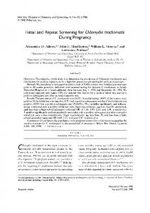
Fig. 1 : Showing X-Ray Chest AP view showing opacity in right lower zone Director, **Associate Chest Physician, ***Clinical and Research Associate, Lung Care and Sleep Centre, ****Head, Critical Care, † Director, Oncology, ‡Clinical Associate, Critical Care, @Consultant Minimal Access Surgeon, Fortis Hiranandani Hospital, Vashi Received: 14.09.2010; Revised: 18.11.2010; Re-revised: 17.01.2011; Accepted: 07.03.2011 *
656
intermittent treatment with antibiotics and bronchodilators. Sputum for acid fast bacilli was negative on three occasions. Symptoms never resolved completely and cough with expectoration was present intermittently. Patient developed streaky hemoptysis seven days prior to presenting to us.During the admission, she developed massive hemoptysis (more than 200 ml in 24 hours). She was shifted to intensive care unit in view of massive hemoptysis, desaturation (SpO 2 88%). She had SpO2 92% on 8 liters of oxygen. Arterial blood gas showed hypoxia with partially compensated respiratory alkalosis (pH – 7.50, pCU2 – 31, pO2 – 68, HCO3 -- 20.1). X-ray chest showed opacity in right lower zone, high standing diaphragm, no signs of bronchopneumonia and no evidence of bronchiectasis (Fig. 1). She was negative for HIV. She was started on intravenous coamoxyclav. The etiological possibilities being considered were pulmonary embolism, pulmonary vasculitis or an endobronchial lesions. 2-D Echocardiography done twice showed normal sized chambers with no evidence of pulmonary hypertension. Venous Doppler of both lower limbs was negative for deep vein thrombosis. Factor V Leiden mutation and D-Dimer values measured in blood was negative. Serum Protein-C (130%), Protein-S (36%) and Antithrombin-III (183%) levels were normal. Computed Tomography-Pulmonary angiography was not done at this time in view of her pregnancy. Also 2D Echo, d-Dimer and venous doppler of both lower limbs did not favor embolism. On the next day in the ICU she developed again massive bouts of hemoptysis. Endotracheal intubation was performed and mechanical ventilation was initiated. Following this it was decided to perform emergency lower section caesarian section for delivery of the baby. The delivery of the baby was uneventful. Post surgery Computed tomography Pulmonary angiography was performed which did not show evidence of pulmonary embolism. There was evidence of well defined, rounded, enhancing endobronchial mass lesion obstructing right main bronchus resulting in collapse, consolidation of right lower lobe (Fig. 2). Fibreoptic bronchoscopy done through endotracheal tube showed blood clot in right main bronchus with evidence of glistening mass just below the clot, occluding right main bronchus. Blood clot in left main bronchus was removed with forceps and suctioning. Sleeve resection of this mass was being considered. However, the patient developed, yet another bout of massive hemoptysis and hence was taken up for emergency surgical resection. Intra-operatively the tumor was extending into right lower lobe and middle lobe bronchus and, sleeve resection was not possible hence, a right pneumonectomy was performed. Post pneumonectomy she required ventilator support for two days and was then extubated. Postoperative course was uneventful. Our patient did not receive chemotherapy or radiotherapy, as there was no evidence of metastasis seen on abdominal ultrasound and Computed tomography upper © JAPI • october 2011 • VOL. 59
Fig. 3 : Showing histopathological picture of bronchial carcinoid stained with hematoxylin and eosin stain under 40X magnification.
as general measures. Positioning the patient with the bleeding side down to protect the uninvolved lung is also advocated. This may present a problem in a pregnant patient, whose venous return may be compromised by the gravid uterus in the right lateral decubitus and supine positions. A large bore endotracheal tube should whenever possible be used in order to perform diagnostic therapeutic bronchoscopy in patients with hemoptysis. The differential diagnosis of hemoptysis depends on patient’s age, smoking history, radiological findings, and accompanying symptoms. History of fever may point towards an infective etiology and hemoptysis in a smoker raises suspicion of malignancy. Our patient was young, pregnant and had history of chronic cough for about 7 months without any fever with sputum negativity for Acid Fast Bacilli. In view of her underlying pregnancy with the above clinic-radiological background pulmonary tuberculosis was still a differential diagnosis and other diagnosis considered were pulmonary vasculitis, pulmonary embolism, and intrabronchial tumor such as carcinoid. Final diagnosis on histopathology was bronchial carcinoid. These tumors are neoplasms arising from bronchial endocrine cells and previously grouped as bronchial adenomas. Their etiology is not very clear and it comprises of less than 2% of all pulmonary tumors.1 90% of these tumors are confined to the bronchus or lung with only 10% having regional lymph node involvement.1,4 Distant metastases occur in 15% of cases and are typically located in the liver, bone, adrenal gland, and brain.4
Fig. 2 : CT Thorax showing obstructed right main bronchus with partial collapse of distal segment.
abdominal sections. Histopathology report of surgical specimen showed round to polygonal tumor cells having centrally placed round nuclei displaying stippled chromatin and abundant eosinophilic cytoplasm suggestive of well-differentiated neuroendocrine tumor (carcinoid tumor) (Fig. 3). Immunohistochemistry report showed that the surgical specimen was positive for LMWK (low molecular weight keratin), Synaptophysin, and Chromogranin, NSF (N-ethylmaleimidesensitive factor) suggestive of neuroendocrine tumor.
Discussion Hemoptysis is defined as expectoration of blood from the lower respiratory tract. Hemoptysis is categorized according to the amount of blood expectorated. Common causes of hemoptysis in pregnancy includes Tuberculosis, Bronchiectasis, Pneumonia, Aspergilosis, lung abscess, endobronchial lesions, tracheobronchitis, pulmonary embolism, bleeding disorders, pulmonary vasculitis, arteriovenous malformations, amniotic fluid embolism. Hemodynamic changes during pregnancy like an increase in blood volume and cardiac output’ and hormonally related vascular changes, which may lead to arteriovenous malformations and can cause hemoptysis. Thrombogenic state induced due to pregnancy can lead to deep venous thrombosis and pulmonary embolism. Abruptio placenta can lead to amniotic fluid embolism.
Carcinoid tumors can be divided into two clinicopathological types, typical and atypical. WHO classification includes typical and atypical carcinoids in the spectrum of neuroendocrine tumors. Typical carcinoid tumors are low-grade tumors, with 10-year survival rates approaching 90%.4 They are capable of local invasion, including invasion of local lymph nodes, but rarely metastasize. The second type of carcinoid is referred to as atypical carcinoid. It is much more aggressive and carries a 5-year survival rate of 25% to 69%. Both subtypes tend to arise from the bronchial tree and spread by local invasion. Typical carcinoid tumors are more commonly found centrally within the major bronchi, whereas the atypical carcinoids tend to arise from the peripheral and central bronchi with equal frequency. But groups them separately from large cell neuroendocrine carcinoma and small cell lung carcinoma on the basis of close histologic and biologic interrelationships.4 The patients with
When a pregnant patient comes with hemoptysis she should be evaluated with CBC, Sputum-Routine microscopy, acid fast bacilli , d-dimer, coagulation profile, X-ray chest with abdominal shield, 2-D echocardiography, Venous Doppler, Factor V Leiden mutation, Serum Protein-C, Protein-S and Antithrombin-III. The optimal management of a pregnant patient with massive hemoptysis depends on the patient’s underlying condition, the cause of the hemoptysis, and the available expertise. Massive hemoptysis in pregnancy needs special attention as it can rapidly cause hemodynamic instability and lead to fetal hypoxia. Bed rest, a regimen of broad-spectrum antibiotics, and the judicious use of cough suppressants are often recommended © JAPI • october 2011 • VOL. 59
657
central lesions are more likely to be symptomatic on presentation. These symptoms include cough, hemoptysis, wheeze, stridor or post obstructive pneumonia. Some patients may present with hemoptysis as these are vascular tumors. Extra-pulmonary symptoms are due to release of various biologically active peptides like serotonin by carcinoid cells. Carcinoid syndrome is characterized by tachycardia, flushing, broncho-constriction, diarrhea, and hypotension. This is due to excessive release of serotonin. Our patient did not exhibit such symptoms relating to the carcinoid syndrome. About 75% of pulmonary carcinoids are visible on bronchoscopy; however, on biopsy chances of bleeding are high. Newer modalities for bronchial carcinoids include positron emission tomography (PET) and octreotide scintigraphy. PET imaging of bronchial carcinoids demonstrate lower uptake than non-small cell lung carcinoma. Octreotide scintigraphy has demonstrated reliable uptake in primary tumors and the ability to detect early recurrences and metastases even in asymptomatic patient. 2,3 Surgery offers definitive treatment for these tumors. In patients with centrally located carcinoid, tumor bronchial sleeve resection or sleeve lobectomy should be considered, when possible, because local recurrence is uncommon. For peripherally located tumors, lobectomy or segmentectomy may be considered.5 The patient in this report underwent pneumonectomy as there was involvement of the entire right bronchial tree. Bronchoscopic resection of an intrabronchial carcinoid is recommended in selected cases for pre-operative management of symptomatic bronchial obstruction prior to formal resection or in patients who are not able to withstand pulmonary resection. Endobronchial laser resection via rigid or flexible bronchoscope can photocoagulate the lesion. The Nd: YAG laser is currently preferred for airway resection because of its predictable effect on living tissue, depending on amount of energy applied. Radiation therapy has a palliative role in disseminated disease.6 Targeted radiotherapy with radiolabeled octreotide is under
658
investigation. Cisplatinum and etoposide based chemotherapy is indicated for unresectable disease as well as metastases. However, the response rate is only 22 percent with a median survival of 20 months. Biotherapy with interferon alpha and octreotide is used for the treatment of carcinoid syndrome with symptomatic relief in 70 percent of patients. Liver embolisation can be used to de-bulk liver metastases in symptomatic patients. This was not required in our patient as there was no evidence of any liver metastases. Recurrence free survival is commonly seen in typical carcinoid. Five-year survival following complete resection of typical carcinoid and atypical carcinoid is 87-100% and 44-77%, respectively. Tumor histology and nodal status are the main predictors of mortality. Our patient is asymptomatic at six-month follow up.
References 1. Thomas R, Christopher DJ, Balamugesh T, Shah A. Clinicopathologic study of pulmonary carcinoid tumours-a retrospective analysis and review of literature. Respir Med 2008;102:1611-4/ Epub2008 Jul 9. 2.
Erasmus JJ, McAdams HP, Patz Jr EF, Coleman RE, Ahuja V, Goodman PC. Evaluation of primary pulmonary carcinoid tumors using FDG PET. Am J Roentgenol 1998;170:1369-73.
3.
Musi M, Carbone RG, Bertocchi C, Cantalupi DP, Michetti G, Pugliese C. Bronchial carcinoid tumors: a study on clinicopathological features and role of octreotide scintigraphy. Lung Cancer 1998;22:97102.
4.
Alessandro B, Jury B, Nicola C, Fabio D, Giampiero D, Francesco S et al.Typical and atypical pulmonary carcinoids: our institutional experience. Interact Cardio Vasc Thorac Surg 2008;7:415-8.
5.
Pier LF, Ottavio R, Giovanni D, Caterina C. Bronchial carcinoid tumors: Surgical management and long term outcome. J Thorac Cadiovasc Surg 2002;123:303-9.
6. R Hage, A Brutel de la Riviere, CA Seldnrijk, JMM van den Bosch. Update in pulmonary carcinoid tumors: A review article. Annals of Surgical Oncology 2003;10:697-704.
© JAPI • october 2011 • VOL. 59