Original Article
Medical Thoracoscopy for Undiagnosed Pleural Effusions: Experience from a Tertiary Care Hospital in North India V.K. Mootha, R. Agarwal, N. Singh, A.N. Aggarwal, D. Gupta and S.K. Jindal Department of Pulmonary Medicine, Postgraduate Institute of Medical Education and Research, Chandigarh, India
ABSTRACT Background and Aims. Medical thoracoscopy, also called pleuroscopy, has received renewed interest in the recent past for diagnostic as well as therapeutic uses. In this study, we describe our experience with thoracoscopy for undiagnosed pleural effusions. Methods. In a retrospective analysis of thoracoscopic procedures we performed between January 2007 and December 2008, yield of thoracoscopic pleural biopsy for achieving a diagnosis in undiagnosed pleural effusions, defined as pleural effusions with adenosine deaminase (ADA) levels less than 70 IU/L and negative pleural fluid cytology for malignancy on three occasions was evaluated. Complications of thoracoscopy were also analysed. Results. Overall diagnostic yield of thoracoscopic pleural biopsy was 74.3% in patients with undiagnosed pleural effusions. Pleural malignancy was diagnosed in 48.6% of patients. There was only one case of mesothelioma and the rest were due to pleural metastasis. Lung cancer and breast cancer were the most common sites of primary malignancy. Tuberculosis was diagnosed with pleural biopsy in 22.8% of patients. We had low complication rate after thoracoscopy. Only two cases of empyema were observed. Conclusion. Medical thoracoscopy is a safe procedure and has good diagnostic yield in patients with undiagnosed pleural effusions. [Indian J Chest Dis Allied Sci 2011;53:21-24] Key words: Malignant pleural effusion, Tuberculosis, Pleuroscopy, VATS.
INTRODUCTION Pulmonary physicians world over have been showing increasing interest in rapidly expanding field of interventional pulmonology. Medical thoracoscopy, also called as pleuroscopy is one of them. This procedure was described for diagnostic purpose in 1910 by Jacobaeus and was subsequently used in the management of tuberculosis (TB), to create pneumothorax (Jacobaeus operation). 1 Medical thoracoscopy has received renewed interest among pulmonary physicians in the recent past because of better instrumentation and simpler sedation protocols. With current techniques, medical thoracoscopy can be done as a day-care procedure under conscious sedation by pulmonary physicians. Medical thoracoscopy is a minimally invasive procedure done in spontaneously breathing patient, unlike video-assisted thoracoscopic surgery (VATS) which is conducted under general anaesthesia with single lung ventilation.2 It allows one to visualise the
entire pleural surface and perform limited diagnostic and therapeutic procedures. The major indication for medical thoracoscopy is evaluation of exudative pleural effusions which remain undiagnosed after pleural fluid analysis, where thoracoscopy is suggested as an alternative to closed pleural biopsy. With thoracoscopy, one can visualise the entire visceral and parietal pleura and take pleural biopsy from suspicious sites under vision. Larger pleural biopsy specimen taken under direct vision allows greater diagnostic yield up to 90 percent.3 Diagnosis of pleural TB can be achieved in 99% of patients with thoracoscopy, which is higher than the 51% yield for closed pleural biopsy. 4 Similarly, yield of thoracoscopic pleural biopsy is higher in patients with suspected pleural malignancy. A diagnosis could be achieved in 95% of patients as against 44% patients using closed pleural biopsy.4 Although thoracoscopy can be used to visualise pleural blebs and bullae in patients with spontaneous pneumothorax, this is seldom the indication for
[Received: December 23, 2009; accepted after revision: April 27, 2010]
Correspondence and reprint requests: Dr Dheeraj Gupta, Additional Professor, Department of Pulmonary Medicine, Postgraduate Institute of Medical Education and Research, Sector-12, Chandigarh-160 012 (India); Phone: 91-172-2756823; Fax: 91-172-2748215; E-mail:
[email protected]
22
Medical Thoracoscopy for Pleural Effusion
thoracoscopy. Medical thoracoscopy can be used for therapeutic procedures, such as adhesiolysis and evacuation of pleural fluid in patients with empyema, pleurodesis in patients with malignant pleural effusion and spontaneous pneumothorax.2 In the present study, we describe our experience with the technique of medical thoracoscopy in patients who underwent thoracoscopy for diagnostic purposes.
MATERIAL AND METHODS This was a retrospective study conducted in the Department of Pulmonary Medicine, PGI, Chandigarh, between January 2007 and December 2008. We performed thoracoscopy for diagnosis of undiagnosed pleural effusions. Undiagnosed pleural effusion was defined as failure to achieve a diagnosis by initial pleural fluid analysis including pleural fluid adenosine deaminase (ADA) levels and at least three pleural fluid analyses negative for malignant cells. All patients underwent detailed clinical evaluation with history and clinical examination. Computed tomography (CT) of the chest was performed to assess feasibility of thoracoscopy. Patients with excess rib crowding with narrow intercostal space and loculated pleural effusion could not undergo thoracoscopy. All patients undergoing thoracoscopy were investigated with complete blood count including prothrombin time (PT), activated plasma thrombin time (aPTT) and platelet count to rule out bleeding diathesis. Patients with platelet count less than 75,000/mm 3 and those with PT or aPTT prolonged by more than four seconds above control were not subjected to thoracoscopy. Other contraindications for thoracoscopy included haemodynamic instability, arrhythmias and intractable cough. Patients were kept fasting for six hours prior to the procedure. Vascular access was achieved with intravenous cannula inserted in the upper limb opposite to the side of thoracoscopy. In patients with small pleural effusion, an artificial pneumothorax was created by injecting approximately one liter of air into pleural cavity just prior to the procedure. This allowed lung to collapse and reduces the chances of lung being injured while introduction of trocar. Patients were positioned in lateral decubitus with diseased side up. Arm on the side of thoracoscopy was positioned above the patient’s head. This allowed better access and widens the intercostal spaces. Thoracoscopy was conducted under conscious sedation. Chest wall was draped with sterile cloth after cleaning the skin with 7.5% povidone iodine. Patients were sedated with intravenous midazolam (0.5mg/kg body weight) and intravenous tramadol
V.K. Mootha et al
5mg was given for analgesia prior to the start of procedure. The skin, subcutaneous tissue, intercostal muscle and parietal pleura were anesthetised with 10mL 2% lignocaine to achieve local anaesthesia. During the procedure intravenous midazolam and tramadol boluses were repeated as required to achieve adequate sedation and analgesia. Intravenous pethidine 25mg was given as bolus to control pain if analgesia could not be achieved with tramadol. We used single port for visualising and taking pleural biopsy. A 1.5cm to 2cm long skin incision along the line of intercostal space was given in 4th or 5th intercostal space in mid-axillary line using sterile surgical blade. After blunt dissection of subcutaneous tissue and the intercostal muscles with curved artery forceps, a cannula of 10mm diameter with blunt trocar is inserted into the pleural cavity. The trocar was then replaced with rigid video thoracoscope (Richard Wolf GmbH, Knettligen, Germany). Pleural fluid was suctioned to enable clear visualisation of entire pleural surface. Thoracoscope was manoeuvered to see visceral, costal, diaphragmatic surface as well as the costophrenic recess. Adhesions were gently lysed using thoracoscope or biopsy forceps to allow visualisation of pleura. After selecting suitable site on parietal pleura for biopsy, biopsy forcep was introduced through working channel of the thoracoscope. Pleura was grasped under vision and biopsy is taken with a shearing movement of the thoracoscope. After the procedure is completed, thoracoscope and the cannula were removed and a 28 to 32 Fr chest tube was inserted. Chest drain was connected to water-seal drainage bag. Once the lung had expanded and drain output had decreased to less than 50mL per 24 hours, chest drain was removed. Demographic characteristics of the patient including the age, gender, clinical diagnosis, pleural fluid analysis, including total and differential count, protein and glucose values, ADA levels, stain for acid-fast bacilli (AFB) and cytology findings and findings of the CT of the chest were recorded. Data are presented in a descriptive fashion.
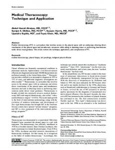
RESULTS During the study period, 35 patients (71.4% men and 28.6% women; mean [SD] age 48.68 [14] years) with undiagnosed pleural effusion underwent thoracoscopy for diagnostic purposes (Table 1). The representative images of pleural abnormalities visualised during thoracoscopy are shown in the figure.
2011;Vol.53
The Indian Journal of Chest Diseases & Allied Sciences
Figure. Thoracoscopic pictures of pleural abnormalities: (A) large pleural nodule in malignant pleural effusion and (B) sago nodules appearance in tubercular pleural effusion.
Of the 35 patients with undiagnosed pleural effusion, the initial clinical diagnosis was malignant pleural effusion in 26 (74.3%) cases. Three patients with clinical suspicion of TB and no diagnosis after initial pleural fluid analysis underwent thoracoscopy. One patient was thought to have Churg-Strauss syndrome with eosinophilic pleural effusion, but no histological diagnosis. In five patients, despite pleural fluid and radiological investigations, no clinical diagnosis could be made. Demographic details and radiological features of these patients are shown in table 1. Pleural fluid ADA was not elevated (>70 IU/L) in any of the patients with suspected malignant pleural effusion or tuberculosis effusion. Table 1. Demographic characteristics and details of investigations of 35 patients undergoing thoracoscopy for undiagnosed pleural effusion Demographic Characteristic
Result
Age (years)
48.68 (14.0)
Male:Female
25:10
Initial clinical diagnosis Malignant pleural effusion Tuberculosis No diagnosis Churg-Strauss syndrome Pleural nodules in CT chest Pleural fluid TLC (/mm 3) Differential count Protein (g/dL) Sugar (mg/dL) AFB ADA (I/L) Malignant cells
26 3 5 1 25.7% 1,525 (1,795) 50% lymphocytic effusions 50% neutrophilic effusions 4.89 (1.21) 72.22 (38.3) 0% 39.1 (19.5) 0
The results are depicted as mean±SD or No. (%) unless otherwise stated CT=Computed tomography; TLC=Total leukocyte count; AFB=Acid-fast bacilli; ADA=Adenosine deaminase activity
Thoracoscopic pleural biopsy could achieve diagnosis in 26 of the 35 patients (74.3%). Final diagnosis of pleural malignancy was made in 17
23
patients and a diagnosis of TB was made in eight patients. Pleural biopsy confirmed eosinophilic inflammation in the patient with Churg-Strauss syndrome. Of 17 patients with proven pleural malignancy, only one had mesothelioma and the remaining had metastatic pleural cancer. Seven patients had metastatic adenocarcinoma, five patients had poorly differentiated metastatic pleural malignancy, two patients had squamous cell lung cancer and one patient each had small cell lung cancer and lymphoma. In eight patients, thoracoscopic pleural biopsy showed granulomatous inflammation consistent with TB. Biopsy revealed AFB in one case. In nine out of 35 (25%) patients with pleural effusion, thoracoscopic pleural biopsy did not reveal any specific diagnosis, and these cases were classified as idiopathic pleural effusions. We analysed the yield of biopsy according to the initial clinical diagnosis and found that 19 of the 26 (73%) patients with initial diagnosis of malignant pleural effusion had a definitive diagnosis on thoracoscopic pleural biopsy. Sixteen of them had pleural malignancy and incidentally three of them had chronic granulomatous inflammation consistent with TB (Table 2). We could confirm diagnosis of TB in two out of three patients with initial diagnosis of TB. Among five patients with no initial clinical diagnosis, three had TB diagnosed on pleural biopsy and one had lymphoma (Table 2). Table 2. Results of diagnostic thoracoscopy Initial Clinical Diagnosis
Yield of Thoracoscopic Pleural Biopsy
Final Diagnosis on Thoracoscopic Pleural Biopsy
Malignant pleural effusion (n=26)
19/26 (73%)
Tuberculosis (n=3)
2/3 (66.6%)
No clinical diagnosis (n=5)
4/5 (80%)
Churg-Strauss syndrome (n=1)
1/1(100%)
Pleural malignancy - 16 (mesothelioma - 1 and metastatic pleural malignancy - 15) Tuberculosis - 3 Idiopathic - 7 Tuberculosis - 2 Idiopathic - 1 Tuberculosis - 3 Lymphoma - 1 Idiopathic - 1 Churg-Strauss syndrome - 1
Total
26/35 (74.3%)
Of the 35 thoracoscopic procedures, two cases developed empyema (5.2%). There were no instances of haemorrhage, shock or subcutaneous emphysema.
DISCUSSION In this study, we have presented the data of 35 consecutive patients who underwent thoracoscopy for the diagnosis of undiagnosed pleural effusions. We included patients with undiagnosed pleural
24
Medical Thoracoscopy for Pleural Effusion
effusions for thoracoscopy in whom initial diagnostic work-up with pleural fluid analysis including pleural fluid ADA and three pleural fluid cytologies were inconclusive. The yield of thoracoscopic pleural biopsy was 74.3% (26/35) patients in this group. Similar experience with medical thoracoscopy has been described from other centers. Kendall et al 5 reported yield of thoracoscopic pleural biopsy to be 83% in their study which included 48 patients. Tscheikuna et al 6 described their experience from Thailand (n=86) and thoracoscopy was diagnostic in 95% of 34 patients. Ng et al7 could achieve diagnosis with thoracoscopic pleural biopsy in 45.5% (10/22) patients with undiagnosed pleural effusions. In a majority of patients in our study, thoracoscopic pleural biopsy yielded diagnosis of pleural malignancy. A significant proportion of patients, 45.7% (16/35) with un-diagnosed pleural effusion had pleural malignancy. Similar observations were made by Tscheikuna et al 6 who found pleural malignancy in 45% of patients with undiagnosed pleural effusions undergoing thoraco-scopy. Ng et al7 found that 45.5% of patients with undiagnosed pleural effusions had pleural malignancy. Pleural metastasis is the more common cause of malignant pleural effusions than mesothelioma. We could diagnose only one case of mesothelioma whereas 16 of the 17 cases were due to pleural metastasis. Among the patients with metastatic pleural malignancy diagnosed with thoracoscopic pleural biopsy, the most common site of primary malignancy was the lung. In fact, in 50% (8/16) of patients the primary cancer was bronchogenic and in 12.5% (2/16) patients, the primary was breast and in 31.2% (5/16) of cases, the primary site remained unidentified. Among the patients with metastatic pleural effusion from lung cancer, adenocarcinoma was the most common diagnosis. Among those with malignant pleural effusion from primary lung cancer, five of the eight patients had adenocarcinoma. Small cell lung cancer and squamous cell lung cancer were less common diagnosis. These findings are in concordance with the findings of others.8,9 Eight out of 35 (22.9%) patients in whom we performed thoracoscopy had pleural TB on pleural biopsy. Only one of the eight patients had AFB in their biopsy specimens. This is in stark contrast to the findings of Kendall et al 5 who did not find any case of TB in their study of 48 patients undergoing thoracoscopy for undiagnosed pleural effusions. This is probably due to low prevalence of TB in the West. Thoracoscopic pleural biopsy is considered gold standard in diagnosis of malignant pleural effusion and TB pleural effusion. Diagnostic yield of thoracoscopic pleural biopsy can be as high as 95% in malignant pleural effusions and 99% in TB pleural effusions which is far superior to that of pleural fluid analysis and closed pleural biopsy. 4 These findings
V.K. Mootha et al
along with results of our study and similar studies mentioned above suggest that thoracoscopic pleural biopsy should be considered in all patients with pleural effusions who remain undiagnosed after initial pleural fluid analysis. A variety of complications associated with thoracoscopy have been described in the literature, 2,6,10-13 such as subcutaneous emphysema (0.6%-4.9%), air leak (0.5%-8.1%), empyema (0.5%-2.7%), haemorrhage (0.3%-0.4 %), shock (0.2%), chest wall seeding by malignancy (0.5%-4.0%). We had only 2 (5%) cases of empyema and noted no other complications.
CONCLUSIONS The results of this study suggest that medical thoracoscopy should be considered in patients with undiagnosed pleural effusions, particularly those lymphocytic exudative effusions where TB and malignant pleural effusion are clinical possibilities and initial pleural fluid analysis is inconclusive.
REFERENCES 1. 2 3 4
5 6 7 8 9 10 11 12 13
Jacobeus HC. The cauterization of adhesions in artificial pneumothorax treatment of pulmonary tuberculosis under thorascopic control. Proc R Soc Med 1923;16:45-62. Casal RF, Eapen GA, Morice RC, Jimenez CA. Medical thoracoscopy. Curr Opin Pulm Med 2009;15:313-20. Loddenkemper R. Thoracoscopy: state of the art. Eur Respir J 1998;11:213-21. Loddenkemper R, Grosser H, Gabler A, Mai J, Presseuler H, Brandt HJ. Prospective evaluation of biopsy methods in diagnosis of malignant pleural effusions: intra patient comparision between pleural fluid cytology, blind needle biopsy and thoracoscopy. Am Rev Respir Dis 1983;127:114. Kendall SW, Bryan AJ, Large SR, Wells FC. Pleural effusions: is thoracoscopy a reliable investigation? A retrospective review. Respir Med 1992;86:437-40. Tscheikuna J, Silairatana S, Sangkeaw S, Nana A. Outcome of medical thoracoscopy. J Med Assoc Thai 2009; 92 (Suppl. 2):S19-S23. Ng TH, How SH, Kuan YC, Hasmah H, Norra H, Fauzi AR. Medical thoracoscopy: Pahang experience. Med J Malaysia 2008;63:298-301. Johnston WW. The malignant pleural effusion: a review of cytopathologic diagnoses of 584 specimens from 472 consecutive patients. Cancer 1985;56:905-9. Chernow B, Sahn SA. Carcinomatous involvement of the pleura: an analysis of 96 patients. Am J Med 1977;63: 695-702. Boutin C, Viallat JR, Cargnino P, Farisse P. Thoracoscopy in malignant pleural effusions. Am Rev Respir Dis 1981; 124:588-92. Viskum K, Enk B. Complications of thoracoscopy. Poumon Coeur 1981;37:25-8. Menzies R, Charbonneau M. Thoracoscopy for the diagnosis of pleural disease. Ann Intern Med 1991;114:271-6. de Campos JR, Vargas FS, de Campos Werebe E, Cardoso P, Teixeira LR, Jatene FB, et al. Thoracoscopy talc poudrage: a 15-year experience. Chest 2001;119:801-6.