1559
Development 128, 1559-1572 (2001) Printed in Great Britain © The Company of Biologists Limited 2001 DEV1631
Mesodermal patterning defect in mice lacking the Ste20 NCK interacting kinase (NIK) Yingzi Xue1, Xiaozhong Wang, Zhai Li1, Noriko Gotoh1, Deborah Chapman2 and Edward Y. Skolnik1,* 1New
York University Medical Center, Skirball Institute of Biomolecular Medicine, Department of Pharmacology, 540 First Avenue, NY, NY10016, USA 2University of Pittsburgh, Department of Biological Sciences, Fifth and Ruskin Avenues, Pittsburgh, PA 15260, USA *Author for correspondence (e-mail:
[email protected])
Accepted 2 February; published on WWW 5 April 2001
SUMMARY We have previously shown that the Drosophila Ste20 kinase encoded by misshapen (msn) is an essential gene in Drosophila development. msn function is required to activate the Drosophila c-Jun N-terminal kinase (JNK), basket (Bsk), to promote dorsal closure of the Drosophila embryo. Later in development, msn expression is required in photoreceptors in order for their axons to project normally. A mammalian homolog of msn, the NCKinteracting kinase (NIK) (recently renamed to mitogenactivated protein kinase kinase kinase kinase 4; Map4k4), has been shown to activate JNK and to bind the SH3 domains of the SH2/SH3 adapter NCK. To determine whether NIK also plays an essential role in mammalian development, we created mice deficient in NIK by homologous recombination at the Nik gene. Nik−/− mice die postgastrulation between embryonic day (E) 9.5 and E10.5. The most striking phenotype in Nik−/− embryos is the failure of mesodermal and endodermal cells that arise from the anterior end of the primitive streak
INTRODUCTION Ste20 kinases constitute a large family of protein kinases that are best known for their roles in activating the JNK MAP kinase pathway and in the regulation of the actin cytoskeleton (Kyriakis, 1999). Based on sequence similarity to the kinase domain of the yeast Ste20 kinase protein, two broad families of Ste20 kinases have been identified in mammalian cells and lower organisms, the p21 activated protein kinase (PAK) and the germinal center kinase (GCK) families (Kyriakis, 1999; Manser and Lim, 1999). However, despite the distant homology between their kinase domains, these two kinase families exhibit many differences in both regulation and function. For example, PAKs, unlike GCK family kinases, possess a CRIB motif and therefore bind and are regulated by Rho family GTPases (Burbelo et al., 1995; Martin et al., 1995). In addition, unlike PAKs which have a C-terminal kinase and an N-terminal regulatory domain, GCK family members contain an N-terminal kinase and a C-terminal regulatory domain whose function is distinct from the regulatory domain
(PS) to migrate to their correct location. As a result Nik−/− embryos fail to develop somites or a hindgut and are truncated posteriorly. Interestingly, chimeric analysis demonstrated that NIK has a cell nonautonomous function in stimulating migration of presomitic mesodermal cells away from the PS and a second cell autonomous function in stimulating the differentiation of presomitic mesoderm into dermomyotome. These findings indicate that despite the large number of Ste20 kinases in mammalian cells, members of this family play essential nonredundant function in regulating specific signaling pathways. In addition, these studies provide evidence that the signaling pathways regulated by these kinases are diverse and not limited to the activation of JNK because mesodermal and somite development are not perturbed in JNK1-, and JNK2-deficient mice. Key words: Ste20 kinase, NCK interacting kinase (NIK), misshapen, Gastrulation, knockout, N-terminal JUN kinase (JNK), Mouse
of PAK (Kyriakis, 1999; Su et al., 1997). As a result, it has now been proposed to classify PAK and GCK family members into two distinct protein kinase families (Kyriakis, 1999). At least 20 different GCK family members have been identified in mammalian cells so far. These GCK members can be further subdivided into 5 subgroups based on similarities in sequence, structure and function (Kyriakis, 1999). Although the specific function served by individual GCK members or by subgroups of GCK in mammalian cells is still poorly defined, most studies have focussed on the role of these kinases in activation of the JNK MAP kinase pathway. With respect to JNK activation, GCK family members can be subdivided into two broad groups, group 1 and group 2 (Kyriakis, 1999). Group 1 kinases activate JNK, and include GCK, GCK related (R), hematopoietic progenitor kinase 1 (HPK1) and NIK (Diener et al., 1997; Hu et al., 1996; Pombo et al., 1995; Su et al., 1997). All group 1 kinases share a conserved C-terminal regulatory domain that may function to couple these kinases to downstream MAP kinase kinase kinase (MAP3Ks) (Su et al., 1997). Less is known about the second group of GCK, which
1560 Y. Xue and others includes Ste20-like oxidant stress activated kinase 1 (SOK1), mammalian sterile20 like 1 and 2 (MST1 and 2) and lymphocyte oriented kinase (LOK) (Creasy and Chernoff, 1995a; Creasy and Chernoff, 1995b; Kuramochi et al., 1997; Pombo et al., 1997). Members of this group contain a Cterminal domain that is significantly different from those of group 1 members and as yet members of this group have not been linked to activation of a signaling pathway (Kyriakis, 1999). Recent experiments in both Drosophila and C. elegans have confirmed an essential role for one member of the GCK family, NIK, in both JNK activation and development in a biologically relevant system (Su et al., 1998; E. Hedgecock and X. Zhu personal communication). We have placed the Ste20 kinase encoded by misshapen (msn), the Drosophila homolog of mammalian NIK, genetically upstream of the JNK MAP kinase pathway in Drosophila (Su et al., 1998). The failure to activate JNK in Drosophila leads to embryonic lethality due to defects in embryonic dorsal closure; in embryos mutant for components of the JNK pathway the lateral epithelial sheets fail to elongate and migrate dorsally (Noselli, 1998). A number of signaling molecules that are critical for stimulating dorsal closure and JNK activation in Drosophila can now be ordered on a signaling pathway. Msn likely functions as a MAP kinase kinase kinase kinase (M4K) and activates the JNK pathway by activating a yet to be defined DM3K. A DM3K would likely phosphorylate and activate the JNK kinase encoded by hemipterous (hep) (Glise, 1995), which in turn phosphorylates and activates Drosophila JNK, encoded by basket (bsk) (Riesgo-Escovar, 1996; Sluss et al., 1996). DJNK phosphorylates and activates DJun, which in turn cooperates with DFos to stimulate transcription of dpp, a member of the transforming growth factor-β family (Glise, 1997; Hou, 1997; Kockel, 1997; Riesgo-Escovar and Hafen, 1997a; RiesgoEscovar and Hafen, 1997b). In addition to its role in dorsal closure, Msn also couples Frizzled (Fz) and Dishevelled (Dsh) to JNK activation in the planar polarity pathway; msn mutants show defects in epithelial planar polarity in both the eyes and wings due to defective JNK activation (Mlodzik, 1999; Paricio et al., 1999). In C. elegans, mig-15, the homolog of mammalian NIK and Drosophila msn, has also been found to be an essential gene in development. mig-15 mutants have a number of developmental defects including defects in Q neuroblast migration and muscle arm targeting, although it is not yet clear whether these defects are due to defective activation of JNK (E. Hedgecock and X. Zhu personal communication). In addition to activating JNK, NIK contains PXXP motifs that mediate its interaction with the SH3 domains of Nck (Nck1; Su et al., 1997). Nck is a ubiquitously expressed protein composed of one SH2 and three SH3 domains (Lehman et al., 1990). Adapter molecules like Nck are proposed to regulate signaling pathways downstream of tyrosine kinases by coupling catalytic subunits, bound to their SH3 domains, to phosphotyrosine-containing proteins that interact with their SH2 domains (Pawson and Nash, 2000; Schlessinger, 1994). Recent experiments in Drosophila have shed light on some functions of Nck in mammalian cells and provide evidence that Ste20 kinases are downstream effectors of Nck. A Drosophila homolog of mammalian Nck encoded by dreadlock (dock) is required for correct targeting of photoreceptor axons (Garrity et al., 1996). Drosophila Pak has been shown to be one of the
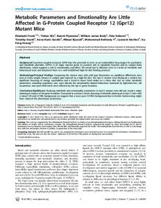
key targets for regulation by dock in this pathway and an interaction between Pak and the SH3 domains of Dock is required for Dock regulation of R cell axon guidance and targeting (Hing et al., 1999). We and others have found that Msn also binds the SH3 domains of Dock and that expression of Msn in photoreceptors is also required for the correct targeting of photoreceptor axons (Ruan et al., 1999; Su et al., 2000). In mammalian cells, NCK couples NIK to tyrosine phosphorylated p62 Dok in Eph-stimulated cells, and this interaction is important for the EphB1 and EphB2 receptors to activate NIK kinase activity as well as JNK and integrins (Becker et al., 2000). Owing to the important roles of msn, dock and mig-15 in Drosophila and C. elegans development, we determined whether NIK also performs an essential role in mammalian development by creating mice with a targeted disruption of the Nik locus. Mice containing a homozygous loss-of-function of Nik die around E9.5. The most obvious defect in Nik−/− embryos is the failure of mesodermal cells derived from the anterior end of the primitive streak to migrate away from the streak. As a result, Nik−/− embryos are truncated posteriorly and somitogenesis is greatly affected. The most affected embryos have no somites, while less affected mutants have 12 pairs of somites, compared to the 10-15 somites found in control littermates. Interestingly, the failure of mesodermal cells to migrate away from the PS is not due to loss of NIK in these cells because chimeric analysis showed that presomitic mesoderm arising from Nik−/− ES cells migrated to the appropriate position in the chimeric embryos. Thus, NIK is most probably required in the PS to regulate the production of factors that are required for proper cell migration. In addition, NIK also acts in presomitic mesoderm to stimulate its differentiation into somites, because Nik−/− ES cells failed to differentiate into dermomyotome in the chimeric embryos. These findings provide the first evidence for an essential function of a Ste20 kinase in mammalian development. MATERIALS AND METHODS Gene targeting A targeted mutation was introduced into the murine Nik gene by homologous recombination in embryonic stem cells as described previously (Joyner, 1993). The Nik genomic DNA was isolated by screening a 129 mouse genomic library (Stratagene) with the Nik cDNA. A 15 kb genomic Nik clone containing the 5′ start ATG of Nik was isolated, subcloned into Bluescript KS, and a restriction map was constructed. The 5′ homology arm of the targeting vector was the 4 kb KpnI-ApaI murine genomic fragment, which was subcloned into targeting vector pPNT (Tybulewicz et al., 1991). The 3′ homology fragment was a 3.5 kb SmaI-KpnI genomic fragment. In the final targeting vector an ApaI fragment in the Nik genomic DNA is deleted and replaced with a PGK.neo cassette. This removes part of exon VI and all of exon VII of the kinase domain is removed. These exons encode subdomain VIII and IX in the kinase domain (at amino acid 165 of Nik) and therefore the targeted Nik allele lacks critical residues that are essential for a functional kinase (Fig. 1). Following electroporation, G418 and gancyclovir-resistant ES clones were selected and successful targeting of the Nik locus was determined by PCR (4/300). The primer pairs used for PCR genotyping the 5′ arm are: Nik exon primer (sense) 5′-GAGATCAGCAGCCTCTGTTCCACA-3′ and an antisense primer to the untranslated region in pGK 5′ GAGATCABCAGCCTCTGTTCCACA 3′; for the 3′ arm:
Mesodermal patterning in Nik mutant mice 1561 sense primer to pGK promoter 5′ GCTACCGGTGGATGTGGAATGTG 3′ and an antisense primer to a Nik intron 5′ GCAAGCGAGTTAAGTTTAGCCTGCAG 3′. Aggregation chimeras between the targeted ES cell and BALB/c morulas were generated and founders were bred to 129Svev mice or outbred to CD-1 mice. Transmission of the targeted ES clone was determined by coat color analysis and genotyping of offspring. The targeted mutation was propagated in a Swiss Webster genetic backgrounds. Mutant Nik−/− homozygous embryos were obtained by crossing NIK+/- mice and Nik−/− mice were determined by their characteristic phenotype or by PCR analysis as described in Fig. 1. In situ hybridization Whole-mount embryo in situ hybridization was performed as described using riboprobes as indicated (Swiatek et al., 1994). To section whole-mount stained embryos, after postfixing in 4.0% formaldehyde overnight, embryos were incubated in 30% sucrose/ PBS for 2 days. The embryos were embedded in paraffin wax and sectioned at between 10 and 20 µm. Sections were then mounted onto glass slides and after dewaxing were photographed. The probes used for whole-mount in situ staining were Shh (Echelard et al., 1993), Brachyury (Herrmann, 1991), Mox1 (Candia et al., 1992), HNF3β (Ang et al., 1993), fgf4, Tbx6 (Chapman et al., 1996), Otx2 (Simeone et al., 1993), En1 (Davis and Joyner, 1988), Gbx2 (Bouillet et al., 1995), and lim1 (Barnes et al., 1994). The NIK probe corresponds to nucleotides 1347-1845 in full length NIK. This region is located between the kinase domain and C-terminal domain and is the region least conserved between NIK and other Ste20 kinases. Generation of Nik−/− ES cells Nik−/− ES cells were generated from Nik K+/− ES cells by culturing in high concentration of G418 (1.4 mg/ml; Mortensen et al., 1992). G418 clones were expanded and genotyped by Southern blot analysis. 2 Nik−/− ES cell lines were obtained out of 35 clones examined. Generation of chimeric mice and β-galactosidase staining Chimeric embryos were generated by injecting 8 or 14 Nik−/− ES cells into ROSA lacZ+/− blastocysts. Injected blastocysts were surgically transferred into the uteri of pseudopregnant CD-1 foster mothers and chimeric embryos were dissected at E9.5, fixed and whole-mount stained for β-galactosidase according to standard protocols (Song et al., 1996). Generation of TIE2/lacZ:Nik−/− mice TIE2/lacZ (Tek-lacZ) transgenic mice were obtained from Jackson Labs and have been described previously (Schlaeger et al., 1997). To generate TIE2-lacZ:Nik−/− mice, TIE2-lacZ/TIE2-lacZ;Nik+/− mice were crossed and embryos were dissected at embryonic day 9.5. Embryos were whole-mount stained for β-galactosidase as described above.
RESULTS Targeted disruption of the Ste20 kinase NIK in ES cells by homologous recombination To create mice with a targeted disruption of the Ste20 kinase NIK, we generated the targeting construct pNIKneo in the vector pPNT (Tybulewicz et al., 1991). This construct contains 9 kb of NIK genomic sequence in which half of exon 6 and all of exon 7 of the kinase domain are replaced by a PGKneo cassette. These exons encode for subdomains VIII and IX of the NIK kinase domain (at amino acid 165 of the NIK protein) and therefore the targeted Nik allele lacks residues that are essential for a functional kinase domain. To prevent potential read through transcription from the PGK promoter and to introduce
stop codons in all 3 reading frames, PGK-neo was cloned in an opposite orientation to that of NIK. This would be predicted to truncate the coding region after subdomain VI of the kinase domain at amino acid 164 of NIK and would disrupt all major isoforms of NIK that have been described (Fig. 1A). We confirmed that Nik−/− mice are unlikely to express either a truncated or alternatively spliced NIK protein because northern analysis did not detect binding of a Nik cDNA probe to RNA derived from Nik−/− fibroblasts or ES cells, whereas NIK expression was easily detected in wild-type cells (figure 1D). Four ES clones containing a targeted NIK mutation were identified by screening for the 5′ and 3′ insertion sites by PCR and by Southern blot analysis (Fig. 1B and data not shown). Morula aggregation was performed for three of the four clones with Balb/c mice. All of the clones generated highdegree chimerisms and when crossed with CD-1 mice generated agouti pups indicating germline transmission. Germline transmission was confirmed by both PCR and Southern blot analysis (Fig. 1C and data not shown). In analysis of the mice, identical results were obtained from all three lines and therefore for simplicity the results described are from pooled data from all 3 lines. Nik−/− mice die embryonically between day 9.5 and day 10.5 All heterozygous progeny were viable and fertile and did not manifest any overt phenotypes. To determine whether Nik−/− mice were viable, Nik+/− mice were crossed and litters were genotyped 3 weeks after birth. No homozygous Nik mutant littermates were present (0/37) as determined by both PCR and Southern blot analysis (Fig. 1C and data not shown). Since no perinatal lethality was observed in these litters, these findings indicate that Nik is a recessive embryonically lethal allele. To determine the point at which Nik−/− mice die, embryos from a cross between Nik heterozygous mice were dissected at different gestational stages. Analysis of E10.5 embryos revealed that about 1/4 of the decidua were much smaller, very necrotic and were partially resorbed whereas the remaining were phenotypically normal. To genotype the normal and abnormal embryos, embryos were dissected away from maternal tissues and genotyped by PCR (data not shown). This analysis demonstrated that all of the abnormal embryos were Nik−/−, while the remaining normal embryos were either Nik+/+ or Nik+/− at the expected ratio of 1:2. Thus, these findings, when coupled with the finding that E9.0 Nik−/− embryos have a beating heart, indicate that homozygous mutant Nik embryos die about 9.5 days after implantation. To determine whether the expression pattern of NIK may give an insight into the lethality of Nik mutants, the expression pattern of Nik mRNA in both E7.5 and E8.5 embryos was determined. Whole-mount in situ hybridization revealed that Nik transcripts are widely expressed at both time points (figure 1E). The staining for Nik mRNA was specific because the same probe did not give a signal in similarly aged Nik−/− embryos and a similarly made sense probe did not give a signal in wildtype embryos (data not shown).
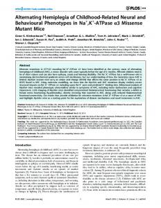
Nik mutant embryos are truncated posteriorly and lack somites and presomitic mesoderm Morphological and histological analysis of E8.5-9.0 Nik−/− embryos demonstrated that Nik−/− embryos exhibited a distinct
1562 Y. Xue and others Fig. 1. Targeted disruption of the Nik gene. (A) Targeting construct. The top line shows a restriction map of a portion of the Nik gene. An ApaI fragment, which includes part of exon 6 and all of exon 7, was replaced with the pGKneo gene in the targeting vector. The oligos used for PCR screening to verify homologous recombination are shown. (B) PCR analysis of one ES clone that was successfully targeted at the Nik locus. Lanes 1 and 2 show that the 5′ arm was correctly targeted and lanes 3 and 4 show that the 3′ arm was correctly targeted. PCR primers shown in A amplified products of 6.8 kb (lane 1) and 4.5 kb (lane 3), both of which were of the size predicted form the restriction map of the Nik gene. To confirm the specificity of the PCR product, the amplified DNA in lane 1 was digested with KpnI (lane 2) and DNA in lane 3 was digested with EcoRI (lane 4). Both restriction digests yielded fragments of the expected size. (C) Southern analysis of tail DNA from a wild-type mouse (lane 1) and a Nik+/− heterozygous mouse (lane 2). Mouse tail DNA was digested with EcoRI, and probed with the 5′ SmaI, KpnI fragment shown in A. Southern analysis yielded bands of the predicted size of 12 kb for the wild type and 8 kb for the mutant. (D) Northern analysis of Nik expression in wild-type (ES+/+) and Nik−/− (ES−/−) ES cells and fibroblasts derived from wild-type (fb+/+) or Nik−/− (fb−/−) mice. Nik mRNA is not expressed in Nik mutant fibrobasts or ES cells. (E) Whole-mount in situ hybridization analysis of E7.5 and E8.5 Nik+/+ embryos stained for Nik.
phenotype. Nik−/− embryos appeared developmentally retarded and were smaller than control embryos (compare Fig. 2A and B). The most obvious defect in the Nik mutants was the absence of somites in most E8.5-9.0 Nik−/− embryos, while comparably staged wild-type and heterozygous Nik littermates developed between 10 and 15 somites (compare Fig. 2C and E with D and F). While occasional Nik mutants (