Metabotropic Glutamate Receptor Activation in Cere~bellar Purkinje. Cells as Substrate for Adaptive Timing of the Classi(:ally. Conditioned Eye-Blink Response.
The Journal of Neuroscience, June 1, 1996,16(11):3760-3774
Metabotropic Glutamate Receptor Activation in Cere~bellar Purkinje Cells as Substrate for Adaptive Timing of the Classi(:ally Conditioned Eye-Blink Response John C. Fiala, Stephen Grossberg, and Daniel Bullock Department of Cognitive and Neural Systems, Boston University, Boston, Massachusetts 02215-2411
To understand how the cerebellum adaptively times the classically conditioned nictitating membrane response (NMR), a model of the metabotropic glutamate receptor (mGluR) second messenger system in cerebellar Purkinje cells is constructed. In the model, slow responses, generated postsynaptically by mGluR-mediated phosphoinositide hydrolysis and calcium release from intracellular stores, bridge the interstimulus interval (ISI) between the onset of parallel fiber activity associated with the conditioned stimulus (CS) and climbing fiber activity associated with unconditioned stimulus (US) onset. Temporal correlation of metabotropic responses and climbing fiber signals produces persistent phosphorylation of both AMPA receptors and Ca2+-dependent K+ channels. This is responsible for longterm depression (L TO) of AMPA receptors. The phosphorylation of Ca2+-dependent K+ channels leads to a reduction in baseline membrane potential and a reduction of Purkinje cell population firing during the CS-US interval. The Purkinje cell firing
decrease disinhibits cerebellar nuclear cells, which then produce an excitatory response corresponding to the learned movement. Purkinje cell learning times the response, whereas nuclear cell learning can calibrate it. The model reproduces key features of the conditioned rabbit NMR: Purkinje cell population response is timed properly; delay conditioning occurs for ISis of up to 4 sec, whereas trace conditioning occurs only at shorter ISis; mixed training at two different ISis produces a doublepeaked response; and ISis of 200-400 msec produce maximal responding. Biochemical similarities between timed cerebellar learning and photoreceptor transduction, and circuit similarities between the timed cerebellar circuit and a timed dentate-CA3 hippocampal circuit, are noted. Key words: classical conditioning;
nictitating membrane
sponse; cerebellum; long-term depression; metabotropic tamate receptors; AMPA receptors; neural network
reglu-
The cerebellum is involved in the learned timing of classically mano and Mank, 1994). Given that eye blinks may be delayed for conditioned eye blinks. Maladaptively timed conditioned reup to 4 sec after onset of the CS (Gormezano, 1966),there do not sponses(CRs) occur after cerebellar cortical lesions (McCormick .seem to be delay lines of sufficient length in cerebellar cortex and Thompson,1984;Perrett et al., 1993).Neural activity patterns (Freeman, 1969). Noise in network activity pattern models seem in cerebellar Purkinje cells and interpositus nuclear cells precede to preclude their operation over these long intervals as well and model the CR (McCormick et al., 1982; Thompson and (Bounomano and Mank, 1994). The most likely candidate mechKrupa, 1994). Direct stimulation of mossy fiber inputs to the anism is a slow neuron response. Given the above evidence that cerebellum can substitute for external conditioned stimulus (CS) timing occurs in cerebellar cortex and the fact that granule cells presentation,whereas direct stimulation of the sourceof climbing seemto have only short latency responses(Thompson and Bower, fibers can serveas the unconditioned stimulus (US) (Steinmetzet ,.1993), the simplest explanation is that slow responsesin Purkinje al., 1989). Classical conditioning with direct brain stimulation cells are the operative mechanismin adaptive timing. We hypothresults in an adaptively timed CR and a correspondinglytimed esize that Purkinje cell slow responsesare produced by activation increase in interpositus activity (Steinmetz, 1990b). of metabotropic glutamate receptors (mGluRs) and that the laA number of mechanismshave beenproposedto explaintiming tency of the mGluR response spans the range of conditionable of eye blinks, including delay lines (Zipser, 1986; Moore et al., eye-blink interstimulus intervals (ISIs). 1989),slowresponsesin neurons (Grossbergand Schmajuk,1989; Experimental study of metabotropic responsesin Purkinje cells Bartha et al., 1991;Grossbergand Merrill, 1992,1996;Jaffe,1992; is difficult. Slow excitatory postsynaptic potentials mediated by Bullock et al., 1994), and temporal evolution of the network mGluRs have beenobservedin somepreparations (Batchelor and activity pattern (Chapeau-Blondeauand Chauvet, 1991; BuonoGarthwaite, 1993; Batchelor et al., 1994) but not in others (Miyakawa et aI., 1992; Midtgaard et aI., 1993; Eilers et aI., 1995). ReceivedOct. 18, 1995; revised March 1, 1996; acceptedMarch 4, 1996. This difficulty may be related to the fact that the endoplasmic This researchwas supported in part by the Advanced ResearchProjects Agency (ONR NOOO14-92-J-4015) and the Office of Naval Research (ONR NOOO14-92-J- reticulum (ER) is reorganized rapidly in Purkinje cells after 1309 and NOOOl4-95-1-0409) to J.C.F.; the Air Force Office of Scientific Research perfusion with artificial media such that normal release of calcium (AFOSR F49620-92-J-0225),the National ScienceFoundation (NSF IRI-90-24877), from intracellular stores is blocked (Takei et al., 1994). In the and the Office of Naval Research (ONR NOOOl4-92-J-1309 and NOOOl4-95-1-0409) present study, the basic hypothesiswas tested by constructing a to S.G.; and the National ScienceFoundation (NSF IRI-90-24877) and the Office of Naval Research (ONR NOOO14-92-J-1309 and NOOO14-95-1-O409) to D.B. mathematical model of the mGluR response in Purkinje cells. Correspondenceshould be addressedto Professor Stephen Grossberg,DepartSimulations of the model demonstrate how adaptive mechanisms ment of Cognitive and Neural Systems,Boston University, 677 Beacon Street, within Purkinje cells can produce a temporal regulation of the Boston, MA 02215-2411. firing rate of these cells that times the disinhibition of interpositus Copyright ~ 1996 Society for Neuroscience 0270-6474/96/163760-15$05.00/0
Fiala et al. .Adaptive Timing in Purkinje Cells
J. Neurosci., June 1, 1996, 16(11):3760-3774 3761
200 MSEC CS TrST TRIALS
700 MSEC CS TEST TRIALS
I
GROU~ tOO F
10 \
)
~---=~'"~ ... ... ...
:I ~ -'
\'
~ ~
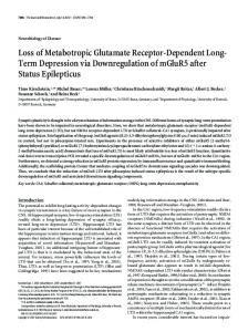
Figure 1. Basic neuronal circuitry of the cerebellumthat forms the basis for the present model of adaptivetiming of eyeblinks. Inhibitory neurons, dark; excitatory neurons, white. PC, Purkinje cell; BA, basket cell; ST, stellate cell; GR, granule cell; PF, parallel fiber; MF, mossy fiber; CF, climbing fiber; N, cerebellar nuclear cell; PN, precerebellar neuron that issues mossy fibers; 10, inferior olive; CS, conditioned stimulus; CR, conditioned response; US,unconditioned stimulus.
... 0 2
)
~ Co :1
GROUI' P 1/2
< ~
v
10
>-
~
servedin Purkinje cells after activationof mGluR at the parallelfiber-Purkinje cell synapse,asdiscussedbelow (Batchelorand Garthwaite,1993;Batchelor et al., 1994).When the
3768 J. Neurosci., June 1,1996,16(11):3760-3774
Fiala et al. .Adaptive Timing in Purkinje Cells
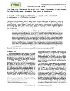
Figure8. Spectrumproduced by variation in Bmaxin responseto sustained [giu] concentration.Bmax= {360,21,4.7,1.73,0.97,0.625,0.458,0.368,0.315, 0.283,0.261,0.245,0.236,0.23,0.226}; these receptor concentrationvalues were chosento give approximatelyequallyspacedresponsesspanning4 sec. With a sustained[giu] input, [Ca2+]spikeresponsecanbe observedout to-5 sec if the Bmaxdistribution is allowed to range down to O.
Figure9. Spectrumproduced by variation in Bmaxin responseto a 50 msec [gIu] application. Bmax= {360, 18, 6.5, 3.9, 3.18, 2.93, 2.87, 2.859, 2.858, 2.8579};valueswithin the indicatedrangewere chosento give equallyspaced responses.The value 2.585is the smallestBmaxfor which the 50 msec [gIu] stimuluswas sufficientto induce a [Ca2+]spike in the mGluR pathway.
Ca2+-dependentK+ current is also activated by the intracellular calcium transient,the net effect on the membranepotential can be hyperpolarizationrather than depolarization(Fig. 7C). Thus, if the input to the Purkinje cell elevatesits membranepotential and establishes a certain rate of simple-spikefiring, the firing rate can be decreasedfrom this level during the calcium transientby the activation of the Ca2+-dependentK+ conductance.This "pause" in Purkinje cell firing will allow an increasein activity in the interpositus cells,which governthe eye-blink response(Bullock et aI., 1994). If the Purkinje cell pause is made adaptive, then a mechanismfor eye-blinkconditioning is realized.
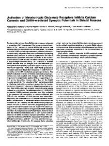
relevant interval for eye blinks of -4 sec is created in Purkinje cells by choosing Bmaxin the range of 0.1-500. The particular values used in generating a given spectrum are given in the associated figure caption. No other parameters are varied in producing these responses.The present model is thus a biochemically derived variant of a spectral timing model (Grossberg and Schmajuk, 1989; Grossberg and Merrill, 1992; Bullock et al., 1994). The spectrum of response times can be used to learn an adaptively timed eye blink, as discussedbelow. Although for purposes of simulation we assumed that the latency variations are attributable wholly to a natural spectrumof Bmaxvalues, it is possible that variance in other mGluR pathway components may contribute to generation of different latencies. For example, variations in IP3R density or in luminal calcium stores, which affect the rate of mGluR-mediated Ca2+ release, will affect responselatency. The model exhibits different calcium response properties to transient versus maintained agonist concentrations. Although maintained parallel fiber inputs produce a spectrum that spans 4 sec,a transient parallel fiber activation of 50 msec duration admits only a spectrum spanning ~2 sec. This is because the dynamics engenderedby a 50 msec stimulus fail to generate a [Ca2+] spike in mGluR pathwayswhose Bmaxvalues are associatedwith longer latencies. Figure 9 showsthe spectral responseproperties of the model to 50 msec parallel fiber activations. This difference in the spectralproperties of transient versus maintained inputs is analogousto the differencesin the maximal ISIs for trace versus delay eye-blink conditioning (Smith et al., 1969; Solomon et al., 1986). Measurementof the potential of a population of Purkinje cells in slice reveals a slow response after brief activation of parallel fibers (Batchelor and Garthwaite, 1993; Batchelor et al., 1994). The responsehas a slow rise-time, with a peak at 300-700 msec and a slow decayover severalseconds(Fig. lOA). The responseis observablein a bath of ionotropic glutamate and GABA antagonists,which suggeststhat the responseis mediated by mGluRs at the parallel fiber-Purkinje cell synapses.This type of responseis the result of the summation of the individual signals in a spectrum, suchas that of Figure 8 or 9. Figure lOB showsthe summationof the potential changesproduced by Na/Ca exchange current in a heterogeneous population of mGluR response pathways. This
Populationresponse Large quantities of glutamate are released presynaptically after activation. It has been estimated that the postsynapticconcentration of glutamate in the center of the synapsereacheslevels> 1 mM (Clements et al., 1992).Metabotropic receptors,however,are located at the periphery of the synapse(Nusser et al., 1994).This means that a much lower level of glutamate will reach these receptors.This concentration at the periphery will exhibit a slower decay than that in the synapticcleft. Therefore, we assumethat in responseto maintained parallel fiber firing of sufficientfrequency, the population of mGluR receptors at the synapsewill be exposed to a 10 ILMlevel of glutamate. Many of the G-protein-coupled receptor types use the second messengersysteminvolving PLC and IP3-mediatedcalcium release (McGonigle and Molinoff, 1994).Responsesmediatedby these receptorscan exhibit a wide range of temporal latencies.Serotonergic and muscarinic receptor responsescan exhibit latenciesof a few secondsto >30 sec (Berridge et al., 1988; Devor et aI., 1991).The photoresponseattributable to activationof rhodopsinin the invertebrate photoreceptorhaslatencieson the order of tensof milliseconds (Fuortes and Hodgkin, 1964).The rapidity of the responsefor a givenreceptortype is dependenton the levelof G-proteinactivation. This, in turn, is dependenton the level of activation of receptors. Assuming a relatively constantglutamate level of 10 ILM,latency of the responsewill be dependenton the numberof availablereceptors in the vicinity of the parallel fiber synapse. Thus, variation in the number of mGluRl receptors,Bmax,at different synapsesproduces intracellular calcium responseswith different latencies.Figure 8 demonstratesthe effect of variation of Bmax.A spectrumof calcium responsesspanning the behaviorally
"" ~
Fiala et al. .Adaptive Timing in Purkinje Cells
J. Neurosci., June 1, 1996, 16(11):3760-3774 3769
.50mV
cs us A
Figure 11. Progress of model population responseduring 30 pairings of CS and US at an ISI of 500 msec. Initially, mGluR activation produces a depolarizing response,but as learning progresses,a timed hyperpolarization is realized. Spectral components are the same as for Figure lOB.
2mvL
msec. Note that the rate of learning is accelerated from that observed in experimentsto decreasesimulation time. Also, learning is asymptotic becauseof the balance between CS-driven dephosphorylation and CS- and US-driven phosphorylation. CSdriven dephosphorylation alone causesextinction of the learned
200ms
--
response.
--L62mv
t
B
Figure 10. A, Metabotropic glutamate response in a slice population of Purkinje cells recorded using the three-chamber grease-gap method. (Reprinted with permission from Batchelor and Garthwaite, 1993.) B, Model population response produced by summation of spectral components in response to a 150 msec agonist application at the a"ow, with a = 0.1, N = 60,Bmax = {360, 170, 100, 65, 42, 29, 21,15.7,12,9.2,7.2,5.8,4.7,3.8, 3.15,2.65,2.25,1.96,1.73,1.55,1.4,1.27,1.15,1.06, 0.97, 0.89, 0.82, 0.763, 0.706,0.66,0.625,0.59,0.555,0.525,0.5,0.478,0.458, 0.44, 0.422, 0.407, 0.393,0.38,0.368,0.357,0.347,0.338,0.33,0.322, 0.315, 0.309, 0.303, 0.298, 0.293, 0.288, 0.283, 0.279, 0.275, 0.271, 0.267, 0.264}; values within the indicated range were chosen to give a smooth population response. This distribution was used for all results except those reported in Figures 8 and 9.
population signal, P(t), is computed by: N
P(t) = a 2: AVi + Vb,
(22)
;=1
where N is number of responsepathways,AVi = (Vi -Vb) is the mGluR-induced potential change in a given pathway, a is a constantscaling factor, and Vb is the baselineresting potential of the population. As shown in Figure 10, even though the individual responses are localized in time, the population signal is broad and smooth becauseof distribution of the localized signals throughout a long interval. This population response phenomenon also is seen in other IP3-mediatedresponsesystems,suchas histamine receptors of HeLa cells (Bootman, 1994). Conditioning Given a CS-activated spectrumof responsesdistributed among a population of response pathways,a US input can select spectral components that will produce the desired behavioral response (Grossbergand Schmajuk,1989). In the present model, the CS is parallel fiber activation of mGluRs, whereas the climbing fibers produce the [cGMP] increase at US onset. Figure 11 showsthe population responseduring 36 pairings of a 600 msec CS and a 100msecUS. The CS and US coterminate, suchthat the ISI is 500
Those mGluR responsepathways that have PKC activity at the time of climbing fiber activation correlated with US onset exhibit a persistent phosphorylation of Ca2+-dependent K+ channels. This increasesthe peak conductanceof thesechannels in response to the intracellular calcium transient, such that these pathways produce a more hyperpolarizing responseafter repeated pairings. Thus, those Purkinje cells whose CS-activated mGluRI pathway has a latency that approximatesthe ISI will exhibit a progressive decreasein simple-spike firing during the CS-US interval. Other Purkinje cells will exhibit increases in simple-spike firing in the CS-US interval attributable to the depolarizing Na/CA exchanger as well as the AMP A receptor input. Those cells exhibiting a decrease in firing will realize a minimum firing rate near the expectedtime of US onset. These characteristicsare in agreement with in vivo recordings of Purkinje cell activity during eye-blink conditioning (Berthier and Moore, 1986; Thompson, 1990). Interpositus nuclear cells receive input from a population of Purkinje cells. Therefore, the population response shown in Figure 11 is responsible for the observed CR-related activity in interpositus. In agreementwith recordings from interpositus (Fig. 12), the population responsepeak occurs before the time of the expected US.. Both the latency of the response peak and the response onset latency decreaseduring learning.
~!.lL~~..,.;. ~:.J,J.L.1..IJ~.~
J..:
,..,...
Figure 12. Average nictitating membrane movement (top) and peristimuIus histogram of interpositus nucleus neural activity (bottom) during classical conditioning of a rabbit with a 25msecpontine stimulation as the CS and an air-puff delivered 225 msec later as the US. (Reprinted with permission from Steinmetz, 1990b.)
3770 J. Neurosci., June 1, 1996,16(11):3760-3774
Fiala et al. .Adaptive Timing in Purkinje Cells
1 Steinmetz (1990a) present model
0.9
-0--+--.
0.8
~~
0.7
i!B,.+~;,Ji{~i:lc:;t£
-5
bI) =
0.6
t/)
0.5
~ u
0.4
~
0.3
0.2
Two sites of learning need to be considered in eye-blink conditioning: cerebellar cortexand interpositus. Although the present model focuseson learning in cortex, the result is compatible with an additional learning site in the interpositus (Fig. 1). Learning at mossyfiber synapseson nuclear cells can provide a learned gain that canbe expressedthrough the interpositus when Purkinje cell activity pauses. In this way, learning at Purkinje cells opens a timed gate that enableslearned gains at the intracerebellar nuclei to control a movement at the appropriate time. We have demonstrated previously that the existence of this type of interpositus learning in conjunction with cortical learning can explain the maladaptivelytimed CRs, which can occur after cortical lesions (Bullock et al., 1994).
0.1 0 ' -,
0
200
,
,
,
,
.,
400
600
800
1000
1200
.,
1400
1600
1800
2000
ISI (ms) Figure 13. Comparison of CR strength-ISI dependencycurves for the model and the behavioral data. Data of Steinmetz (1990a)is normalized to 86% CRs. Model data are the magnitude of the learned hyperpolarization below -50 mY, normalized to the amount of hyperpolarization obtained at asymptote during training with an ISI of 250 msec.
The strength of the CR depends on ISI in a characteristicway. CR strength is maximal at ISIs of 200-400 msecand is reduced at shorter or longer ISIs (Smith et al., 1969; Steinmetz, 1990a). By taking the depth of the population responseas a measureof CR strength, it is possible to reconstruct.the CR strength-ISI dependency curve produced by the model. Figure 13 showsthe curve for the model in comparison with the experimental data obtained by Steinmetz(1990a). Strength of CR in the experimentis calculated as percentageCRs over test trials. For the model, CR strength is calculated by,the magnitude of hyperpolarization below the baseline value of-50 mY. As shown in the figure, the model reproducesthe characteristicISI dependencyas measuredbehaviorally. A spectral timing model is able to produce double-responding after conditioning with two different ISIs in alternation. Figure 14 depicts the effectof conditioning with alternating ISIs of 350msec and 1000msec.A double-peakedCR is produced with peaks near the expected times of the US, and with the Weber law property (compare Fig. 2) whereby the earlier peak is narrower and the later peak broader. The figure also demonstrates extinction of a learned responsewith repeated presentation of the CS alone. .
-.J
L
r--L
CSI
US1 CS2
US2 Figure 14. Progress of population response during first 10 extinction trials after 30 CS-US pairings with alternating ISIs of 350 and 1000 msec. After conditioning, the 1100msec CS2 is used to elicit a doublepeaked CR.
DISCUSSION As described in the introduction, the most parsimonious explanation for direct mossyfiber stimulation producing a timed response in interpositus is that a timing function is present in the cerebel-
lum. The present model demonstratesthat the mGluR1phosphoinositide hydrolysis second messengersystem in cerebellar Purkinje cells can perform a timing function, both in maintaining a CS trace for associationwith a temporally remote US and in the delayed onset of the CR. The basicschemeof Figure 6 for control of phosphorylation has been recognized for many years (Nestler and Greengard, 1984, their Fig. 9.3). It is important to realize, however, that the cGMP signal that increaseslevels of phosphorylation is antagonized by the parallel fiber-mediated intracellular calcium signal, which decreasesphosphorylation. The fact that the cGMP signal correspondsto a US signal,whereasthe mGluR activation corresponds to a CS signal,makes it clear that conditioning is obtainable only when activation of thesepathwaysoccurs in temporal conjunction. Activation of the mGluR pathway alone gradually reverses the effects of any previous conjunctive activation. L TD of AMPA receptors Although the phosphorylation of AMP A receptors is not crucial to behavioral learning in the model, it certainly has some bearing in vivo. The exactrole played by the AMP A receptor in eye-blink conditioning remains an unresolved issue. It is not even clear whether AMP A receptor activation is necessaryfor AMP A receptor LTD (Linden et al., 1993). Nonetheless,it seems that the mechanismsinducing AMP A receptor LTD are also responsible for behavioral learning, possibly through the phosphorylation of Ca2+-dependentK+ channels.The fact that mGluR1 is critical for induction of AMP A receptor LTD (Aiba et al., 1994; Shigemoto et al., 1994)motivates our hypothesis that it is temporal correlation of the mGluR1-mediated second messengersand the climbing fiber-evoked cGMP signal that produce behavioral learning. Aiba et al. (1994) reported loss of AMP A receptor LTD and diminished but extant eye-blink conditioning in mice lacking mGluR1. Furthermore, the eye blink seemsto be timed correctly, although a detailed study over various ISIs was not conducted. This would seemto argue that mGluR1 is not involved in timing; however, another possible explanation for this finding is that mGluR5, which also couples to phosphoinositide hydrolysis, is able to partially replacemGluR1 functionally in the mutant mice. The mGluRS subtypeis present in Purkinje cells of immature rat brain (Abe et al., 1992), but during development is normally supplanted by a proliferation of mGluR1 (Shigemoto et al., 1992). To completely rule out a role for mGluR-mediated phosphoino-
Fiala et al. .Adaptive Timing in Purkinje Cells
sitide hydrolysis in timing, both subtypes would need to be eliminated. A recent report by Linden et al. (1995) demonstrates that activation of the NO/cGMP pathway is not required for Lill in culture. It sufficesto depolarize Purkinje cells (3 sec of + 10mV) significantly in conjunction with application of glutamate. Kasono and Hirano (1994) found that the depolarization can be replaced by an artificial elevation of intracellular calcium to 6 ILMin L ill induction. Linden et al. (1995) reported that the Lill theyobserved is blocked by PKC inhibitors but not inhibitors of PKG. According to the model of Figure 6, a large enough [Ca2+]cytrise canevoke L ill in the absenceof cGMP. This is attributable to the fact that calcium activates PLC, which producesDAG. The combination of high levels of calcium and DAG could drive PKC phosphorylation beyond that recoverable by baseline protein phosphatase activity. This can be realized in Equation 21 by assumingnonzero resting levels of cGMP. Our hypothesis,however, is that this situation is not occurring-in vivo. Both mGluR1activated PKC and climbing fiber-activated PKG must be present for LTD. A possible role for AMPA receptor Lill could be to unblock the mGluR-mediated response, which is inhibited by AMPA receptor stimulation (Lonart et al., 1993).The mechanismstudied by Lonart et al. (1993) seems to involve AMPA activation of voltage-dependentcalcium channels and subsequentactivation of a calcium-dependentprotein kinase. In the present model, significant calcium influx would result in activation of calciumdependentPLC and thus would invariably stimulate, rather than inhibit, formation of IP3. Therefore, Lill does not seem to unblock mGluR responsesin the Purkinje cell. The manner of interaction between mGluR and AMP A receptors in cerebellar Purkinje cells awaits further investigation. Purkinje cell and invertebrate photoreceptor The biochemistry of the invertebrate photoresponseis similar to the biochemistry of the Purkinje cell mGluR response. In the invertebrate photoreceptor light activates rhodopsin. Activated rhodopsin stimulates PLC through a G-protein (Yarfitz and Hurley, 1994), as described above for mGluR. In both invertebrate photoreceptors and Purkinje cells, activated PLC catalyzes the production of the second-messengersIP3 and DAG from PIP2, and IP 3 subsequentlyreleases calcium from intracellular stores. The rapid increase of the cytosolic calcium concentration in the invertebrate photoreceptor activates a plasma membrane Na+ conductance,which producesa depolarizing photoresponse(Shin et al., 1993). The specific mechanisms that activate this conductance in the invertebrate photoreceptor are not well understood, but they may involve Ca2+-stimulated increasesin cGMP (Bacigalupo et al., 1991; Richard et al., 1995). The photoreceptor is a site of associativeconditioning in marine mollusks such as Hel11lissenda(Atkon, 1984; Crow, 1988). Repeated pairings of light and rotation with a forward ISI results in a persistent suppressionof photokinesis in these animals (Matzel et al., 1990). This behavioral change is affected by a modification of voltage-dependent and Ca2+-dependent K+ conductances within the photoreceptor (Atkon, 1986). Similarly, the Purkinje cell seemsto play an essentialrole in certain forms of classicalconditioning. Our theory proposes that, like the invertebrate photoreceptor,behavioralleaming in the cerebellum canbe produced by persistent modification of a Ca2+-dependent K+ conductance. The biochemical cascadeproducing the invertebrate photore-
J. Neurosci., June 1,1996,16(11):3760-3774
3771
sponseis designedto remain sensitive to light over a wide range of stimulus intensities and durations. Weak signals are amplified and prolonged by the positive feedback in the biochemical cascade. This amplification results in a single absorbed photon opening 1000plasma membrane channels and eliciting a current transient of several nanoamps in Limu/us ventral photoreceptors (Nagy, 1991). The photocurrent in response to a maintained stimulus is reduced through negative feedback in the second messengerpathway,ensuring that a transient photoresponse can be produced even at high background intensities (Fuortes and Hodgkin, 1964). A similar mechanism seems to occur in turtle cones (Baylor and Hodgkin, 1974) and has been modeled by a Ca2+-mediatedgating function (Carpenter and Grossberg,1981). Our theory suggeststhat the Purkinje cell usessomething very similar to the robust signal transduction mechanismof photoreception for the specialized purpose of forming associationsbetween temporally separated stimuli. In both cases,there is a functional need to respond reliably to signalswhose intensity and duration may vary over a wide range. In the photoreceptor, this variation is attributable to changes in photon density. In the cerebellar cortex, it is attributable to variations in the number of convergentCS-activatedcells. The mechanismsin question may have evolved to improve the signal-to-noise ratio in response to weak signals by amplifying and prolonging them without losing sensitivity or temporal resolution to more intense signals. Whether this relationship betweenPurkinje cell and invertebrate photoreceptor represents convergent evolution or a true homology is an open question. Homology is possiblebecauseassociative learning arises in the invertebrates and probably postdates the evolution of photoreceptors,whereascerebellarand Purkinje cells are not found until the vertebrates, for which the cerebellum is virtually a defining feature. Data on protochordates may be able to shed light on this question. Another link warranting exploration is with the dentate-CA3 circuit in hippocampus,which exhibits adaptive timing (Hoehler and Thompson, 1980; Berger et al., 1986) and seems to use mechanismson the circuit level that are similar in many respects to those used here. Grossberg and Merrill (in press) have discussed how the hippocampal circuit may fit into a larger model neural architecture for timed reinforcement learning, attention, and movement control through interactions between the hippocampal system and cerebellum, among other brain regions. Taken together, these functional similarities suggestthat learned timing in the cerebellum uses a specialized version of neural mechanismsthat are of much broader occurrence and functional significance. REFERENCES Abe T, SugiharaH, Nawa H, Shigemoto R, Mizuno N, Nakanishi S (1992) Molecular characterization of a novel metabotropic glutamate receptor mGLUR5 coupled to inositol phosphate/Ca2+ signal transduction. J Bioi Chern267:13361-13368. Aiba A, Kano M, Chen C, Stanton ME, Fox GD, Herrup K, Zwingman TA, TonegawaS (1994) Deficient cerebellar long-term depressionand impaired motor learning in mGluRI mutant mice. Cell 79:377-388. Ajima A, Ito M (1995) A unique role of protein phosphatasesin cerebellar long-term depression. NeuroReport 6:297-300. Alkon DL (1984) Calcium-mediated reduction of ionic currents: a biophysical memory trace. Science226:1037-1045. Alkon DL (1986) Changes of membrane currents and calciumdependent phosphorylation during associativeconditioning. In: Neural mechanismsof conditioning (Alkon DL, Woody CD, eds), pp. 3-18. New York: Plenum.
Fiala et al. .Adaptive Timing in Purkinje Cells
3772 J. Neurosci., June 1, 1996, 16(11):3760-3774
Bacigalupo J, Johnson EC, Vergara C, Lisman JE (1991) Cyclic GMP opens light-dependent channels in excised patches of Limulus ventral photoreceptors (Abstr). Biophys J 59:530A. Baraban JM, Snyder SH, Alger BE (1985) Protein kinase C regulates ionic conductancein hippocampalpyramidal neurons: electrophysiological effects of phorbol esters.Proc Natl Acad Sci USA 82:2538-2542. Bartha GT, Thompson RF (1992a) Control of rabbit nictitating membrane movements.I. A computer model of the retractor bulbi muscle and the associatedorbital mechanics.Bioi Cybern 68:135-143. Bartha GT, Thompson RF (1992b) Control of rabbit nictitating membrane movements.II. Analysis of the relation of motoneuron activity to behavior. Bioi Cybern 68:145-154. Bartha GT, Thompson RF, Gluck MA (1991) Sensorimotorlearning and the cerebellum. In: Visual structures and integrated functions (Arbib M, Ewert J, eds), pp 381-396. Berlin: Springer. Batchelor AM, Garthwaite J (1993) Novel synapticpotentials in cerebellar Purkinje cells: probable mediation by metabotropic glutamate receptors. Neuropharmacology32:11-20. Batchelor AM, Madge DJ, Garthwaite J (1994) Synaptic activation of metabotropic glutamate receptors in the parallel fiber-Purkinje cell pathway in rat cerebellar slices.Neuroscience63:911-915. Baumann 0, Walz B, Somlyo AV, Somlyo AP (1991) Electron probe microanalysisof calcium releaseand magnesiumuptake by endoplasmic reticulum in bee photoreceptors. Proc Natl Acad Sci USA 88:741-744. Baylor DA, Hodgkin AL (1974) Changesin time scale and sensitivity in turtle photoreceptors. J Physiol (Lond) 242:729-758. Berger TW, Berry SD, Thompson RF (1986) Role of the hippocampus in classical conditioning of aversive and appetitive behaviors. In: The hippocampus,Vol 4 (IsaacsonRL, Pribram KH, eds), pp 203-239. New York: Plenum. Berstein G, Blanks JL, Smrcka AV, Higashijima T, Sternweis PC, Exton JH, Ross EM (1992) Reconstitution of agonist-stimulated phosphatidylinositol 4,s-bisphosphate hydrolysis using purified m1 muscarinic receptor, Gqilb and phospholipaseC-{31.J Bioi Chem 267:8081-8088. Berridge MJ, Cobbold PH, Cuthbertson KSR (1988) Spatial and temporal aspects of cell signalling. Philos Trans R Soc Lond [Bioi] 320:325-343. Berthier NE, Moore JW (1986) Cerebellar Purkinje cell activity related to the classicallyconditioned nictitating membrane response.Brain Res 63:341-350. " Bezprozvanny I, Watras J, Ehrlich BE (1991) Bell-shaped calciumresponsecurves of Ins(1,4,5)P3-and calcium-gated channelsfrom endoplasmic reticulum of cerebellum. Nature 351:751-754. Bielefeldt K, Jackson MB (1994) Phosphorylation and dephosphorylation modulate a Ca2+-activatedK+ channel in rat peptidergic nerve terminals. J Physiol (Lond) 475.2:241-254. ' Blackstone CD, Supattapone S, Snyder SH (1989) Inositolphospholipidlinked glutamate receptors mediate cerebellar parallel fiber-Purkinje cell synaptic transmission.Proc Natl Acad Sci USA 86:4316-4320. Bootman MD (1994) Quantal Ca2+release from Ins P3-sensitive intracellular Ca2+stores. Mol Cell Endocrinol 98:157-166. Bredt DS, Hwang PM, Snyder SH (1990) Localization of nitric oxide synthaseindicating a neural role for nitric oxide. Nature 347:768-770. Bullock D, Fiala JC, Grossberg S (1994) A neural model of timed responselearning in the cerebellum. Neural Networks 7:1101':'1114. Buonomano DV, Mauk MD (1994) Neural network model of the cerebellum: temporal discrimination and the timing of motor responses. Neural Comput 6:38-55. Burroughs SE, Horrocks WD, Ren H, Klee CB (1994) Characterization of the lanthanide ion-binding properties of calcineurin-B using laserinduced luminescencespectroscopy.Biochemistry 33:10428-10436. Callaway JC, Lasser-Ross N, Ross WN (1995) IPSPs strongly inhibit climbing fiber-activated [Ca2+);increasesin the dendrites of cerebellar Purkinje neurons. J Neurosci 15:2777-2787. Carafoli E (1987) Intracellular calciumhomeostasis.Annu Rev Biochem 56:395-433. Carpenter GA, Grossberg S (1981) Adaptation and transmitter gating in vertebrate photoreceptors. J Theor Neurobiol1:1-42. Casey PJ, Fong HKW, Simon MI, Gilman AG (1990) Gz, a guanine nucleotide-binding protein with unique biochemical properties. J Bioi Chem 265:2383-2390. Catania MV, Aronica E, Sortino MA, Canonico PL, Nicoletti F (1991) Desensitization of metabotropic glutamate receptors in neuronal cul-
tures. J Neurochem 56:1329-1335.
Chapeau-BlondeauF, Chauvet G (1991) A neural network model of the cerebellar cortex performing dynamic associations. Bioi Cybern
65:267-279. Chen SRW, MacLennan DH (1994) Identification of calmodulin-, Ca2+-, and ruthenium red-binding domains in the Ca2+release channel (ryanodine receptor) of rabbit skeletalmuscle sarcoplasmicreticulum. J Bioi Chem 269:22698-22704. Chen SR, Zhang L, MacLennan DH (1992) Characterization of a Ca2+ binding and regulatory site in the Ca2+ release channel (ryanodine receptor) of rabbit skeletalmusclesarcoplasmicreticulum. J Bioi Chem 267:23318-23326. Clements JD, Lester RAJ, Tong G, Jahr CE, Westbrook GL (1992) The time course of glutamate in the synaptic cleft. Science258:1496-1501. Coleman SR, Gormezano I (1971) Classicalconditioning of the rabbit's (Oryctolaguscuniculus) nictitating membrane response under symmetrical CS-US interval shifts. J Comp Physiol Psychol77:447-455. Crepel F, Jaillard D (1990) Protein kinases, nitric oxide and long-term depressionof synapsesin the cerebellum. NeuroReport 1:133-136. Crow T (1988) Cellular and molecular analysis of associativelearning and memory in Hermissenda.Trends Neurosci 11:136-142. Daniel H, Hemart N, Jaillard D, Crepel F (1992) Coactivation of metabotropic glutamate receptors and of voltage-gated calcium channels induces long-term depressionin cerebellar Purkinje cells in vitro. Exp Brain Res 90:327-331. De Meis L, Inesi G (1982) The transport of calcium by sacroplasmic reticulum and various microsomal preparations. In: Membrane transport of calcium (Carafoli E, ed), pp 141-186. London: Academic. De Schutter E, Bower JM (1994a) An active membrane model of the cerebellar Purkinje cell. I. Simulation of current clamps in slice. J Neurophysiol 71:375-400. De Schutter E, Bower JM (1994b) An active membrane model of the cerebellar Purkinje cell. II. Simulation of synaptic responses.J Neurophysiol 71:401-419. Devor DC, Ahmed Z, Duffey ME (1991) Cholinergic stimulation produces oscillations of cytosolic Ca2+ in a secretory epithelial cell line, T84. Am J Physiol260:C598-608. Eilers J, Augustine GJ, Konnerth A (1995) Subthreshold synaptic Ca2+ signalling in fine dendrites and spines of cerebellar Purkinje neurons.
Nature373:155-158. Fagni L, Bossu JL, Bockaert J (1991) Activation of a large-conductance Ca2+-dependent K+ channel by stimulation of glutamate phosphoinositide-coupledreceptors in cultured cerebellargranule cells. Eur J Neurosci 3:778-789. Freeman JA (1969) The cerebellum as a timing device: an experimental study in the frog. In: Neurobiology of cerebellar evolution and development (Llinas R, ed), pp 397-420. Chicago: American Medical Association. Fuortes MGF, Hodgkin AL (1964) Changesin time scale and sensitivity in the ommatidia of Limulus. J Physiol (Lond) 172:239-263. Furuichi T, Mikoshiba K (1995) Inositol 1,4,5-trisphosphate receptormediated Ca2+signaling in the brain. J Neurochem 64:953-960. Glaum SR, Slater NT, Rossi DJ, Miller RJ (1992) Role of metabotropic glutamate (ACPD) receptors at the parallel fiber-Purkinje cell synapse. J Neurophysiol68:1453-1462. Gormezano I (1966) Classical conditioning. In: Experimental methods and instrumentation in psychology (Sidowski JB, ed), pp 385-420. New York: McGraw-Hili. Griffith LC, Wang J, Zhong Y, Wu C-F, GreenspanRJ (1994) Calcium! calmodulin-dependentprotein kinase II and potassium channel subunit Eag similarly affect plasticity in Drosophila. Proc Natl Acad Sci USA 91:10044-10048. Grossberg S, Merrill JWL (1992) A neural network model of adaptively timed reinforcementlearning and hippocampaldynamics. Cognit Brain Res 1:3-38. Grossberg S, Merrill JWL (1996) The hippocampus and cerebellum in adaptively timed learning, recognition, and movement. J Cognit Neurosci, in press. Grossberg S, Schmajuk NA (1989) Neural dynamics of adaptive timing and temporal discrimination during associativelearning. Neural Networks 2:79-102. Hartell NA (1994) Induction of cerebellar long-term depressionrequires activation of glutamate metabotropic receptors. NeuroReport
5:913-916.
Fiala et al. .Adaptive
Timing in Purkinje Cells
Herrero I, Miras-Portugal MT, Sanchez-Prieto J (1994) Rapid desensitization of the metabotropic glutamate receptor that facilitate glutamate release in rat cerebrocorticalnerve terminals. Eur J Neurosci6:115-120. Hirano T (1990) Depression and potentiation of the synaptic transmission betweena granule cell and a Purkinje cell in rat cerebellarculture. Neurosci Lett 119:141-144. Hodgkin AL, Nunn BJ (1987) The effect of ions on sodium-calcium exchangein salamanderrods. J Physiol (Lond) 391:371-398. Hoehler FK, Leonard DW (1976) Double responding in classical nictitating membrane conditioning with single-CSdual-ISI training. PavlovJ BioI Sci 11:180-190. Hoehler FK, Thompson RF (1980) Effects of the interstimulus (CSUCS) interval on hippocampal unit activity during classicalconditioning of the nictitating membrane response of the rabbit (Oryctolaguscuniculus). J Comp Physiol Psychol94:201-215. Homma Y, Imaki J, Nakanishi 0, Takenawa T (1988) Isolation of characterization of two different forms of inositol phospholipid-specific phospholipaseC from rat brain. J BioI Chern 263:6592-6598. Iino M (1990) Biphasic Ca2+dependenceof inositoI1,4,5-trisphosphateinduced Ca release in smooth muscle cells of the guinea pig Taenia caeci. J Gen Physiol 95:1103-1122. Ito M (1984) The cerebellum and neural control. New York: Raven. Ito M (1991) The cellular basisof cerebellar plasticity. Curr Opin Neurobiol 1:616-620. Ito M, Karachot L (1992) Protein kinases and phosphatase inhibitors mediating long-term desensitizationof glutamate receptors in cerebellar Purkinje cells. Neurosci Res 14:27-38. Jaffe S (1992) A neuronal model for variable latencyresponse.In: Analysis and modeling of neural systems (Eeckman FH, ed), pp 405-410. Boston: KIuwer Academic Publishers. Joseph SK, Rice HL, Williamson JR (1989) The effect of external calcium and pH on inositol trisphosphate-mediatedcalcium release from cerebellum microsomal fractions. Biochem J 258:261-265. Kasono K, Hirano T (1994) Critical role of postsynaptic calcium in cerebellar long-term depression. NeuroReport 6:17-20. Kasono K, Hirano T (1995) Involvement of inositol trisphosphate in cerebellar long-term depression. NeuroReport 6:569-572. Khodakhah K, Ogden D (1993) Functional heterogeneity of calcium release by inositol trisphosphate in single Purkinje neurones, cultured cerebellar astrocytes,and peripheral tissues.Proc Natl Acad Sci USA 90:4976-4980. King MM, Huang CY, Chock PB, Nairn AC, Hemmings Jr HC, Chan K-FJ, Greengard P (1984) Mammalian brain phosphoproteinsas substrates for calcineurin. J BioI Chern 259:8080-8083. Konnerth A, Dreessen J, Augustine GJ (1992) Brief dendritic calcium signals initiate long-lasting synaptic depression in cerebellar Purkinje cells. Proc Natl Acad Sci USA 89:7051-7055. Lamb W, Pugh Jr EN (1992) A quantitative account of the activation steps involved in phototransduction in amphibian photoreceptors. J Physiol (Lond) 449:719-758. Levitan I, Hillman P, PayneR (1993) Fast desensitizationof the response to InsP3 in Limulus ventral photoreceptors. Biophys J 64:1354-1360. Linden DJ, Connor JA (1991) Participation of postsynaptic PKC in cerebellar long-term depressionin culture. Science254:1656-1659. Linden DJ, Connor JA (1993) Cellular mechanismsof long-term depression in the cerebellum. Curr Opin Neurobiol 3:401-406. Linden DJ, Dawson TM, Dawson VL (1995) An evaluation of the nitric oxide/cGMP/cGMP-dependentprotein kinasecascadein the induction of cerebellar long-term depressionin culture. J Neurosci 15:5098-5105. Linden DJ, Dickinson MH, SmeyneM, Conner JA (1991) A long-term depressionof AMP A currents in cultured cerebellar Purkinje neurons. Neuron 7:81-89. Linden DJ, Smeyne M, Connor JA (1993) Induction of cerebellar longterm depressionin culture requires postsynaptic action of sodium ions. Neuron 11:1093-1100. Llinas R, Sugimori M (1992) The electrophysiology of the cerebellar Purkinje cell revisited. In: The cerebellum revisited(Llinas R, Sotelo C, eds), pp 167-181. New York: Springer. Lonart G, AIagarsamyS, Johnson KM (1993) (R,S)-a-amino-3-hydroxy5-methylisoxazole-4-propionic acid (AMPA) receptors mediate a calcium-dependentinhibition of the metabotropic glutamate receptorstimulated formation of inositol 1,4,5-trisphosphate. J Neurochem
60:1739-1745.
J. Neurosci., June 1, 1996,16(11):3760-3774
3773
Matzel LD, SchreursBG, Lederhendler I, Alkon DL (1990) Acquisition of conditioned associationsin Helmj,\"senda:additive effectsof contiguity and the folWard interstirnulus interval. Bchav Neurosci 104:597-606. McCormick DA, Thompson RF (1984) Neuronal responsesof the rabbit cerebellum during acquisition and performance of a classically conditioned nictitating membrane-eyelidresponse. J Neurosci 4:2811-2822. McCormick DA, Clark GA, Lavond DG, Thompson RF (1982) Initial localization of the memory trace for a basic form of learning. Proc Natl Acad Sci USA 79:2731-2735. McGonigle P, Molinoff PB (1994) Receptors and signal transduction: classification and quantitation. In: Basic neurochemistry: molecular, cellular, and medical aspects,5th ed (Siegel GJ, Agranoff BW, Albers RW, Molinoff PB, eds), pp 417-428. New York: Raven. Meissner G, Darling E, Eveleth J (1986) Kinetics of rapid Ca2+ release by sarcoplasmicreticulum. Effects of Ca2+: Mg2+, and adenine nucleotides. Biochemistry 25:236-244. Midtgaard J, Lasser-Ross N, Ros.~WN (1993) Spatial distribution of Ca2+influx in turtle Purkinje cell dendrites in vitro: Role of a transient outward current. J Neurophysiol 70:2455-2469. Mignery GA, Johnston PA, Siidhof TC (1992) Mechanism of Ca2+ inhibition of inositol 1,4,5-trisphosphate(InsPJ binding to the cerebellar InsP3receptor. J BioI Chem 267:7450-7455. Millenson JR, Kehoe EJ, Gormenzano I (1977) Classicalconditioning of the rabbit's nictitating membrane response under fixed and mixed CS-US intervals. Learn Motiv 8:351-366. Missiaen L, Parys JB, De Smedt H, Oike M, Casteels R (1994) Partial calcium release in response to submaximal inositol 1,4,5-trisphosphate receptor activation. Mol Cell Endocrinol 98:147-156. Miyakawa H, Lev-Ram V, Lasser-Ross N, Ross WN (1992) Calcium transientsevoked by climbing fiber and parallel fiber synaptic inputs in guinea pig cerebellar Purkinje neurons. J NeurophysioI68:1178-1189. Moore JW, Desmond JE, Berthier NE (1989) Adaptively timed conditioned responsesand the cerebellum: a neural network approach. Bioi Cybern 62:17-28. Nagy K (1991) Biophysical processesin invertebrate photoreceptors: recent progressand a critical overviewbased on Limulus photoreceptors. Q Rev Biophys 24:165-226. Nakanishi Y (1988) The molecular heterogeneity of protein kinase C and its implications for cellular regulation. Nature 334:661-665. Nestler EJ, Duman RS (1994) Gproteins and cyclic nucleotides in the nervoussystem.In: Basicneurochemistry: molecular, cellular, and medical aspects,5th ed (Siegel GJ, Agranoff BW, Albers RW, Molinoff PB, cds), pp 429-448. New York: Raven. Nestler EJ, Greengard P (1984) Protein phosphorylation in the nervous system.New York: Wiley. Nishizuka Y (1986) Studiesand perspectivesof protein kinaseC. Science 233:305-310. Nishizuka Y (1988) The molecular heterogeneity of protein kinase C and its implications for cellular regulation. Nature 334:661-665. Nusser Z, Mulvihill E, Streit P, Somogyi P (1994) Subsynaptic segregation of metabotropic and ionotropic glutamate receptors as revealed by immunogold localization. Neuroscience61:421-427. Palay SL, Chan-Palay V (1974) Cerebellar cortex: cytology and organization. New York: Springer. Perrett SP, Ruiz BP, Mauk MD (1993) Cerebellar cortex lesions disrupt learning-dependenttiming of conditioned eyelid responses.J Neurosci 13:1708-1718. Reinhart PH, Levitan IB (1995) Kinase and phosphataseactivities intimately associated with a reconsqtuted calcium-dependent potassium channel. J Neurosci 15:4572-4579. Richard EA, Sampat P, LismanJE (1995) Distinguishing between roles for calcium in Limulus photoreceptor excitation. Cell Calcium 18:330-340. Sakurai M (1989) Depression and potentiation of parallel fiber-Purkinje cell transmissionin in vitro cerebellar slices.In: Olivo-cerebellar system in motor control (Strata P, ed), pp 221-230. Berlin: Springer. Sakurai M (1990) Calcium is an iQtracellular mediator of the climbing fiber in induction of cerebellar long-term depression. Proc Natl Acad Sci USA 87:3383-3385. SchwartzJH, Kandel ER (1991) Synaptic transmissionmediated by second messengers.In: Principles of: neurul science. 3rd ed (Kandel ER, SchwartzJH, Jessell TM, eds), pp 173-193. New York: Elsevier. Shibuki K, Okada D (1991) Endogenousnitric oxide releaserequired for long-term synaptic depressionin the cerebellum. Nature 349:326-328.
3774 J. Neurosci., June 1, 1996, 16(11):3760-3774
Shibuki K, Okada D (1992) Cerebellar long-term potentiation under suppressedpostsynaptic Ca2+activity. NeuroReport 3:231-234. Shigemoto R, Abe T, Nomura S, Nakanishi S, Hirano T (1994) Antibodies inactivating mGluRI metabotropic glutamate receptor block long-term depressionin cultured Purkinje cells. Neuron 12:1245-1255. Shigemoto R, Nakanishi S, Mizuno N (1992) Distribution of the mRNA for a metabotropic glutamate receptor (mGluRl) in the central nervous system: an in situ hybridization study in adult and developing rat. J Comp Neurol 322:121-135. Shin J, Richard E;A, LismanJE (1993) Ca2+is an obligatoryintermediatein the excitationcascadeof Limu/us photoreceptors.Neuron 11:845-855. Smith MC, Coleman SR, Gormezano I (1969) Classicalconditioning of the rabbit's nictitating membrane responseat backward, simultaneous, and forward CS-US intervals. J Comp Physiol Psychol69:226-231. Solomon PR, Vander Schaaf ER, Thompson RF, Weisz DJ (1986) Hippocampus and trace conditioning of the rabbit's classicallyconditioned nictitating membrane response. Behav Neurosci 100:729-744. Staub C, Vranesic I, Knopfel T (1992) Responsesto metabotropic glutamate receptor activation in cerebellar Purkinje cells: induction of an inward current. Eur J Neurosci 4:832-839. SteinmetzJE (1990a) Classicalnictitating membraneconditioning in rabbits with varying interstimulus intervals and direct activation of cerebellar mossyfibers as the CS. Behav Brain Res 38:97-108. Steinmetz JE (1990b) Neuronal activity in the rabbit interpositus nucleus during classical NM-conditioning with a pontine-nucleus-stimulation CS. Psychol Sci 1:378-382. SteinmetzJE, Lavond DG, ThompsonRF (1989) Oassicalconditioning in rabbits using ontine nucleus stimulation as a conditioned stimulus and inferior olive stimulationas anunconditionedstimulus.Synapse 3:225-233. Steinmetz JE, Logan CG, Rosen DJ, ThompsonJK, Lavond DO, Thompson RF (1987) Initial localization of the acoustic conditioned stimulus projection to the cerebellum essentialfor classicaleyelid conditioning. Proc Natl Acad Sci USA 84:3531-3535. Steinmetz JE, Rosen DJ, Chapman PF, Lavond DG, Thompson RF (1986) Classicalconditioning of the rabbit eyelid responsewith a mossy fiber stimulation CS: I. Pontine nuclei and middle cerebellarpeduncle stimulation. Behav Neurosci 100:878-887. Stemmer PM, Klee CB (1994) Dual calcium ion regulation of calcineurin by calmodulin and calcineurin B. Biochemistry 33:6859-6866.
Fiala et al. .Adaptive Timing in Purkinje Cells
Takei K, Mignery GA, Mugnaini E, Siidhof TC, De Camilli P (1994) Inositol 1,4,5-trisphosphatereceptor causes formation of ER cisternal stacks in transfectedfibroblasts and in cerebellar Purkinje cells. Neuron 12:327-342. Takei K, Stukenbrok H, Metcalf A, Mignery GA, Siidhof TC, Volpe P, De Camilli P (1992) Ca2+ stores in Purkinje neurons: endoplasmic reticulum subcompartments demonstrated by the heterogeneous distribution of the InsP3 receptor, Ca2+-ATPase,and calsequestrin.J Neurosci 12:489-505. Thompson JH, Bower JM (1993) Electrophysiological dissection of the excitatory inputs to Purkinje cells. In: Computation and neural systems (Eeckman FH, Bower JM, eds), pp 349-353. Norwell, MA: KIuwer Academic Publishers. Thompson RF (1990) Neural mechanisms of classical conditioning in mammals. Philos Trans R Soc Lond [Bioi] 329:161-170. Thompson RF, Krupa DJ (1994) Organization of memory traces in the mammalian brain. Annu Rev Neurosci 17:519-549. Thomsen C, Mulvihill ER, Haldeman B, Pickering DS, Hampson DR, Suzdak PD (1993) A pharmacological characterization of the mGluR1a subtype of the metabotropic glutamate receptor expressedin a cloned baby hamster kidney cell line. Brain Res 619:22-28. Villa A, Podini P, Clegg DO, POllan T, Meldolesi J (1991) Intracellular Ca2+stores in chicken Purkinje neurons: differential distribution of the low affinity-high capacity Ca2+binding protein, calsequestrin,of Ca2+ ATPase and of the ER lumenal protein, Bip. J Cell Bioi 113:779-791. Wang SS-H, Alousi AA, Thompson SH (1995) The lifetime of inositol 1,4,5-trisphosphatein single cells. J Gen PhysioI105:149-171. Watras J, BezprozvannyI, Ehrlich BE (1991) Inositol 1,4,5-trisphosphategated channelsin cerebellum: presenceof multiple conductancestates. J Neurosci11:3239-3245. Yamada WM, Koch C, Adams PR (1989) Multiple channelsand calcium dynamics.In: Methods in neuronal modeling: from synapseto networks (Koch C, SegevI, eds), pp 97-133. Boston: MIT. Yarfitz S, Hurley JB (1994) Transduction mechanismsof vertebrate and invertebrate photoreceptors. J Bioi Chern 269:14329-14332. Zipser D (1986) A model of hippocampalleaming during classicalconditioning. Behav Neurosci 100:764-776.