Radiology_39_2
04.07.2005
10:30
Page 141
Radiol Oncol 2005; 39(2): 141-6.
case report Metastatic thymoma: a case report of an isolated, intra-abdominal metastasis causing asymptomatic spinal cord compression
Douglas G. Gold, Robert C. Miller Division of Radiation Oncology, Mayo Clinic, Rochester, Minnesota, USA
Background. Although thymomas are characterized histologically by a benign appearance, they have the potential for aggressive local invasion, and occasionally they metastasize. Case report. We describe a 47-year-old woman who recently presented to our clinic with asymptomatic spinal cord compression due to an intra-abdominal metastasis of a thymoma arising as the first site of metastasis 21 years after the primary tumour was resected. Conclusions. For the patient presented here, radiotherapy and surgery were chosen over systemic therapy as the primary treatment modalities at the time of recurrence for two reasons. First, the patient had a single, isolated metastasis that occurred after a 2-decade disease-free interval; thus, preoperative radiotherapy followed by resection was potentially curative. Second, it was thought, on the basis of the retroperitoneal location of the recurrent tumour immediately below the diaphragm, that it possibly was not a haematogenously disseminated metastasis but a local pleural and lymphatic migration. Key words: thymoma; neoplasm metastasis – radiotherapy; spinal cord compression
Introduction Thymomas are unusual tumours that typically arise in the anterior mediastinum and are derived from thymic epithelial cells. Although the tumours are characterized histologically by a benign appearance, they have the potential for aggressive local invasion,
and occasionally they metastasize.1 A 47year-old woman recently presented to our clinic with asymptomatic spinal cord compression due to an intra-abdominal metastasis of a thymoma arising as the first site of metastasis 21 years after the primary tumour was resected.
Case report Received 15 April 2005 Accepted 22 April 2005 Correspondence to: Robert C. Miller, MD, MSc, Division of Radiation Oncology, Mayo Clinic, 200 First Street SW, Rochester, MN 55905, USA; E-mail:
[email protected] ©2005 Association of Radiolog and Oncology
In March 1983, when the patient was 25 years old, invasive thymoma of the anterior mediastinum was diagnosed. At her most recent evaluation in 2004, only partial records were available about the evaluation and treatment in 1983. However, according to the existing
Radiology_39_2
04.07.2005
10:30
142
Page 142
Gold DG and Miller RC / Metastatic thymoma
medical records, the primary tumour, which involved the anterior mediastinum, had been resected piecemeal. She was referred for postoperative external beam radiotherapy because of concern about residual tumour within the operative bed. A total radiation dose of 39.6 Gy in 22 fractions was administered to the mediastinum using opposed photon beams delivered by a 10-MV linear accelerator. A boost was delivered to a smaller volume within the mediastinum for a total dose in that area of 54.0 Gy in 30 fractions. The maximum spinal cord dose was 40.2 Gy. The patient tolerated radiotherapy well and had no evidence of recurrence or treatment toxicity for more than 20 years. In March 2003, she noted a lower abdominal mass after a year of menorrhagia and sought medical evaluation. She subsequently underwent simple hysterectomy. At the time of hysterectomy, an omental mass, 18×12×9 cm, was resected along with the uterus. Pathologic evaluation of the uterus demonstrated a subserosal, 0.5-cm leiomyoma; no other abnormality was noted. The omental mass was found to be an inflammatory myofibroblastic tumour. Immunohistochemical staining of the omental mass for anaplastic lymphoma kinase and smooth muscle actin was negative.
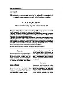
Figure 1. Magnetic resonance image of the metastatic lesion at the level of vertebral body T12 before radiotherapy. Note the proximity of left kidney to the mass and the relation of the mass to the spinal cord. Radiol Oncol 2005; 39(2): 141-6.
Progressive left hip pain developed in early 2003. This worsened in 2004, and in October 2004, magnetic resonance imaging (MRI) showed an epidural mass that extended from vertebral body T11 to L1, with compression of the left lateral thecal sac. A left paraspinal mass was also present, extending from the T11-12 interspace to L2. The two lesions were connected at the T12-L1 neural foramen. There were no abnormalities of the vertebral bodies. The intimate relation between the tumour and spinal cord is shown in Figure 1. Subsequent computed tomography (CT) of the chest, abdomen, and pelvis did not show abnormalities of concern other than a soft tissue mass arising in the left retroperitoneum near the origin of the left psoas muscle and contiguous with an epidural mass extending through the left T12 neural foramen. No bony destruction was apparent. The state of the tumour before radiotherapy is shown in Figure 2a. No evidence of local recurrence or new primary tumour was detected in the thorax. A nuclear bone scan did not show any abnormality. An incidental MRI finding was an incompletely imaged T2 hyperintense thyroid nodule, 1.8×2.2×2.7 cm. A CT-guided needle biopsy was performed in October 2004 at an outside institution and repeated at a different site within the tumour at Mayo Clinic in early November 2004. Both biopsy specimens revealed metastatic thymoma. Immunohistochemical stains showed that the tumour contained a mixture of cytokeratin-positive epithelial cells and CD3positive T cells. CD20 staining showed only a few reactive lymphocytes. Staining for S100 protein was negative. This staining pattern was thought to be consistent with the diagnosis of metastatic thymoma, presumably related to the tumour resected from the chest in 1983. The original pathology slides from 1983 were not available for comparison with the metastatic lesion found in 2004. The omental tumour identified in 2003 was compared with the new lesion and the two tumours were his-
Radiology_39_2
04.07.2005
10:30
Page 143
Gold DG and Miller RC / Metastatic thymoma
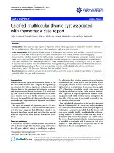
Figure 2a. Computed tomographic images showing the paraspinal mass before radiotherapy.
tologically different. A biopsy specimen from the thyroid lesion demonstrated a benign thyroid nodule. The patient was in excellent health, with no major symptoms other than occasional mild left hip pain. Her past medical history was unremarkable. A detailed neurologic evaluation did not document a clinical myelopathy. No other neurologic deficits were present except for minimal loss of sensation over the left iliac crest in the region where she was experiencing pain. Optimal surgical management of the recurrent thymoma would entail an en bloc resection. However, because of the location of the tumour, its apparent adherence to the spinal cord, and its local invasion of surrounding bony and muscular structures, the patient was referred for preoperative radiotherapy. After a medical oncology evaluation, it was thought that chemotherapy was not indicated at that time. The patient received 50.4 Gy in 28 fractions of external beam radiation delivered with intensity-modulated radiotherapy. The gross target volume was determined from the patient’s MRI, which was fused with a treatment-planning CT scan. The clinical target volume was considered the gross tumour volume as demonstrated on MRI plus areas where bone invasion was suspected on the basis of the CT scan. The patient’s previous radiotherapy fields were reconstructed to en-
143
Figure 2b. Computed tomographic images showing the paraspinal mass approximately 4 weeks after radiotherapy.
sure no overlap between those fields and the current treatment. The patient was carefully counselled about the potential risk of myelopathy from overlap of the two radiotherapy treatments. Intensity-modulated planning priorities were assigned to minimize the dose to the kidneys, followed by the small intestine (Figure 3). The spinal cord was not avoided because of the proximity of the tumour to the cord as well as the convex shape of the tumour, which surrounded one-half of the circumference of the spinal cord in some areas. Ninety-nine percent of the planning
Figure 3. Isodose distribution from the intensity-modulated radiotherapy plan showing the gross target volume (shaded structure, which indicates macroscopically evident tumor by fusion of the computed tomographic and magnetic resonance imaging data sets) and the 49.5-Gy, 40-Gy, and 20-Gy isodose lines. (The clinical and planning target volumes have been deleted for clarity.) Radiol Oncol 2005; 39(2): 141-6.
Radiology_39_2
04.07.2005
10:30
Page 144
Gold DG and Miller RC / Metastatic thymoma
144
target volume, consisting of the clinical target volume plus a 1.0-cm margin, received a dose of 48.5 Gy or greater. The minimal planning target volume dose was 43.2 Gy. The maximal spinal cord dose was 50.9 Gy. Also, 17.6% of the right kidney and 37.3% of the left kidney were treated beyond the normal tissue tolerance limit of 20 Gy. The patient tolerated the treatment well, without experiencing acute gastrointestinal tract or other toxic effects. Her hip pain resolved during the final weeks of radiotherapy, suggesting an early response to radiotherapy as the left neural foramen at the T12-L1 level was decompressed. In January 2005, restaging studies were performed preoperatively, including MRI of the thoracic and lumbar spine and CT of the chest and abdomen. MRI demonstrated a marked decrease in the size of the paraspinal mass. CT also showed a decrease in the size of the lesion, with less encroachment on the spinal canal as compared with the imaging studies before radiotherapy (Figure 2b). No evidence of malignant disease was found elsewhere. In February 2005, laminectomy of T11-L1, posterior instrumented fusion of T5-L4, left thoracotomy with en bloc resection of the paraspinal metastasis, and T11-L1 corpectomies with titanium cage strut grafting were successfully performed. Pathologic evaluation of the resected specimen showed a 5.0×4.0×2.5-cm mass containing extensive fibrosis and metastatic thymoma. All surgical margins were negative for tumour.
Discussion The histologic classification of thymomas, which are derived from thymic epithelial cells, has been debated, and several classification systems have been proposed. However, it is generally accepted that the clinical degree of invasion of the tumour, not Radiol Oncol 2005; 39(2): 141-6.
the presence of benign or malignant histologic features, determines prognosis. This observation led to the formulation of the Masaoka staging system, currently the most commonly used staging system.2 Masaoka stage I consists of encapsulated tumours. Stage II includes tumours with macroscopic invasion of the surrounding mediastinal tissues or microscopic invasion of the capsule. Stage III includes tumours with macroscopic invasion of nearby organs. Stage IVA includes pleural or pericardial dissemination, and stage IVB includes lymphatic or haematogenously disseminated metastases.1,2 Surgical resection is the treatment of choice for most thymomas confined to the thoracic cavity, where the success of surgery and adjuvant therapy depends on the extent of resection and stage of disease.3 According to one report, the frequency of recurrence for stage I disease may be less than 5% after complete resection, whereas for stages II and III disease, the frequency of recurrence is 7% and 16%, respectively.4 Postoperative radiotherapy is often administered for stages II and III tumours because of the apparent local control and survival benefit reported in retrospective series.5-7 Preoperative radiotherapy has been used for stage III disease to facilitate total or subtotal resection.8 Our current practice is to consider adjuvant radiotherapy for resected Masaoka stage II tumours that penetrate the capsule and for resected stage III tumours. However the decision depends on several factors, including the potential sites of tumour adherence or invasion, surgical expertise and technique, and the patient’s underlying medical condition. Thymomas typically spread by direct invasion of nearby organs. Metastases may occur in the thorax as pulmonary nodules, pleuralbased implants, diaphragmatic masses, or malignant pericardial or pleural effusions. Extrathoracic metastases are rare but may involve the kidney, bone, liver, and brain. Disease may spread directly from the thorax
Radiology_39_2
04.07.2005
10:30
Page 145
Gold DG and Miller RC / Metastatic thymoma
to the abdomen or retroperitoneum, as has been documented for mesotheliomas.1,3,4,9 Chemotherapy may be considered for unresectable and metastatic thymomas. Singleagent chemotherapy, with various agents, has been studied, with ifosfamide appearing to be a promising agent. Multiagent chemotherapy regimens, with combinations of cisplatin, doxorubicin, cyclophosphamide, and vincristine, have produced response rates in excess of 50%. Chemotherapy has also been given neoadjuvantly as part of combined modality therapy involving surgery and radiotherapy.10-13 For the patient presented here, radiotherapy and surgery were chosen over systemic therapy as the primary treatment modalities at the time of recurrence for two reasons. First, the patient had a single, isolated metastasis that occurred after a 2-decade diseasefree interval; thus, preoperative radiotherapy followed by resection was potentially curative, whereas systemic therapy would not offer the possibility for a durable cure. Second, it was thought, on the basis of the retroperitoneal location of the recurrent tumour immediately below the diaphragm, that it possibly was not a haematogenously disseminated metastasis. It was hypothesized that the spread of the tumour from the mediastinum to the retroperitoneum occurred through very slow-growing microscopic tumour deposited in the pleural space before or at the time of surgery in 1983. These microscopic tumour cells eventually migrated through lymphatic channels across the diaphragm, in a manner similar to that described for mesotheliomas. The absence of other identifiable sites of distant metastatic disease lends support to this hypothesis. However, even if the underlying method of spread in this case was from an isolated, blood-borne metastasis rather than through local pleural and lymphatic migration, the treatment strategy would have been the same because of the very long diseasefree interval between the initial diagnosis and
145
the development of the metastasis. Postoperatively, this patient did well. Adjuvant systemic therapy will not be administered because of the absence of known residual malignant disease. Close observation with serial history and physical examinations and periodic CT of the chest and abdomen are planned for follow-up.
References 1. Thomas CR, Wright CD, Loehrer PJ. Thymoma: state of the art. J Clin Oncol 1999; 17: 2280-9. 2. Masaoka A, Monden Y, Nakahara K, Tanioka T. Follow-up study of thymomas with special reference to their clinical stages. Cancer 1981; 48: 248592. 3. Zhu G, He S, Fu X, Jiang G, Liu T. Radiotherapy and prognostic factors for thymoma: a retrospective study of 175 patients. Int J Radiat Oncol Biol Phys 2004; 60: 1113-9. 4. Regnard JF, Magdeleinat P, Dromer C, Dulmet E, de Montpreville V, Levi JF, et al. Prognostic factors and long-term results after thymoma resection: a series of 307 patients. J Thorac Cardiovasc Surg 1996; 112: 376-84. 5. Curran WJ Jr, Kornstein MJ, Brooks JJ, Turrisi AT III. Invasive thymoma: the role of mediastinal irradiation following complete or incomplete surgical resection. J Clin Oncol 1988; 6: 1722-7. 6. Urgesi A, Monetti U, Rossi G, Ricardi U, Casadio C. Role of radiation therapy in locally advanced thymoma. Radiother Oncol 1990; 19: 273-80. 7. Pollack A, Komaki R, Cox JD, Ro JY, Oswald MJ, Shin DM, et al. Thymoma: treatment and prognosis. Int J Radiat Oncol Biol Phys 1992; 23: 1037-43. 8. Ohara K, Okumura T, Sugahara S, Akisada M, Yokose T, Ogata T, et al. The role of preoperative radiotherapy for invasive thymoma. Acta Oncol 1990; 29: 425-9. 9. Shin MS, Bailey WC. Computed tomography of invasive pleural mesothelioma. J Comput Tomogr 1983; 7: 389-94. 10. Highley MS, Underhill CR, Parnis FX, Karapetis C, Rankin E, Dussek J, et al. Treatment of invasive thymoma with single-agent ifosfamide. J Clin Oncol 1999; 17: 2737-44. Radiol Oncol 2005; 39(2): 141-6.
Radiology_39_2
04.07.2005
10:30
146
Page 146
Gold DG and Miller RC / Metastatic thymoma
11. Fornasiero A, Daniele O, Ghiotto C, Piazza M, Fiore-Donati L, Calabro F, et al. Chemotherapy for invasive thymoma: a 13-year experience. Cancer 1991; 68: 30-3. 12. Loehrer PJ Sr, Chen M, Kim K, Aisner SC, Einhorn LH, Livingston R, et al. Cisplatin, doxorubicin, and cyclophosphamide plus thoracic radiation therapy for limited-stage unresectable thymoma: an intergroup trial. J Clin Oncol 1997; 15: 3093-9. 13. Shin DM, Walsh GL, Komaki R, Putnam JB, Nesbitt J, Ro JY, et al. A multidisciplinary approach to therapy for unresectable malignant thymoma. Ann Intern Med 1998; 129: 100-4.
Radiol Oncol 2005; 39(2): 141-6.