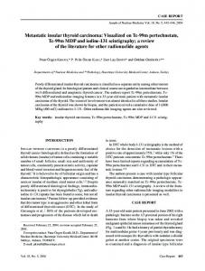
Case Rep Oncol 2017;10:1057–1064 DOI: 10.1159/000484597 Published online: November 27, 2017
© 2017 The Author(s) Published by S. Karger AG, Basel www.karger.com/cro
This article is licensed under the Creative Commons Attribution-NonCommercial 4.0 International License (CC BY-NC) (http://www.karger.com/Services/OpenAccessLicense). Usage and distribution for commercial purposes requires written permission.
Case Report
Metastatic Urothelial Carcinoma with Glandular Differentiation That Confirmed the Response by Autopsy Specimen to Second-Line mFOLFOX6 (Fluorouracil, Oxaliplatin, and Leucovorin) plus Bevacizumab Chemotherapy Taku Naiki a Toshiki Etania Aya Naiki-Itob Kana Fujiic Ryosuke Andoa Keitaro Iidaa Takashi Nagaia Yosuke Sugiyama d Motoo Nakagawa e Noriyasu Kawaia Takahiro Yasuia a
Department of Nephro-urology, Nagoya City University Graduate School of Medical b Sciences, Nagoya, Japan; Department of Experimental Pathology and Tumor Biology, c Nagoya City University Graduate School of Medical Sciences, Nagoya, Japan; Department of Clinical Pathology and Molecular Diagnostics, Nagoya City University Graduate School d of Medical Sciences, Nagoya, Japan; Department of Pharmacy, Nagoya City University e Hospital, Nagoya, Japan; Department of Radiology, Nagoya City University Graduate School of Medical Sciences, Nagoya, Japan
Keywords Urothelial carcinoma with glandular differentiation · Bladder · mFOLFOX6 plus bevacizumab chemotherapy
Taku Naiki, MD, PhD Department of Nephro-urology Nagoya City University Graduate School of Medical Sciences Kawasumi 1, Mizuho-cho, Mizuho-ku, Nagoya 467-8601 (Japan) E-Mail
[email protected] or
[email protected]
Downloaded by: 191.96.252.211 - 11/28/2017 5:04:54 AM
Abstract The prognostic significance of glandular differentiation in urothelial carcinoma (UC) is controversial, and thus far there is no established treatment strategy against metastasis of glandular component. We describe here a case of metastatic UC with glandular differentia-
1058
Case Rep Oncol 2017;10:1057–1064 DOI: 10.1159/000484597
© 2017 The Author(s). Published by S. Karger AG, Basel www.karger.com/cro
Naiki et al.: A Case of Urothelial Carcinoma with Glandular Differentiation of the Urinary Bladder
tion that had histological disappearance of adenocarcinoma components at autopsy after sequential chemotherapy with S-1 and cisplatin (CDDP) and with mFOLFOX6 (fluorouracil, oxaliplatin, and leucovorin) plus bevacizumab (mFOLFOX6+Bev). A 62-year-old Asian male was diagnosed with invasive UC with glandular differentiation (T2N0M0) by radical cystectomy and ileal conduit, and careful follow-up observation was made. Eight years after radical operation, peritoneal metastases occurred, and a biopsy specimen using colon fiber revealed high-grade adenocarcinomas with an immunohistochemical profile that included positivity for cytokeratin 7 (CK7) and negativity for cytokeratin 20 (CK20) and uroplakin, which was identical to the radical cystectomy specimen. Thus, he received combination chemotherapy consisting of S-1 and CDDP; however, the peritoneal metastasis worsened after 2 cycles. Therefore, second-line mFOLFOX6+Bev chemotherapy was performed for a total of 5 courses. In spite of this, the patient died, and the final diagnosis by autopsy was multiple metastases of infiltrating pure UC to the lung, bone, and peritoneum. Interestingly, there were no pathological findings of adenocarcinoma, and the immunohistochemical profile of the metastatic lesions was identical to that of the previous specimens from the bladder and colon. This suggests that sequential chemotherapy of S-1 and CDDP and second-line mFOLFOX6+Bev might be a feasible option in metastatic UC with glandular differentiation. © 2017 The Author(s) Published by S. Karger AG, Basel
Introduction
Urothelial carcinoma (UC) is often a comorbidity of various phenotypes, and the most common variant is squamous cell differentiation and glandular differentiation. The prognostic significance of glandular differentiation of UC is controversial; it was described as having poorer responsiveness to systemic chemotherapy in some studies, while others reported that the cancer-specific survival was similar to that of pure UC [1–4]. Several studies have described the utility of systemic combination chemotherapy with S-1 and cisplatin (CDDP) for metastatic adenocarcinoma with pure glandular differentiation originating from the bladder [5, 6], as well as regimens based on gastric malignancy. However, therapeutic regimens for metastatic adenocarcinoma originating from mixed UC with glandular differentiation have not yet been established. We report here a case of metastatic UC with glandular differentiation that had histological disappearance of adenocarcinoma components at autopsy after sequential chemotherapy with S-1 and CDDP, and mFOLFOX6 (fluorouracil, oxaliplatin, and leucovorin) plus bevacizumab (mFOLFOX6+Bev).
A 62-year-old Asian male visited our hospital with asymptomatic gross hematuria. Enhanced computed tomography (CT), magnetic resonance imaging (MRI), and bone scintigraphy revealed an advanced bladder tumor, and transurethral biopsy was achieved. Pathological findings revealed invasive UC; thus, radical cystectomy and ileal conduit were performed. The final diagnosis was invasive UC with glandular differentiation (Fig. 1a, c) confirmed by periodic acid-Schiff stain (Fig. 1b), and the pathological stage was T2N0M0. Careful followup observation and monitoring of the patient were made on an outpatient basis. Six years after the radical operation, CT revealed a solitary nodule, approximately 2 cm in size, in the S5 area of the liver (Fig. 2a, b), which was revealed as a defect in the hepatobiliary phase of
Downloaded by: 191.96.252.211 - 11/28/2017 5:04:54 AM
Case Report
Case Rep Oncol 2017;10:1057–1064 DOI: 10.1159/000484597
1059
© 2017 The Author(s). Published by S. Karger AG, Basel www.karger.com/cro
Naiki et al.: A Case of Urothelial Carcinoma with Glandular Differentiation of the Urinary Bladder
gadolinium-ethoxybenzyl-diethylenetriamine pentaacetic acid-enhanced MRI (Fig. 2c). Since this lesion was detected with slightly high signal intensity in diffusion-weighted images (Fig. 2d), we considered it a metastasis. Pathological findings obtained by fine-needle biopsy revealed a high-grade adenocarcinoma (Fig. 3a) that had the same immunohistochemical profile, which included positivity for cytokeratin 7 (CK7) and negativity for cytokeratin 20 (CK20) and uroplakin, as the radical cystectomy specimen (Fig. 1c–f, 3b–e). Therefore, based on a diagnosis of metastasis of UC with glandular differentiation originating from the bladder, a total of 3 cycles of combination induction chemotherapy using S-1 and CDDP (S-1 was administered orally at a dose of 80 mg/m2 on days 1–14 and CDDP was intravenously infused at a dose of 70 mg/m2 on day 8 of each 28-day cycle) was performed. After the treatment, the patient had complete response (Fig. 2e, f); thus, careful observation and monitoring were once again performed on an outpatient basis. Two years after the treatment, CT revealed that peritoneal metastases had occurred, and a biopsy specimen using colon fiber revealed high-grade adenocarcinomas that also had the same immunohistochemical profiles of CK7, CK20, and uroplakin as before (Fig. 3e–h). He was readministered combination chemotherapy with S-1 and CDDP; however, the peritoneal metastases worsened after 2 cycles. Therefore, with approval of the institutional review board of Nagoya City University Hospital, he received second-line mFOLFOX6+Bev chemotherapy (bevacizumab was administered as a 30- to 90-min intravenous infusion before oxaliplatin at a dose of 5 mg/kg, leucovorin at 200 mg/m2, oxaliplatin at 85 mg/m2, and bolus fluorouracil at 400 mg/m2 all on day 1, followed by 2,400 mL/m2/48 h, each 14-day cycle). After 2 cycles, the tumor response was stable disease, so an additional 3 courses of the same regimen were added. However, the efficacy was limited as new metastatic lesions in the lung and bone were detected, and after 6 months of palliative care, the patient finally succumbed to the disease. With permission from the bereaved family, an autopsy was performed. The final diagnosis was multiple metastases of infiltrating pure UC to the lung, bone, and peritoneum. There were no pathological findings of adenocarcinoma; however, the immunohistochemical profile of the metastatic lesions was identical to that of the previous specimens from the bladder, liver metastasis, and colon (Fig. 3i–l). Discussion
Downloaded by: 191.96.252.211 - 11/28/2017 5:04:54 AM
Glandular differentiation is defined by the presence of glandular formation within UC and has been reported in as many as 18% of invasive UC. Glandular differentiation resembles glands of colorectal adenocarcinoma. Immunohistochemical analysis of lesions with glandular differentiation shows a high rate of acquisition of an enteric expression pattern, including positivity for CK20 and CDX2, the latter being 100% positive in colorectal adenocarcinoma. However, the expressions of these markers were found to be completely lost in several previous cases, except for the expression of CK7, which is usually 100% positive in UC, that often remained high as in our case. The clinical course of our case depended on the differential diagnosis between metastasis of UC with glandular differentiation and adenocarcinoma originating from the bile duct of the liver tumor 6 years after the radical operation because the chemotherapy regimen would have been different. On the basis of the expression profile pattern of CK7, CK20, and uroplakin, a combination regimen with S-1 and CDDP was performed which resulted in the achievement of radiological complete response for 2 years. Considering the clinical course of
Case Rep Oncol 2017;10:1057–1064 DOI: 10.1159/000484597
1060
© 2017 The Author(s). Published by S. Karger AG, Basel www.karger.com/cro
Naiki et al.: A Case of Urothelial Carcinoma with Glandular Differentiation of the Urinary Bladder
this case, the needle biopsy of the liver tumor and subsequent immunohistochemical analyses were beneficial. Several reports have described the chemotherapy regimens for pure adenocarcinoma originating from the bladder [5–7]. We previously reported the efficacy of the combination regimen with S-1 and CDDP in urachal adenocarcinoma and signet ring adenocarcinoma patients [5, 6], and recently, the efficacy of mFOLFOX6 and FOLFIRI (irinotecan in combination with 5-fluorouracil and leucovorin) regimens with or without bevacizumab for metastatic pure colorectal adenocarcinoma originating from the bladder was also reported [8, 9]. However, the adoption of those regimens for pure adenocarcinoma in patients harboring UC with glandular differentiation is difficult because the guidelines recommend combination regimen with gemcitabine and CDDP as first-line chemotherapy for metastatic UC [10]. In this case, considering the confirmation of metastasis of pure glandular component in the liver by needle biopsy specimen, the S-1 and CDDP combination regimen was selected, and complete response was obtained for 2 years. There is currently no established sequential chemotherapy regimen specifically for metastatic UC with glandular differentiation. In our case, after the failure of the first-line S-1 and CDDP regimen, second-line mFOLFOX6+Bev therapy was effective because radiological stable disease was achieved after 2 cycles of chemotherapy and glandular components were absent in the autopsy specimen, suggesting that this sequential chemotherapy might be a potent tool for controlling metastatic UC with glandular differentiation. Our conclusion is limited by the fact that it is based on only one case; however, second-line chemotherapy with mFOLFOX6+Bev can be considered an option for patients with metastatic UC with glandular differentiation. Conclusion
We report here a case of metastatic UC with glandular differentiation that responded to second-line mFOLFOX6+Bev chemotherapy. Repeated biopsy of metastatic lesions was important in determination of the treatment strategy for selection of systemic chemotherapy. Statement of Ethics
Written informed consent was obtained from the patient for publication of this case report and accompanying images. This study was conducted according to the Declaration of Helsinki. Disclosure Statement
Downloaded by: 191.96.252.211 - 11/28/2017 5:04:54 AM
There are no potential conflicts of interest. The authors received no financial support for the research, authorship, and/or publication of this article.
Case Rep Oncol 2017;10:1057–1064 DOI: 10.1159/000484597
1061
© 2017 The Author(s). Published by S. Karger AG, Basel www.karger.com/cro
Naiki et al.: A Case of Urothelial Carcinoma with Glandular Differentiation of the Urinary Bladder
References
2 3 4 5 6 7 8 9 10
Ehdaie B, Maschino A, Shariat SF, Rioja J, Hamilton RJ, Lowrance WT, et al: Comparative outcomes of pure squamous cell carcinoma and urothelial carcinoma with squamous differentiation in patients treated with radical cystectomy. J Urol 2012;187:74–79. Lopez-Beltran A, Cheng L: Histologic variants of urothelial carcinoma: differential diagnosis and clinical implications. Hum Pathol 2006;37:1371–1388. Wasco MJ, Daignault S, Zhang Y, Kunju LP, Kinnaman M, Braun T, et al: Urothelial carcinoma with divergent histologic differentiation (mixed histologic features) predicts the presence of locally advanced bladder cancer when detected at transurethral resection. Urology 2007;70:69–74. Gofrit ON, Yutkin V, Shapiro A, Pizov G, Zorn KC, Hidas G, et al: The response of variant histology bladder cancer to intravesical immunotherapy compared to conventional cancer. Front Oncol 2016;6: 43. Kojima Y, Yamada Y, Kamisawa H, Sasaki S, Hayashi Y, Kohri K: Complete response of a recurrent advanced urachal carcinoma treated by S-1/cisplatin combination chemotherapy. Int J Urol 2006;13: 1123–1125. Hamakawa T, Kojima Y, Naiki T, Kubota Y, Yasui T, Tozawa K, et al: Long-term survival of a patient with invasive signet-ring cell carcinoma of the urinary bladder managed by combined S-1 and cisplatin adjuvant chemotherapy. Case Rep Urol 2013;2013:915874. Tatokoro M, Kawakami S, Yonese J, Fujii Y, Okubo Y, Yamamoto S, et al: Preliminary report of multimodal treatment with ifosfamide, 5-fluorouracil, etoposide and cisplatin (IFEP chemotherapy) against metastatic adenocarcinoma of the urachus. Int J Urol 2008;15:851–853. Tran B, McKendrick J: Metastatic urachal cancer responding to FOLFOX chemotherapy. Can J Urol 2010;17:5120–5123. Kanamaru T, Iguchi T, Yukimatsu N, Shimizu Y, Kohyama Y, Tachibana H, et al: A case of metastatic urachal carcinoma treated with FOLFIRI (irinotecan and 5-fluorouracil/leucovorin) plus bevacizumab. Urol Case Rep 2015;3:9–11. Clark PE, Spiess PE, Agarwal N, Bangs R, Boorjian SA, Buyyounouski MK, et al: NCCN Guidelines Insights: Bladder Cancer, Version 2.2016. J Natl Compr Canc Netw 2016;14:1213–1224.
Downloaded by: 191.96.252.211 - 11/28/2017 5:04:54 AM
1
Case Rep Oncol 2017;10:1057–1064 DOI: 10.1159/000484597
1062
© 2017 The Author(s). Published by S. Karger AG, Basel www.karger.com/cro
Naiki et al.: A Case of Urothelial Carcinoma with Glandular Differentiation of the Urinary Bladder
Fig. 1. a HE staining of the radical cystectomy specimen. By gross examination, glandular differentiation
Downloaded by: 191.96.252.211 - 11/28/2017 5:04:54 AM
was recognized in the infiltrating urothelial carcinoma (UC). b Periodic acid-Schiff staining found evidence of mucinous secretion. c HE staining of the UC lesion in the radical cystectomy specimen. d–f Immunohistochemical staining of the UC lesion in the radical cystectomy specimen showed positivity for cytokeratin 7 (d) and negativity for cytokeratin 20 (e) and uroplakin (f).
Case Rep Oncol 2017;10:1057–1064 DOI: 10.1159/000484597
1063
© 2017 The Author(s). Published by S. Karger AG, Basel www.karger.com/cro
Naiki et al.: A Case of Urothelial Carcinoma with Glandular Differentiation of the Urinary Bladder
Fig. 2. a Abdominal plain computed tomography (CT) showed a 2-cm-sized lesion (arrows) in the S5 area of
Downloaded by: 191.96.252.211 - 11/28/2017 5:04:54 AM
the liver. b Abdominal enhanced CT showed a slight enhancement at late phase (arrows). c Gadoliniumethoxybenzyl-diethylenetriamine pentaacetic acid-enhanced magnetic resonance imaging showed the nodule in the S5 area revealed as a defect in hepatobiliary phase (arrows). d Diffusion-weighted images of the S5 nodule in the liver. The lesion showed slightly high signal intensity (arrows). e Abdominal plain CT showed disappearance of the liver tumor after treatment. f Abdominal enhanced CT showed disappearance of the liver tumor after treatment.
Case Rep Oncol 2017;10:1057–1064 DOI: 10.1159/000484597
1064
© 2017 The Author(s). Published by S. Karger AG, Basel www.karger.com/cro
Naiki et al.: A Case of Urothelial Carcinoma with Glandular Differentiation of the Urinary Bladder
Fig. 3. HE staining and immunohistochemical staining of the fine-needle biopsy of the liver (a–d), the biop-
Downloaded by: 191.96.252.211 - 11/28/2017 5:04:54 AM
sy specimen obtained by colon fiber (e–h), and the liver metastasis obtained at autopsy (i–l). These specimens demonstrated the same immunohistochemical profile, including positivity for cytokeratin 7 (b, f, j) and negativity for cytokeratin 20 (c, g, k) and uroplakin (d, h, l). Glandular differentiation was detected in the fine-needle biopsy specimen of the liver metastasis and the peritoneal metastases specimen obtained by colon fiber, which completely disappeared at autopsy.