Molecules 2012, 17, 9818-9834; doi:10.3390/molecules17089818 OPEN ACCESS
molecules ISSN 1420-3049 www.mdpi.com/journal/molecules Review
Microfluidic Approaches to Bacterial Biofilm Formation Junghyun Kim 1, Hee-Deung Park 2,* and Seok Chung 1,* 1 2
School of Mechanical Engineering, Korea University, Seoul 136-713, Korea School of Civil, Environmental and Architectural Engineering, Korea University, Seoul 136-713, Korea
* Authors to whom correspondence should be addressed; E-Mails:
[email protected] (H.-D.P.);
[email protected] (S.C.); Tel.: +82-2-3290-3352 (S.C.); Fax: +82-2-926-9290 (S.C.). Received: 30 May 2012; in revised form: 27 July 2012 / Accepted: 9 August 2012 / Published: 15 August 2012
Abstract: Bacterial biofilms—aggregations of bacterial cells and extracellular polymeric substrates (EPS)—are an important subject of research in the fields of biology and medical science. Under aquatic conditions, bacterial cells form biofilms as a mechanism for improving survival and dispersion. In this review, we discuss bacterial biofilm development as a structurally and dynamically complex biological system and propose microfluidic approaches for the study of bacterial biofilms. Biofilms develop through a series of steps as bacteria interact with their environment. Gene expression and environmental conditions, including surface properties, hydrodynamic conditions, quorum sensing signals, and the characteristics of the medium, can have positive or negative influences on bacterial biofilm formation. The influences of each factor and the combined effects of multiple factors may be addressed using microfluidic approaches, which provide a promising means for controlling the hydrodynamic conditions, establishing stable chemical gradients, performing measurement in a high-throughput manner, providing real-time monitoring, and providing in vivo-like in vitro culture devices. An increased understanding of biofilms derived from microfluidic approaches may be relevant to improving our understanding of the contributions of determinants to bacterial biofilm development. Keywords: microfluidics; biofilms; bacteria; bacterial biofilm
Molecules 2012, 17
9819
1. Introduction Aquatic bacteria preferentially adhere to solid surfaces and form bacterial community [1]. Bacterial aggregates form biofilms, which are structured microbial communities supported by an extracellular polymeric substrate (EPS). Biofilm development is not simply a passive aggregation of cells. The biological ecology dynamics involve physical, chemical, and biological interactions with the microenvironment. The biofilm matrix provides microorganisms with a protective shield that contributes significantly to several clinical challenges, including symptomatic inflammation, antibiotic resistance, recurrence, and the spread of infectious emboli [2–6]. In biofilms, cells can survive under harsh environments, for example, at high temperatures or in the presence of antibiotics. The biofilm matrix improves a microbe’s opportunities for proliferation inside the body. These considerations may explain the correlation between dental hygiene and nosocomial diseases [7,8]. Many studies have examined the effects of genetic and environmental factors on biofilm development, including shear stress [9], surface topography [10], quorum sensing signals [11,12], temperature, and nutrient concentration [13,14]; however, a detailed understanding of the mechanism associated with biofilm development and the compounding effects of each factor have not been well characterized. Under these circumstances, microfluidic approaches present a promising platform for bacterial biofilm research. Microfluidics provide unprecedented control over the flow conditions, accessibility to real-time observation, high-throughput testing, and in vivo like biological environments. An understanding of the mechanism underlying biofilm formation and the design of advanced experiments using microfluidic could help identify solutions to biofilm-related problems, such as biofilm infections. In this review, we discuss bacterial biofilm development in terms of the structurally and dynamically complex interactions between the biological system and its environment. Microfluidic approaches provide promising tools, and their use in investigations of the fundamental mechanism of biofilm development will be introduced. 2. Biofilm Development 2.1. Why Do Planktonic Cells form Biofilms? What are the advantages of biofilm formation for the survival and proliferation of bacteria? Several factors contribute to the success of biofilm-forming bacteria [15,16]. The biofilm matrix shields the microbes from harsh external conditions, such as shear stress, nutrient deprivation, pH changes, oxygen radicals, disinfectants, and antibiotics. Bacterial cells within biofilms can withstand these stressful conditions because the EPS neutralize or bind antimicrobial agents, thereby protecting cells from physical stress [17]. Delay on cell maturation in the biofilm matrix and induction of rpoS-mediated stress response could cause resistance to antibiotics [18]. Adhesion to surfaces and biofilm formation provide favorable habitats for cells. Biofilms offer a degree of stability and function catalytically in the sense that cells remain localized in close proximity. The microbial communities in biofilms consist of a variety of cells and tend to behave as multicellular organisms. The bacterial cell types cooperatively survive as a whole community [19,20].
Molecules 2012, 17
9820
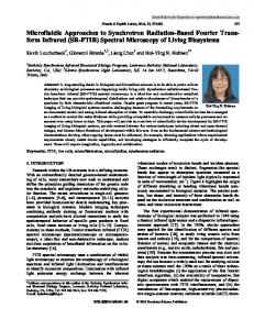
2.2. Biofilm Development Sequence Studies have revealed that bacterial biofilm development by Pseudomonas aeruginosa proceeds through a series of steps (Figure 1) [21]. The initial two steps are characterized by the loose adhesion of planktonic cells to a surface and the production of EPS. Planktonic bacterial cells can approach surfaces under bacterial motility, or under physical forces, such as Brownian motion, van der Waals attraction forces, gravitational forces, surface electrostatic charge, and hydrophobic interactions. As bacterial cells approach other cells or come within 50 nm of a surface, specific interactions between the two entities become significant. The interactions are a direct function of the free energy characteristics of and distance between the two entities [22]. Steps three and four of biofilm development entail the cellular aggregation and the subsequent growth and maturation processes. Depending on the nutrient conditions, biofilm structures can be slab or mushroom-like in shape [23]. Step five involves the dispersion of single cells from the biofilm matrix and film detachment by erosion [24,25]. Although several studies have investigated the initial steps of biofilm formation, more intensive investigations of biofilm detachment, which is a major cause of biofilm-related disease, would be valuable and inform therapeutic strategies. Figure 1. Pseudomonas aeruginosa biofilm development sequence; step 1 initial adhesion of a bacterial cell to a surface; step 2 induce irreversible adhesion by EPS generation; step 3 early structural development; step 4 maturation of the biofilm; step 5 dispersion of cells from the biofilm matrix.
2.3. Determinants of Biofilm Development Biofilms have been studied by many researchers, and a variety of determinants of biofilm development have been revealed. Biofilm formation is influenced by both gene expression and environmental conditions, including surface properties, shear stress, quorum sensing signals, and the characteristics of the aqueous medium. These factors are not the only important considerations, but
Molecules 2012, 17
9821
they provide significant contributions to biofilm development. The most dominant factor and the magnitude of the relative contributions of other factors remain under debate among biofilm researchers. 2.3.1. Gene Expression Molecular techniques, such as random transposon mutagenesis and knockout mutant studies, have revealed that biofilm-specific gene expression is involved in biofilm formation [16]. Jefferson et al. organized the knowledge of bacterial genes and their functionalities in the context of biofilm development [15]. In the case of P. aeruginosa, crc regulates global carbon metabolism, algC promotes alginate synthesis, and PA2128 reduces biofilm development by producing a probable fimbrial protein [26–28]. Although many studies have revealed biofilm-specific genes and their effects, genetic-based approaches have their limits [29–46]. These studies compare biofilm formation by mutant and wild-type strains in high-throughput micro wells. Such studies limit the examination of biofilm development to particular stable conditions with low shear flow and no nutrient exchange. Comparative biofilm growth studies are assessed within short periods of time and consider only the early stages of biofilm development. These limitations may be compensated by revealing interaction between gene expression and environmental conditions. 2.3.2. Surface Properties Surface properties, including the surface composition, morphology, structure, and material properties, affect biofilm development from the earliest stages of adhesion to the final stages of dispersion. Studies have shown that biofilm formation and colonization is promoted by rough surfaces due to the presence of beneficial local environments and the increased opportunities for biofilm formation. For example, the pockets in rough surfaces provide a protective habitat with reduced shear stress [47]. Bacterial cells more tend to adhere more strongly to hydrophobic and non-polar surfaces than to hydrophilic surfaces [48]. Additionally, porous materials are associated with a higher degree of biofilm formation compared with dense and smooth materials. The attachment of bacterial cells to porous substrates is affected by the degree of porosity, the pore size, and the permeability distribution [49,50]. After bacterial cells adhere to a surface, the biofilm matrix is influenced by the architecture and electronic properties of the solid surface. Cations on a surface, such as magnesium and calcium, actively contribute to biofilm cohesion and development. They act as lipopolysaccharide cross-linkers by promoting the integrity of the outer cell membrane [51]. Modified titanium surfaces were used to exam the effects of the solid surface tension on biofilm adhesion and cohesion forces. Pseudomonas fluorescens biofilms endured and thrived under a high shear stress when cultured over a chloropropyl-terminated surface, whereas biofilm formation was less extensive on an alkyl-terminated surface [52]. The surface architecture of the abiotic target was demonstrated to affect the metabolism and morphology of the colonizer. Nanometer sized topographical features on titanium surfaces reduced the bacterial adhesion forces and promoted selected target cells (e.g., osteoblasts). The size and shape of the nanostructure either positively or negatively influenced biofilm formation [10].
Molecules 2012, 17
9822
2.3.3. Hydrodynamic Conditions Hydrodynamic conditions significantly influence biofilm development [53]; whether shear stress, for example, enhances or hinders biofilm development remains under debate. During the initial stages of biofilm formation, shear stress can increase the residence time of the bacterial cells at the interface, providing more opportunities for bacterial cells to adhere and disperse [24]; however, many experimental studies have reported that shear stress acts as an inhibitor of biofilm development. Bacterial cells subject to high shear stress tend to form thin monolayer biofilms [54,55]. Shear stress can slow down maturation, maintaining biofilms in a “young” or early stage, and decrease bacterial diversity in a biofilm [9]. Under low shear stress conditions, biofilms develop thick multilayer structures. These structural adaptations can affect the bacterial susceptibility to antibiotics. Under high shear stress conditions, the viability of biofilms to gentamicin was observed to decrease. Two explanations for this effect were presented: high flow rates promote molecular diffusion, and thin biofilm structures easily establish a significant antibiotic gradient through their matrix [54]. Additionally, the time-dependent stress profile influenced the success of initial biofilm colonization. For a given mean shear stress, non-uniform shear stress more effectively prevented biofilm formation than a uniform stress distribution [25]. Although the shear stress interrupted biofilm formation and covered a large space, it assisted with the clumping and dispersion of the biofilms. Under a high flow rate, the biofilm area and thickness was reduced, but the total number of bacterial cells in the fluid was high [56]. Clumped biofilm debris migrated with the flow and provided opportunities for colonizing new niches [16]. Hydrodynamic conditions can have contradictory influences on biofilm development. Shear stress suppresses the development of a biofilm matrix but provides more opportunities for new biofilm formation by increasing the residence time and improving motility. A balance between the two opposing effects may be struck to provide optimal shear stress conditions for promoting biofilm growth. 2.3.4. Quorum Sensing Signals Quorum sensing is the regulation of gene expression in response to changes in the cell population density. Bacterial cells produce and release chemical signaling molecules called autoinducers, the concentration of which increases as a function of the cell density [57]. For example, Pseudomonas aeruginosa cells require lasI to develop mature biofilms. lasI-Mutant cells were observed to terminate biofilm formation at the flat structure, micro-colony state, rather than forming mature biofilm colonies that has mushroom-like shape [58]. Other studies have shown that the induction of quorum sensing is related to a critical biofilm depth. Once biofilm thickness exceeds a critical depth, quorum sensing is induced. The value of the critical depth varies with the pH of the surrounding fluid [59]. Biofilms structures are mainly composed of EPS. In almost all bacterial biofilms, the biosynthesis of EPS is a quorum sensing-dependent process involving auto-inducer molecules. In Gram-negative bacteria, N-acyl-L-homoserine lactone (AHL) autoinducers mediate quorum sensing and biofilm formation. The AHL autoinducer type depends on the bacterial species. Dickschat provided a summary and categorization of all known AHL autoinducers produced by bacterial species [11].
Molecules 2012, 17
9823
2.3.5. Characteristics of the Aqueous Medium Autoinducers do not act independently from other influence factors, such as nutrient concentration, pH, or the concentration of carbon dioxide or oxygen. The role of quorum sensing in Pseudomonas aeruginosa biofilm formation depends on the nutritional conditions. Depending on the nutrient sources, quorum sensing can regulate or not regulate the swarming motility of the bacterial cells, thereby influencing the biofilm structure. Cells with a low motility form aggregates, leading to more structured biofilms, whereas cells with a high motility spread across a surface, leading to flat biofilms [60]. Hunt et al. proposed a hypothesis governing the role of nutrient starvation in biofilm detachment. Under a nutrient-starved environment, biofilm erosion detachment occurs under homogenous hydrodynamic conditions. Computer models of biofilm dynamics suggested a starvation-dependent detachment mechanism. Insufficient nutrition causes void areas toward the centers of biofilm colonies that induce biofilm sloughing detachment [61]. Oxygen and carbon dioxide are important components and determinants of biofilm development. The concentration of dissolved oxygen influences the initial attachment of cells and EPS production [13,62]. Experimental results showed that the adhesion of Pseudomonas aeruginosa to a substrate changed with the oxygen gradient. Planktonic cells prefer to adhere to surfaces in locations that include higher levels of dissolved oxygen [13]. A comparison of the effects of carbon and oxygen concentration showed that oxygen-limited biofilms contain more extracellular polymer carbon than carbon-limited biofilms. High extracellular polymer carbon enhances the structural strength of a biofilm matrix and decreases erosion detachment under shear stress [62]. The concentration of carbon in the fluid determines the proportion of extracellular polymer carbon in the biofilm matrix, which directly affects the susceptibility of a biofilm to shear stress. Dense phase carbon dioxide (DPCD) was shown to inactivate biofilms that were completely wet but not immersed in water. DPCD is one of the most promising techniques available for controlling microorganisms that display a high antimicrobial resistance [63]. Table 1 summarizes the effects of the environmental conditions, including the surface properties, hydrodynamic conditions, quorum sensing signals, and characteristics of the medium. Table 1. Effects of the environmental conditions on biofilm development. Environmental conditions
Effect on biofilms
Surface properties surface roughness hydrophobicity non-polar surface
Positive Positive Positive
porosity
Positive
cations on the surface chloropropyl-terminated surface alkyl-terminated surface
Positive Positive Negative
nanostructure of the surface
Positive/Negative
Species
Reference
P. aeruginosa Pseudomonas sp. Pseudomonas sp. Corynebacterium, Rhodococcus, Gordona P. fluorescens P. fluorescens P. fluorescens S. aureus, S. epidermidis, P. aeruginosa
[47] [47] [48] [49,50] [51] [52] [52] [10]
Molecules 2012, 17
9824 Table 1. Cont.
Environmental conditions
Effect on biofilms
Hydrodynamic conditions residence time Positive shear stress at the interface Positive/Negative hetero-stress distribution at the Negative interface Quorum sensing signals quorum sensing autoinducers Positive Characteristics of the aqueous medium nutrient source Positive/Negative nutrient starvation Negative oxygen concentration in the fluid Positive carbon dioxide concentration in the Positive fluid dense phase carbon dioxide Negative
Species
Reference
P. aeruginosa P. aeruginosa, P. fluorescens
[24] [54–56]
P. aeruginosa
[25]
Gram-negative bacteria
[57]
P. aeruginosa P. aeruginosa P. aeruginosa
[60] [61] [13]
P. putida
[62]
P. aeruginosa
[63]
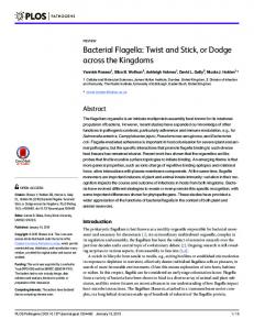
3. Microfluidic Approach 3.1. Advantages of Microfluidics Microfluidic devices manipulate fluids that generally constrained to a small environment, sub-millimeter scale. They are easily fabricated of wafers, plastics, elastomers, papers, and glass. Many studies have applied microfluidic technology due to its remarkable potentials; small liquid volume control, confining cells and molecules in a spatial geometry, temperature control and precise gradient generation, enabling low cost, rapid and precise analysis. The microfluidic devices present a promising platform for bacterial biofilm studies (Figure 2). They provide closed system where bacterial biofilms could interact with hydrodynamic environments. It allows developing mathematical models that account influences of these interactions and revealing the effects of hydrodynamic conditions (e.g., shear stress) on development of biofilms [64]. Fluid flows in these devices are very stable and yield fast response times due to the low Reynolds number, to generate gradient of chemical attractant and monitor bacterial chemotaxis. Their compactness and transparency permit observation of biofilm development in real time using high-throughput arrays. The short diffusion time and small scale facilitate bacteria culture and biofilm formation because it is easy to set up a variety of favorable conditions. The environments in microfluidic devices can be used to create models of the in vivo conditions in 3D culture platforms. These features were beneficial not only for assessing the contributions of each influencing factor to biofilm growth, but also for revealing the compounded effects. Microfluidic approach can potentially reveal the mechanism underlying biofilm formation and resolve a number of biofilm-related problems.
Molecules 2012, 17
9825
Figure 2. Advantages of microfluidics approach to bacterial biofilm studies. Microfluidics and micro-fabricated platforms have various characteristics as shown in the box that are suitable for biofilm studies. These characteristics allow developing micro-platforms for evaluating the interaction with hydrodynamic environment and bacterial chemotaxis, high throughput biofilm array, real-time monitoring, and in vivo like biological environments.
3.2. Microfluidics Approaches in Bacterial Biofilm Studies 3.2.1. Interaction with Hydrodynamic Environment Microfluidic approaches enable us to access the effects of the hydrodynamic conditions, such as the shear stress, antibiotics under flow conditions, and stress distributions. Polydimethylsiloxane (PDMS) chips are used to prepare micro-channels that permit control over the hydrodynamic conditions under which bacterial cells are cultured. Bahar et al. used microfluidic flow chambers to assess bacterial adhesion of acidovorax citrulli [65]. Lee et al. characterized the structural changes displayed by biofilms under shear stress imposed in a PDMS microfluidic device with a simple straight channel [55]. Shear stress negatively impacts biofilm development in mature biofilms or during the dispersion stages; however, during the initial and adhesion stages, shear stress promotes biofilm formation by offering more nutrient and opportunity for dispersion [66] (Figure 3a). In the absence of stress and the presence of conditions favorable for bacterial cell growth, biofilms form slowly relative to biofilm formation under stressful conditions. The flow channel geometry may be used to modulate the distribution of shear stress on a biofilm interface [54]. At different locations in a channel, bacteria cells can experience different degrees of shear stress, which changes the biofilm coverage, thickness, and viability. Microfluidic channels may be designed to elucidate the combined effects of several influencing factors on biofilm formation. Multi-channel devices have been used to assess the effects of a poly-hydrolyzing enzyme (dispersin B) and/or an antibiotic (rifampicin) on biofilm detachment. Dispersin B and rifampicin treatment induced the removal of most biofilms; however, at the corner,
Molecules 2012, 17
9826
the biofilm remained intact due to an insufficient shear flow. The combined effects of the hydrodynamics and antibiotics provide an effective tool for biofilm removal [55]. With these experimental works, microfluidic approach could be valuable way to developing mathematical models. Janakiraman et al. introduced a mathematical model based on biofilm growth in a closed system, where biofilm development and hydrodynamic environments are interlinked [64]. To verify prediction of model, they used microfluidic chambers as closed system. Based on interaction among biofilm development, mass transport, and hydrodynamics the results of model was well matched with experimental results. When bacterial cells develop biofilms, they continuously interact with their environment and this interaction finally influence to growth of biofilms. Providing closed environment, microfluidic approach allow researchers to understanding a biofilm as a colony of live cells that altered continuously with its environments. 3.2.2. Bacterial Chemotaxis Stable flow conditions in microfluidic devices facilitate the generation of flow-free, steady gradients of arbitrary shape. The chemotaxis of free-swimming or surface-adhered bacterial cells plays a fundamental role in biofilm formation and dispersion. Many examples of gradient generation in microfluidic channels for the purpose of bacterial chemotaxis study rely on flow conditions. Parallel flow devices called T-sensors operate based on the confluence of three streams, which join into a single micro-channel [67–69]. The distribution of bacteria is then measured at the end of the channel, yielding a cumulative response to the evolving nonlinear gradient experienced along the micro-channel [70]. Flow-free chemotaxis generators set up a gradient based on molecular diffusion only, and no flow is present. In this case, a flow structure is used to set up an initial gradient. The flow is then turned off, thereby allowing the gradient to evolve by diffusion alone [71–73]. When operated on a short timescale, the gradient in this type of device can be approximated as being steady-state. Such gradients are useful for quantifying chemotaxis if the timescale of the gradient relaxation is much longer than the characteristic times of the sensing and behavioral processes [70]. The incorporation of porous materials, such as hydrogels or porous membranes, in microfluidic devices has enabled the creation of steady chemical gradients in an environment completely free of flow or shear [71]. Skolimowski et al. showed that the attachment of Pseudomonas aeruginosa to a substrate varied depending on the oxygen concentration, which was modulated using a gas-permeable membrane (Figure 3b) [13]. 3.2.3. High-Throughput Analysis The compactness of microfluidic devices makes them suitable for high-throughput arrays and in situ monitoring. Eun et al. examined arrays of biofilm islands with a variety of shapes using thin polymer stencils as scaffolds [74]. PDMS stencils induce stable biofilm formation at a desired position. The diversity of the biofilm shapes makes it possible to address the effects of colony geometry on the organization, physiology, and homeostasis of biofilms. Kim et al. developed a PDMS-based microfluidic flow cell device with a dual-layer structure for investigating biofilm formation and organization in response to different concentrations of soluble signals (Figure 3c) [75]. It revealed biofilm formation in response to soluble gradients of chemical signals (e.g., 7-hydroxyindole and
Molecules 2012, 17
9827
isatin). It could also be used to screen antibiotics and biofilm inhibitory cocktails. Benoit et al. developed a microfluidic device for the high-throughput screening of biofilm viability under flow on 96 individual biofilm islands [76]. Peng Sun et al. revealed the effect of loading density of bacterial cells on biofilm formation using long-term culture arrays. They evaluated antibiotics under static (no-flow) condition [77], and found that the growth rate of biofilm was constant regardless of initial loading density when bacterial cells entered long-phase proliferation. It is important to maintain uniform bacterial cell density in microfluidic based high-throughput arrays. From simple arrays to multi-chemotaxis generators, microfluidic technology has offered a first step toward investigating the compound effects of physical, chemical, and biological factors on biofilm formation in high-throughput. Figure 3. Microfluidic devices used in the bacterial biofilm studies. (a) Schematic diagram of a microfluidic device used for bacterial biofilm formation. The effects of shear stress were quantified by analyzing the biofilm area in the microfluidic channel [66]; (b) Multi-layer microfluidic device for generating an oxygen gradient. Blue dye was injected into the channel and yellow dye was injected into the chamber. Simulation results modeled the oxygen saturation gradient in the growth chamber [13]; (c) Microfluidic flow cell for high throughput bacterial biofilm studies. The device included a glass coverslip and two PDMS layers. A bacterial biofilm developed in the microfluidic channel upon exposure to the signaling molecules [75]; (d) Model for the co-culture of epithelial cells and bacterial biofilms. A 3D rendering image showed the pneumatically-actuated trapping regions for producing biofilm islands among the epithelial cells. The colored dyes show the different regions of the co-culture device [78].
Molecules 2012, 17
9828
3.2.4. Real-Time Monitoring Microfluidic devices may be combined with existing quantification tools, such as confocal and fluorescence microscopy, to enable real-time monitoring of biofilm developments [79,80] (Table 2). In this application, microfluidic device was used as a flow-cell with precise controllability in compact space. Holman et al. introduced an open-channel microfluidic system for the in situ chemical imaging of bacterial activity in biofilms using synchrotron radiation-based Fourier transform infrared (SR-FTIR) spectroscopy. This system directly monitored the bacterial activity and biochemistry at a molecular level within a biofilm over a long period of time [81]. Meyer et al. designed a microfluidic platform that enabled the simple optical monitoring of bacterial biofilms. Biofilm formation could be monitored during growth by measuring the changes in the optical density or electrical residence using off-the-shelf electrical components [82]. Richter et al. developed a biochip for the online monitoring of biofilm dynamics. This system used high-density integrated capacitors for non-invasive measurements. Biofilm development in the channel induced changes in the voltage at the capacitors, yielding electrical signals [83]. These works made possible that researchers monitored biofilm developments in real-time, only with simple microchannel as a flow-cell, remaining much to be improved with complicated microfluidic circuits. Table 2. Real-time methods to monitor biofilm development. Analysis techniques Fluorescence microscopy Confocal reflection microscopy
Microfluidic approach generating chemical (antibiotic) gradient micro scale culture chamber
Acquired information antibiotic susceptibility of bacterial biofilms
Reference [79]
biofilm growth with time
[80]
SR-FTIR spectroscopy
circumventing water-absorption barrier
molecular level within biofilms over a long time biomolecule synthesis during biofilm development
[81]
Optical density (LED array & photodiodes)
transparent culture chamber
change in biofilm optical density over the growth period
[82]
dielectric micro-sensors integrated transparent biochip
changes of optical density and impedance caused by biofilm growth dynamic responses of biofilms to shear stress and antimicrobial agent concentration
[83]
High-density interdigitated capacitors (µIDES)
3.2.5. Mimicking Biological Environments In the environment or, for example, the human body, bacterial cells are exposed to diverse environmental conditions and must react in order to survive. Microfluidics and micro-fabricated tools make it possible to produce biofilm culture platforms that mimic the in vivo conditions experienced by bacterial cells. In the buccal cavity, Streptococcus mutans is the primary etiological agent responsible for dental caries. Using a microfluidic device with glass beads, Shumi et al. simulated the
Molecules 2012, 17
9829
interproximal space of teeth [84]. They quantified the effects of sucrose and metal ions on biofilm formation in the gaps between glass beads. In the human gastrointestinal (G1) tract, intestinal epithelial cells and non-pathogenic bacteria co-exist as a form of biofilm. In the event that pathogens invade the G1 tract, the biofilm equilibrium becomes perturbed, and commensal bacterial cells from the biofilm matrix navigate toward the pathogens. This is a key step in the infection processes. Microfluidic co-culture models enable the independent culturing of epithelial cells and bacterial biofilms in an effort to exam the role of the commensal microenvironment in pathogen colonization. A pneumatically-actuated system was used to form reversible islands that allow for the development of bacterial biofilms along with epithelial cell monolayer culturing (Figure 3d) [78]. As three-dimensional culture systems, microfluidic approaches provide a more in vivo-like environment for bacterial cells in in vitro experiments. Lee et al. examined the connection between biofilms and infection mechanisms in orthopedic implants using a three-dimensional culture device. A multi-channel microfluidic device was used to observe the development of osteoblasts in three-dimensional tissue-like structures. This study revealed how osteoblast development was influenced by the phenotype of the Staphylococcus epidermidis [85]. Planktonic cells forming biofilms communicate with one another using quorum sensing signals. Micro-encapsulation technologies may be used to study the quorum sensing mechanisms and growth of bacterial biofilms by confining one to three cells in microcapsules. A few cells confined in a very small volume results in the accumulation of auto-inducers, which induce quorum sensing growth. This strategy demonstrated that quorum sensing is a function of the measure of biomass per unit volume [86]. 4. Conclusions Bacterial biofilms are highly dynamic and sensitive to their environments, which make their analysis and control more challenging. Biomedical and bioengineering research studies of the various determinants of biofilm formation have been conducted, from studying the genetic expression patterns to the environmental conditions; however, little is known about the relative contributions of the genetic and environmental factors. A single-species bacterial biofilm had been well studied in various ways however studies about multi-species bacterial biofilms still stay at the beginning stage, remaining a lot of unknowns in bacterial communication. Biofilm engineering like biofilm catalysts and array is in trouble due to uncontrollable characteristics of bacteria. Understanding of microfluidic approach in bacterial biofilm studies could help researches to overcome these hurdles and solve the problems. The improved analyzing ability makes it possible to reveal compounded effects of genetic and environment factors. Lab-on-a-chip system could allow to culture multi-species biofilms without confusing and misunderstanding. Highly improved controllability of microfluidics helps to regulate biofilm growth in catalysts and high-throughput arrays. The knowledge gained from applying microfluidic approaches has significantly improved our understanding of biofilm formation and function. Acknowledgments This work was supported by grants from the National Research Foundation of Korea (2011-0005508) (to H.-D. Park) and from the Korea Ministry of Environment as part of the “Projects for Developing Eco-Innovation Technologies” (GT-11-G-02-001-1).
Molecules 2012, 17
9830
References 1. 2.
3.
4.
5.
6.
7. 8. 9. 10. 11. 12. 13.
14. 15. 16. 17.
18.
Zobell, C.E. The effect of solid surfaces upon bacterial activity. J. Bacteriol. 1943, 46, 39–56. Prince, A.A.; Steiger, J.D.; Khalid, A.N.; Dogrhamji, L.; Reger, C.; Claire, S.E.; Chiu, A.G.; Kennedy, D.W.; Palmer, J.N.; Cohen, N.A. Prevalence of biofilm-forming bacteria in chronic rhinosinusitis. Am. J. Rhinol. 2008, 22, 239–245. Nett, J.; Lincoln, L.; Marchillo, K.; Massey, R.; Holoyda, K.; Hoff, B.; VanHandel, M.; Andes, D. Putative role of beta-1,3 glucans in Candida albicans biofilm resistance. Antimicrob. Agents Chemother. 2007, 51, 510–520. Deligianni, E.; Pattison, S.; Berrar, D.; Ternan, N.G.; Haylock, R.W.; Moore, J.E.; Elborn, S.J.; Dooley, J.S.G. Pseudomonas aeruginosa Cystic Fibrosis isolates of similar RAPD genotype exhibit diversity in biofilm forming ability in vitro. BMC Microbiol. 2010, 10, 38. Sawasdidoln, C.; Taweechaisupapong, S.; Sermswan, R.W.; Tattawasart, U.; Tungpradabkul, S.; Wongratanacheewin, S. Growing Burkholderia pseudomallei in biofilm stimulating conditions significantly induces antimicrobial resistance. PLoS One 2010, 5, e9196. Caliskan, S.; Ozcan, S.K.; Cinar, S.; Corakci, A.; Caliskan, E. In vitro biofilm formation and relationship with antifungal resistance of Candida spp. isolated from vaginal and intrauterine device string samples of women with vaginal complaints. Mikrobiyol. Bul. 2011, 45, 697–706. Thomas, J.G.; Nakaishi, L.A. Managing the complexity of a dynamic biofilm. J. Am. Dent. Assoc. 2006, 137, 10S–15S. Donlan, R.M. Biofilms and device-associated infections. Emerging Infect. Dis. 2001, 7, 277–281. Rochex, A.; Godon, J.J.; Bernet, N.; Escudie, R. Role of shear stress on composition, diversity and dynamics of biofilm bacterial communities. Water Res. 2008, 42, 4915–4922. Puckett, S.D.; Taylor, E.; Raimondo, T.; Webster, T.J. The relationship between the nanostructure of titanium surfaces and bacterial attachment. Biomaterials 2010, 31, 706–713. Dickschat, J.S. Quorum sensing and bacterial biofilms. Nat. Prod. Rep. 2010, 27, 343–369. de Kievit, T.R. Quorum sensing in Pseudomonas aeruginosa biofilms. Environ. Microbiol. 2009, 11, 279–288. Skolimowski, M.; Nielsen, M.W.; Emneus, J.; Molin, S.; Taboryski, R.; Sternberg, C.; Dufva, M.; Geschke, O. Microfluidic dissolved oxygen gradient generator biochip as a useful tool in bacterial biofilm studies. Lab Chip 2010, 10, 2162–2169. Sawyer, L.K.; Hermanowicz, S.W. Detachment of biofilm bacteria due to variations in nutrient supply. Water Sci. Technol. 1998, 37, 211–214. Jefferson, K.K. What drives bacteria to produce a biofilm? FEMS Microbiol. Lett. 2004, 236, 163–173. Hall-Stoodley, L.; Costerton, J.W.; Stoodley, P. Bacterial biofilms: From the natural environment to infectious diseases. Nat. Rev. Microbiol. 2004, 2, 95–108. Dibdin, G.H.; Assinder, S.J.; Nichols, W.W.; Lambert, P.A. Mathematical model of beta-lactam penetration into a biofilm of Pseudomonas aeruginosa while undergoing simultaneous inactivation by released beta-lactamases. J. Antimicrob. Chemother. 1996, 38, 757–769. Mah, T.-F.C.; O’Toole, G.A. Mechanisms of biofilm resistance to antimicrobial agents. Trends Microbiol. 2001, 9, 34–39.
Molecules 2012, 17
9831
19. Shapiro, J.A. Thinking about bacterial populations as multicellular organisms. Annu. Rev. Microbiol. 1998, 52, 81–104. 20. Caldwell, D.E. Post-modern ecology is the environment the organism? Environ. Microbiol. 1999, 1, 279–281. 21. Stoodley, P.; Sauer, K.; Davies, D.G.; Costerton, J.W. Biofilms as complex differentiated communities. Annu. Rev. Microbiol. 2002, 56, 187–209. 22. Gottenbos, B.; van der Mei, H.C.; Klatter, F.; Nieuwenhuis, P.; Busscher, H.J. In vitro and in vivo antimicrobial activity of covalently coupled quaternary ammonium silane coatings on silicone rubber. Biomaterials 2002, 23, 1417–1423. 23. Klausen, M.; Heydorn, A.; Ragas, P.; Lambertsen, L.; Aaes-Jorgensen, A.; Molin, S.; Tolker-Nielsen, T. Biofilm formation by Pseudomonas aeruginosa wild type, flagella and type IV pili mutants. Mol. Microbiol. 2003, 48, 1511–1524. 24. Lecuyer, S.; Rusconi, R.; Shen, Y.; Forsyth, A.; Vlamakis, H.; Kolter, R.; Stone, H.A. Shear stress increases the residence time of adhesion of Pseudomonas aeruginosa. Biophys. J. 2011, 100, 341–350. 25. Ochoa, J.C.; Coufort, C.; Escudie, R.; Line, A.; Paul, E. Influence of non-uniform distribution of shear stress on aerobic biofilms. Chem. Eng. Sci. 2007, 62, 3672–3684. 26. O’Toole, G.A.; Gibbs, K.A.; Hager, P.W.; Phibbs, P.V.; Kolter, R. The global carbon metabolism regulator Crc is a component of a signal transduction pathway required for biofilm development by Pseudomonas aeruginosa. J. Bacteriol. 2000, 182, 425–431. 27. Davies, D.G.; Chakrabarty, A.M.; Geesey, G.G. Exopolysaccharide production in biofilms— Substratum activation of alginate gene-expression by Pseudomonas aeruginosa. Appl. Environ. Microbiol. 1993, 59, 1181–1186. 28. Whiteley, M.; Bangera, M.G.; Bumgarner, R.E.; Parsek, M.R.; Teitzel, G.M.; Lory, S.; Greenberg, E.P. Gene expression in Pseudomonas aeruginosa biofilms. Nature 2001, 413, 860–864. 29. Knobloch, J.K.M.; Nedelmann, M.; Kiel, K.; Bartscht, K.; Horstkotte, M.A.; Dobinsky, S.; Rohde, H.; Mack, D. Establishment of an arbitrary PCR for rapid identification of Tn917 insertion sites in Staphylococcus epidermidis: Characterization of biofilm-negative and nonmucoid mutants. Appl. Environ. Microbiol. 2003, 69, 5812–5818. 30. Corona-Izquierdo, F.P.; Membrillo-Hernandez, J. A mutation in rpoS enhances biofilm formation in Escherichia coli during exponential phase of growth. FEMS Microbiol. Lett. 2002, 211, 105–110. 31. Rachid, S.; Ohlsen, K.; Wallner, U.; Hacker, J.; Hecker, M.; Ziebuhr, W. Alternative transcription factor sigma(B) is involved in regulation of biofilm expression in a Staphylococcus aureus mucosal isolate. J. Bacteriol. 2000, 182, 6824–6826. 32. Knobloch, J.K.M.; Bartscht, K.; Sabottke, A.; Rohde, H.; Feucht, H.H.; Mack, D. Biofilm formation by Staphylococcus epidermidis depends on functional RsbU, an activator of the sigB operon: Differential activation mechanisms due to ethanol and salt stress. J. Bacteriol. 2001, 183, 2624–2633. 33. Yoshida, A.; Kuramitsu, H.K. Multiple Streptococcus mutans genes are involved in biofilm formation. Appl. Environ. Microbiol. 2002, 68, 6283–6291. 34. Wen, Z.Z.T.; Burne, R.A. Functional genomics approach to identifying genes required for biofilm development by Streptococcus mutans. Appl. Environ. Microbiol. 2002, 68, 1196–1203.
Molecules 2012, 17
9832
35. Merritt, J.; Qi, F.X.; Goodman, S.D.; Anderson, M.H.; Shi, W.Y. Mutation of luxS affects biofilm formation in Streptococcus mutans. Infect. Immun. 2003, 71, 1972–1979. 36. Lunsford, R.D.; London, J. Natural genetic transformation in Streptococcus gordonii: comX Imparts spontaneous competence on strain wicky. J. Bacteriol. 1996, 178, 5831–5835. 37. Kjaergaard, K.; Schembri, M.A.; Ramos, C.; Molin, S.; Klemm, P. Antigen 43 facilitates formation of multispecies biofilms. Environ. Microbiol. 2000, 2, 695–702. 38. Toledo-Arana, A.; Valle, J.; Solano, C.; Arrizubieta, M.J.; Cucarella, C.; Lamata, M.; Amorena, B.; Leiva, J.; Penades, J.R.; Lasa, I. The enterococcal surface protein, Esp, is involved in Enterococcus faecalis biofilm formation. Appl. Environ. Microbiol. 2001, 67, 4538–4545. 39. Hufnagel, M.; Koch, S.; Creti, R.; Baldassarri, L.; Huebner, J. A putative sugar-binding transcriptional regulator in a novel gene locus in Enterococcus faecalis contributes to production of Biofilm and prolonged bacteremia in mice. J. Infect. Dis. 2004, 189, 420–430. 40. Hussain, M.; Herrmann, M.; von Eiff, C.; Perdreau Remington, F.; Peters, G. A 140-kilodalton extracellular protein is essential for the accumulation of Staphylococcus epidermidis strains on surfaces. Infect. Immun. 1997, 65, 519–524. 41. Gross, M.; Cramton, S.E.; Gotz, F.; Peschel, A. Key role of teichoic acid net charge in Staphylococcus aureus colonization of artificial surfaces. Infect. Immun. 2001, 69, 3423–3426. 42. Vaudaux, P.E.; Francois, P.; Proctor, R.A.; McDevitt, D.; Foster, T.J.; Albrecht, R.M.; Lew, D.P.; Wabers, H.; Cooper, S.L. Use of adhesion-defective mutants of Staphylococcus aureus to define the role of specific plasma-proteins in promoting bacterial adhesion to canine arteriovenous shunts. Infect. Immun. 1995, 63, 585–590. 43. Caiazza, N.C.; O’Toole, G.A. Alpha-toxin is required for biofilm formation by Staphylococcus aureus. J. Bacteriol. 2003, 185, 3214–3217. 44. Idone, V.; Brendtro, S.; Gillespie, R.; Kocaj, S.; Peterson, E.; Rendi, M.; Warren, W.; Michalek, S.; Krastel, K.; Cvitkovitch, D.; et al. Effect of an orphan response regulator on Streptococcus mutans sucrose-dependent adherence and cariogenesis. Infect. Immun. 2003, 71, 4351–4360. 45. Heilmann, C.; Schweitzer, O.; Gerke, C.; Vanittanakom, N.; Mack, D.; Götz, F. Molecular basis of intercellular adhesion in the biofilm‐forming Staphylococcus epidermidis. Mol. Microbiol. 1996, 20, 1083–1091. 46. Loo, C. Oral streptococcal genes that encode biofilm formation. In Medical Implications of Biofilms; Cambridge University Press: Cambridge, UK, 2003; pp. 189–211. 47. Donlan, R.M. Biofilms: Microbial life on surfaces. Emerging Infect. Dis. 2002, 8, 881–890. 48. Flemming, H.C.; Wingender, J. Relevance of microbial extracellular polymeric substances (EPSs). Part I: Structural and ecological aspects. Water Sci. Technol. 2001, 43, 1–8. 49. Kinnari, T.J.; Esteban, J.; Martin-De-Hijas, N.Z.; Sanchez-Munoz, O.; Sanchez-Salcedo, S.; Colilla, M.; Vallet-Regi, M.; Gomez-Barrena, E. Influence of surface porosity and pH on bacterial adherence to hydroxyapatite and biphasic calcium phosphate bloceramics. J. Med. Microbiol. 2009, 58, 132–137. 50. Kapellos, G.E.; Alexiou, T.S.; Payatakes, A.C. Hierarchical simulator of biofilm growth and dynamics in granular porous materials. Adv. Water Resour. 2007, 30, 1648–1667. 51. Song, B.; Leff, L.G. Influence of magnesium ions on biofilm formation by Pseudomonas fluorescens. Microbiol. Res. 2006, 161, 355–361.
Molecules 2012, 17
9833
52. Brizzolara, R.A.; Holm, E.R. The effect of solid surface tension and exposure to elevated hydrodynamic shear on Pseudomonas fluorescens biofilms grown on modified titanium surfaces. Biofouling 2006, 22, 431–440. 53. Garny, K.; Horn, H.; Neu, T.R. Interaction between biofilm development, structure and detachment in rotating annular reactors. Bioprocess Biosyst. Eng. 2008, 31, 619–629. 54. Salek, M.M.; Jones, S.M.; Martinuzzi, R.J. The influence of flow cell geometry related shear stresses on the distribution, structure and susceptibility of Pseudomonas aeruginosa 01 biofilms. Biofouling 2009, 25, 711–725. 55. Lee, J.H.; Kaplan, J.B.; Lee, W.Y. Microfluidic devices for studying growth and detachment of Staphylococcus epidermidis biofilms. Biomed. Microdevices 2008, 10, 489–498. 56. Tsai, Y.P. Impact of flow velocity on the dynamic behaviour of biofilm bacteria. Biofouling 2005, 21, 267–277. 57. Miller, M.B.; Bassler, B.L. Quorum sensing in bacteria. Annu. Rev. Microbiol. 2001, 55, 165–199. 58. Davies, D.G.; Parsek, M.R.; Pearson, J.P.; Iglewski, B.H.; Costerton, J.W.; Greenberg, E.P. The involvement of cell-to-cell signals in the development of a bacterial biofilm. Science 1998, 280, 295–298. 59. Chopp, D.L.; Kirisits, M.J.; Moran, B.; Parsek, M.R. The dependence of quorum sensing on the depth of a growing biofilm. Bull. Math. Biol. 2003, 65, 1053–1079. 60. Shrout, J.D.; Chopp, D.L.; Just, C.L.; Hentzer, M.; Givskov, M.; Parsek, M.R. The impact of quorum sensing and swarming motility on Pseudomonas aeruginosa biofilm formation is nutritionally conditional. Mol. Microbiol. 2006, 62, 1264–1277. 61. Hunt, S.M.; Werner, E.M.; Huang, B.C.; Hamilton, M.A.; Stewart, P.S. Hypothesis for the role of nutrient starvation in biofilm detachment. Appl. Environ. Microbiol. 2004, 70, 7418–7425. 62. Applegate, D.H.; Bryers, J.D. Effects of carbon and oxygen limitations and calcium concentrations on biofilm removal processes. Biotechnol. Bioeng. 1991, 37, 17–25. 63. Mun, S.M.; Jeong, J.S.; Kim, J.E.; Lee, Y.W.; Yoon, J.Y. Inactivation of Pseudomonas aeruginosa biofilm by dense phase carbon dioxide. Biofouling 2009, 25, 473–479. 64. Janakiraman, V.; Englert, D.; Jayaraman, A.; Baskaran, H. Modeling Growth and Quorum Sensing in Biofilms Grown in Microfluidic Chambers. Ann. Biomed. Eng. 2009, 37, 1206–1216. 65. Bahar, O.; de la Fuente, L.; Burdman, S. Assessing adhesion, biofilm formation and motility of Acidovorax citrulli using microfluidic flow chambers. FEMS Microbiol. Lett. 2010, 312, 33–39. 66. Park, A.; Jeong, H.H.; Lee, J.; Kim, K.P.; Lee, C.S. Effect of shear stress on the formation of bacterial biofilm in a microfluidic channel. BioChip J. 2011, 5, 236–241. 67. Mao, H.B.; Cremer, P.S.; Manson, M.D. A sensitive, versatile microfluidic assay for bacterial chemotaxis. Proc. Natl. Acad. Sci. USA 2003, 100, 5449–5454. 68. Keenan, T.M.; Folch, A. Biomolecular gradients in cell culture systems. Lab Chip 2008, 8, 34–57. 69. Long, T.; Ford, R.M. Enhanced transverse migration of bacteria by chemotaxis in a porous T-sensor. Environ. Sci. Technol. 2009, 43, 1546–1552. 70. Ahmed, T.; Shimizu, T.S.; Stocker, R. Microfluidics for bacterial chemotaxis. Integr. Biol. 2010, 2, 604–629. 71. Diao, J.; Young, L.; Kim, S.; Fogarty, E.A.; Heilman, S.M.; Zhou, P.; Shuler, M.L.; Wu, M.; DeLisa, M.P. A three-channel microfluidic device for generating static linear gradients and its application to the quantitative analysis of bacterial chemotaxis. Lab Chip 2006, 6, 381–388.
Molecules 2012, 17
9834
72. Cheng, S.-Y.; Heilman, S.; Wasserman, M.; Archer, S.; Shuler, M.L.; Wu, M. A hydrogel-based microfluidic device for the studies of directed cell migration. Lab Chip 2007, 7, 763–769. 73. Ahmed, T.; Shimizu, T.S.; Stocker, R. Bacterial chemotaxis in linear and nonlinear steady microfluidic gradients. Nano Lett. 2010, 10, 3379–3385. 74. Eun, Y.J.; Weibel, D.B. Fabrication of microbial biofilm arrays by geometric control of cell adhesion. Langmuir 2009, 25, 4643–4654. 75. Kim, J.; Hegde, M.; Kim, S.H.; Wood, T.K. Jayaraman, A. A microfluidic device for high throughput bacterial biofilm studies. Lab Chip 2012, 12, 1157–1163. 76. Benoit, M.R.; Conant, C.G.; Ionescu-Zanetti, C.; Schwartz, M.; Matin, A. New device for high-throughput viability screening of flow biofilms. Appl. Environ. Microbiol. 2010, 76, 4136–4142. 77. Sun, P.; Liu, Y.; Sha, J.; Zhang, Z.Y.; Tu, Q.; Chen, P.; Wang, J.Y. High-throughput microfluidic system for long-term bacterial colony monitoring and antibiotic testing in zero-flow environments. Biosens. Bioelectron. 2011, 26, 1993–1999. 78. Kim, J.; Hegde, M.; Jayaraman, A. Co-culture of epithelial cells and bacteria for investigating host-pathogen interactions. Lab Chip 2010, 10, 43–50. 79. Kim, K.P.; Kim, Y.G.; Choi, C.H.; Kim, H.E.; Lee, S.H.; Chang, W.S.; Lee, C.S. In situ monitoring of antibiotic susceptibility of bacterial biofilms in a microfluidic device. Lab Chip 2010, 10, 3296–3299. 80. Yawata, Y.; Toda, K.; Setoyama, E.; Fukuda, J.; Suzuki, H.; Uchiyama, H.; Nomura, N. Monitoring biofilm development in a microfluidic device using modified confocal reflection microscopy. J. Biosci. Bioeng. 2010, 110, 377–380. 81. Holman, H.Y.N.; Miles, R.; Hao, Z.; Wozei, E.; Anderson, L.M.; Yang, H. Real-time chemical imaging of bacterial activity in biofilms using open-channel microfluidics and synchrotron FTIR spectromicroscopy. Anal. Chem. 2009, 81, 8564–8570. 82. Meyer, M.T.; Roy, V.; Bentley, W.E.; Ghodssi, R. Development and validation of a microfluidic reactor for biofilm monitoring via optical methods. J. Micromech. Microeng. 2011, 21, 054023. 83. Richter, L.; Stepper, C.; Mak, A.; Reinthaler, A.; Heer, R.; Kast, M.; Bruckl, H.; Ertl, P. Development of a microfluidic biochip for online monitoring of fungal biofilm dynamics. Lab Chip 2007, 7, 1723–1731. 84. Shumi, W.; Lim, J.; Nam, S.W.; Lee, K.; Kim, S.H.; Kim, M.H.; Cho, K.S.; Park, S. Environmental factors that affect Streptococcus mutans biofilm formation in a microfluidic device mimicking teeth. BioChip J. 2010, 4, 257–263. 85. Lee, J.H.; Wang, H.J.; Kaplan, J.B.; Lee, W.Y. Microfluidic approach to create three-dimensional tissue models for biofilm-related infection of orthopaedic implants. Tissue Eng. C Methods 2011, 17, 39–48. 86. Boedicker, J.Q.; Vincent, M.E.; Ismagilov, R.F. Microfluidic confinement of single cells of bacteria in small volumes initiates high-density behavior of quorum sensing and growth and reveals its variability. Angew. Chem. Int. Ed. Engl. 2009, 48, 5908–5911. © 2012 by the authors; licensee MDPI, Basel, Switzerland. This article is an open access article distributed under the terms and conditions of the Creative Commons Attribution license (http://creativecommons.org/licenses/by/3.0/).