Aug 18, 1970 - PDP-8) was used to control experiments, collect data, sort, average, and plot results. The computer delivered stimulus-trigger pulses at intervals ...
J. Exp. Biol. (1971), 54. 373-39° With 7 text-figures Printed in Great Britain
373
MID-BRAIN RESPONSES OF THE AUDITORY AND SOMATIC VIBRATION SYSTEMS IN SNAKES BY PETER H. HARTLINE Department of Neurosciences, School of Medicine, University of California, San Diego, La Jolla, Calif. g20j/ (Received 18 August 1970) INTRODUCTION
This paper presents data on the effects of the parameters of temperature, stimulus intensity and frequency, and stimulus pairing interval, on characteristics of mid-brain responses of snakes to sound and vibration. The results are relevant not only to the understanding of the biology of snakes but also to general aspects of central sensory physiology. The paper also fills a gap in our comparative knowledge of central representation of sensory information in the lower vertebrates, and may shed light on fundamental processes underlying responses to vibration and to sound, and on their phylogenetic development. Evidence concerning the sensory physiology of snakes has only recently become available (Wever & Vernon, i960; Bullock & Cowles, 1952; Bullock & Diecke, 1956; Proske, 1969). Particularly as regards vibratory senses, this may be due to the dearth of behavioural information about the sensory capabilities of these reptiles and the consequent lack of interest in their sensory physiology. In a separate communication, Hartline (1971) demonstrated that in snakes vibration-sensitive organs in the ear and in the skin respond to both air-borne sound and substrate vibration. Effective stimuli for the auditory system can be delivered as sound through the air or as vibration through the substrate, and can be localized at the head or delivered to the body excluding the head. As shown by frequency-response curves, the auditory system (VIII nerve system) was reported to be sensitive to a narrow band of frequencies (150-600 Hz) with greatest sensitivity at a frequency between 200 and 400 Hz. The somatic system (spinal system) responded to air-borne sound or substrate-borne vibrations if either was delivered to the body but not if delivered to the head. It was reported to have a greater frequency range, from less than 100 Hz to greater than 800 Hz in most snakes. The frequency-response curve of the spinal system lacked a distinct peak of sensitivity and was c. 20 dB less sensitive to either air-borne sound or substrate vibration than the auditory system. The responses of the two systems were found to be differently localized in the mid-brain, the spinal system showing a larger area of responsiveness. The peripheral mechanisms and origins of these responses are covered in the above-mentioned report. In this paper, stimuli previously found to be appropriate are used to explore central physiology and to characterize the responses of the two systems.
24-2
374
PETER H. HARTLINE METHODS
Snakes of the family Crotalidae (rattlesnakes) were used for most experiments but the main results were verified qualitatively on species of the families Colubridae and Boidae. Snakes were anaesthetized (15-30 mg pentobarbital sodium I.P. per kilogram of body weight), and often administered artificial respiration. One or both lobes of the tectum were exposed; metal-filled glass micropipettes (Dowben & Rose, 1953) were used for extracellular recording of compound, evoked potentials or spikes from the midbrain. Clicks or sinusoidal stimuli with trapezoidal envelopes were employed. Sound was delivered with an 8-inch loudspeaker and monitored by a calibrated microphone. Vibrations, calibrated in the range 4-500 m/t by optical means and extrapolated to o-i m/i (1 A) by assuming piezoelectric linearity, could be delivered by piezoelectric ceramic elements which drove either a 3 x 3 cm 'vibrating platform' or a hypodermic needle 'vibrating probe'. A general-purpose digital computer (Digital Equipment Corp. PDP-8) was used to control experiments, collect data, sort, average, and plot results. The computer delivered stimulus-trigger pulses at intervals with a random jitter \ to \ the mean interval. This provided averaged responses free from the effects of rhythmic signals nearly synchronous with the stimulus. The computer also permitted several different stimuli to be presented in sequence, the response to each stimulus being computed concurrently with the others. The effects of drift in the responses were thus minimized. Details pertaining to the methods and materials are presented elsewhere (Hartline, 1971). RESULTS
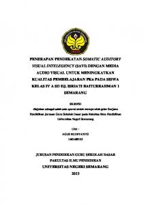
Representative averaged responses are presented in Fig. 1. The amplitude of the averaged response could be measured objectively by the computer; it was defined as the maximum of the average less the minimum of the average over a time interval chosen appropriately for the response. As is illustrated in the figure (line 1, somatic), the amplitude measured could be different depending on the choice of P and Q. For most records the amplitude chosen was from the negative peak to the highest point on the following positive wave {A' in the example) therefore limits such as P' and Q' were specified to the computer. Latency of a response was measured on the X-Y plotter output as indicated in the figure; the width of the negative peak was measured as shown, and used as one indicator of response shape (lines 1 and S, auditory). As in many averaging situations, neither amplitude, latency, nor shape of the average necessarily reflects the characteristics of a single response, since all of these may differ for different repetitions of the stimulus (Brazier, 1964). Variability of latency of individual responses to a given stimulus is among the factors affecting characteristics of averaged responses. The averaging process makes an increase of latency variability show up as an increase in response width and decrease in response amplitude. Increases in latency variability occur especially near threshold values of stimulus intensity ; the effect is striking, but no quantitative measure has been applied. A digital smoothing technique was used to simulate visual estimates of the response shape in cases where high-frequency noise was present in the averaged response. This improved the computer measurement of amplitudes by reducing the high-frequency
Auditory and somatic vibration systems in snakes
375
noise of the baseline. Comparison of the plotter output for smoothed and unsmoothed data shows that the responses with which this paper deals were not much changed by the smoothing process (Fig. 1, lines 2 and 3). The usual form for responses of both systems is that shown in Fig. 1 (auditory): an initial large negative peak followed by a slow positive wave. Preceding positive peaks were sometimes seen (same figure, somatic) in either system. Inversion of individual peaks or of the entire response was observed and depended on electrode depth (a phenomenon which is observed in other animals: see Amassian, Waller & Macey Jr 1964). In general, the response width was as shown; however, in some sites, the
Auditory
Somatic
Fig. 1. Techniques of averaging, smoothing, and amplitude measurement: Each trace is the average of ten repetitions of the stimulus, the envelope of which is shown (line S). Unless specified to the contrary, this envelope was used for all stimuli in all figures: rise and fall times, 20 msec; total duration, ioo msec. Traces (i): measurement of latency, width, and amplitude. Latency, L, and width, W, were measured by hand on the X- Y plotter output. The clock governing sampling was accurate to 5 %. For amplitude, the computer calculated maximum minus minimum values within a portion of the averaged record specified by the operator. If the operator chose P as the lower limit and Q as the upper limit, the resulting amplitude was the value A; if the limits P' and Q' were chosen, the corresponding measurement was A'. Usually the limits P' and Q' were used, adjusted to select the magnitude of the response from the first negative peak to the following positive peak or level. Traces (2) and (3): Smoothing technique. Prior to making computer measurements of amplitude, a digital smoothing technique using a binomial weighting function of order 5 was used to ' filter' high frequencies and thus reduce baseline noise contribution to the measurement. It can be seen that smoothed data (3) do not differ fundamentally from unsmoothed dafa (2) in shape. Records were taken from specimens of Crotalus cerastes in this and all other figures. Negative is downward in all figures where records are shown.
376
PETER H. HARTLINE
somatic system exhibited a negative or positive peak of half the width shown here (see response widths plotted in Fig. 2B for an example). No systematic study was made of the response shape as a function of electrode position. A
50 ms
•o 200 ms
Auditory
§
Somatic
100-
h80
80-
- 60
60-
t-40
I 40H
- 20 a D n
2020
D
I I 0 60 Rise time (ms)
Fig. 2. For legend see facing page.
80
100
Auditory and somatic vibration systems in snakes
377
Effect of stimulus duration and rise time
A significant number of recording sites showed components of the averaged response associated with the onset of a tone, with the continuing tone stimulus, and with the cessation of the stimulus (Fig. 2 A). Particularly of note is the decreased coherence of the two superimposed averaged somatic responses for long-duration stimuli in this figure. The decrease in coherence was due to potentials which had the general shape of an onset response, but occurred more or less at random during the continued tone stimulation. Such potentials did not occur after the tone ceased; the resulting return of coherence in the two traces indicates this. The origin and significance of these potentials during prolonged steady stimulation is not known. Short stimuli (20-50 msec total duration) could be used when responses with the least complicated shapes and with the shortest total duration were desired. Decrease in rise time led to decrease in latency, decrease in response width and increase in amplitude (Fig. 2B). The changes in duration, and to some extent the changes in amplitude, may be explained by the observed decrease in dispersion of latencies of individual responses in addition to any changes in shape of the individual responses. Changes in response characteristics with rise time were most evident for stimuli near threshold amplitude. Frequency and intensity parameters
The intensity-amplitude curves of the auditory system were of fairly constant shape from animal to animal (Fig. 3A); for 20-25 dB above threshold, amplitude rises logarithmically with intensity, then reaches a plateau or even decreases with further Fig. 2. (A) Effect of duration. Auditory stimulus: supramaximal head vibration, 900 A peak to peak (p-p), 100 Hz. Similar effects of duration were seen below 30 A and were dependent on electrode site. Somatic stimulus: vibration to body, 100 A p-p, 300 Hz (both inner ears destroyed). Number averaged for each trace (abbreviated «), n = 25. The interval between stimuli during averaging was variable (see text) and its mean value, the repetition period (abbreviated RP) for these responses was RP = 2 sec. These abbreviations will be used for all figures. Two different specimens of C. cerastes were used for measurements on somatic and auditory systems. (B) Effect of rise time on auditory system (filled symbols and solid lines) and on somatic system (open symbols and broken lines) is shown. The same snake was used for all measurements; the ears were destroyed before measuring somatic responses. For each point on the amplitude curves, 2 measurements of amplitude (ci and cz) of a response to a standard (control) stimulus, and 2 amplitude measurements (ti, tz) of the response to a test stimulus with the same duration and intensity were made. The ratio, is the ordinate (i.e. test response amplitude is expressed as % of the standard response amplitude). Auditory system, • ; somatic system, O. The standard stimulus (auditory: vibration under the head, 8 A p-p, 300 Hz; somatic: vibration under body, 120 A p-p, 300 Hz, both systems, 200 msec tones with 20 msec rise time) was presented in alternation with an appropriate test stimulus of the same amplitude and duration but with rise time specified on the abscissa. The computer's data area, in this and most other experiments, was divided into 4 parts; two were devoted to control averages (alternate control responses were read into segments C1 and C2 of the memory) and two were similarly devoted to test averages. C i , C2, T i , T2, were thus filled concurrently with appropriate averages. This has the advantage of reducing problems of drift in the preparation's response. Latencies (auditory, A ; somatic, A) and response widths (auditory, • ; somatic, • ) were measured as illustrated in Fig. 1. The difference between widths of auditory and somatic responses was not always found to be as great as is shown in this figure (see text). For each average, C i, C2, T i, T2, n = 40; RP = 2 sec.
378
PETER H. HARTLINE
increase in intensity. The method of calculating ordinate values (response as % of control amplitude) meant that as intensity was increased, control response and test response were both affected by failure to recover fully between stimuli, so that there is partial compensation for incomplete recovery. This paradigm was extremely valuable since at high intensities of stimulus, very long times (more than 10 sec) would have been required to allow full recovery after each response. Only crotalids were critically studied, but the general phenomenon was also observed in colubrids and boids. The same shape of intensity-amplitude curve is found in other animals for evoked slow potentials, so the finding, while not itself surprising, extends species uniformity of auditory responses to an animal with a primitive ear (cat colliculus, Kemp, Kopee & Robinson, 1937; Thurlow et al. 1951; cat medial geniculate, Galambos & Davis, 1943; bat inferior colliculus, Grinnell, 1963a; lizards, round window response, Campbell, 1969). The reason for a plateau in response amplitude might be found in the existence, in snakes, of single units whose spike count (number of spikes in response to a single stimulus) achieves a maximum at 10-30 dB above threshold, and then declines. Similar units are reported in other animals (lizard cochlear nucleus, Suga & Campbell, 1967; frog mid-brain, Potter, 1965; cat, Rose, Galambos & Hughes, 1959, and others). But the relation between spike count and these slow responses is not clear; the gross response of a population of units with different thresholds and very small dynamic range (e.g. dorsal column units for somatic stimulation, Kruger & Witkowsky, 1961) would depend more on the distribution of thresholds than on the individual unit spike counts. The phenomenon of crossing of the curves for 300 and 400 Hz (Fig. 3 A) is also reflected in the equal intensity contour plot (Fig. 3 B). Part of this phenomenon may be due to selective adaptation of a population of receptors and neurones which respond to the 'best frequency' (frequency at which the lowest threshold is obtained), 250 Hz, since this frequency was used as the control in all measurements. The points taken at 250 Hz may thus represent a less ' recovered' state than those at other frequencies, since both control and experimental stimuli were of the same frequency (see temporal parameters). This explanation probably cannot be invoked to account for the entire effect observed. There is probably a greater response at frequencies higher than the best frequency, in addition to the adaptation effect just mentioned. This might indicate that in a population of frequency-specialized units, there are greater numbers sensitive to frequencies higher than the best frequency, whereas the units specialized for the best frequency are most sensitive. In general, the somatic intensity-amplitude curves increased over the available intensity range and showed little tendency to reach a plateau. The curves were more variable in shape than the auditory curves; shape depended on the animal and on the recording locus in a given animal. In Fig. 3C the curves for 100, 300, and 600 Hz illustrate the usual increase of amplitude with intensity (insufficient intensity of vibration was available to test the dynamic range at 800 Hz). There is a slight tendency toward decrease in slope in the 300 Hz curve (representative of a ' best frequency range', 200-400 Hz in this specimen), but no snakes showed a distinct plateau such as was found in the auditory system, within the 40-50 dB range of intensities available. Comparison of snakes with other animals is hampered by the absence of data in the
Auditory and somatic vibration systems in snakes
379
latter on central representation of vibration in somatic systems. Tactile and electrical stimulation have been used to explore mammalian cortical responses (see Rosner & Goff, 1967; Keidel, 1968, for reviews). The intensity-amplitude curves for snakes seem best to resemble the data of Keidel (natural click stimulation of cat vibrissae), which showed a logarithmic increase over a 40 dB intensity range. Kruger & Witkowsky A. Auditory
=? 150
B. Auditory
100
a.
100 -
v\ - /// /A^ \ &
50
\
R
i 5
15
25
35
45
200
Stimulus amplitude (dB RE 4-OA p-p)
200 r
•
400
i
i 800
600
Frequency (Hz)
200 r
O. Somatic
C. Somatic
£ 150 o 100
50
2.
J
10 20 30 40 50 Stimulus amplitude (dB RE 40 A p- p)
200
400
600 800 Frequency (Hz)
1000
1200
Fig. 3. (A) Auditory intensity-amplitude curves. Amplitude is plotted as a function of intensity at constant frequency. Auditory system; head vibration; spinal cord severed. All amplitudes are referred to control (standard) at 35 dB, 250 Hz (the best frequency). Frequencies: 150 Hz, A; 250 Hz, 9 ; 400 Hz, O. Note plateaus at 20-30 dB above threshold, and note the crossing of the curves for 250 Hz and 400 Hz. These curves are from an unusually insensitive specimen. Measurements are as in Figs. 1 and 2. n = 25;RP = 2-5 sec. (B) Auditory frequency-amplitude curves. Amplitude is plotted as a function of frequency at constant stimulus intensity. Auditory system: head vibration, spinal cord severed. Intensities: 15 dB, O; 25 dB, O; 35 dB, A; all relative to 4 0 A p-p. Amplitude of response was measured relative to control value at 35 dB, -!-. Note that the greatest response does not occur at the best frequency (see text). Same snake as in (A), n = 25; RP = 2 5 sec. (C) Somatic intensity-amplitude curves. Amplitude is plotted as a function of intensity at constant frequency. Somatic system; ears destroyed, body vibration. All amplitudes are referred to control at 30 dB, 300 Hz (best frequency range: 200—400 Hz). Frequencies: 100 Hz, A; 300 Hz, • ; 600 Hz, O; 800 Hz, A. These curves are from an unusually sensitive specimen. Measurements are as in Fig. 1 and 2. n = 25; RP = 2 5 sec. (D) Somatic frequency-amplitude curves. Amplitude is plotted as a function of frequency at constant intensity. Somatic system; ears destroyed; body vibration. All amplitudes are referred to control at 30 dB, 300 Hz (the best frequency range: 200-400 Hz). Intensities: 20 dB, O; 30 dB, • ; 40 dB, A; 50 dB, A. The frequency axis is broken at 1000 Hz. Responses above this frequency may be due to harmonic distortion; they disappeared at 4000 Hz. Measurements are as in Figs. 1 and 2. Same snake as in (C); n = 25; RP = 2 5 sec.
2000
380
PETER H. HARTLINE
reported dorsal column units in alligators with 6 dB dynamic range for electrical stimulation of the skin, but the population response is not known. It is clear that more comparative data are needed. Overlapping ranges
There is considerable overlap between the range of stimulus intensity effective for the auditory system and that effective for the somatic system. This poses a question about the advantage gained by the snake in having two systems. This question was considered by Hartline (1971), but some relevant data bears notice here. In snakes for which both auditory and somatic sensitivity were measured, the auditory system was 20-30 dB more sensitive than the somatic system at the former's best frequency. This held for either sound or substrate vibration. Much of the range of overlap of the two systems is therefore at intensities for which the response amplitude of the auditory system is nearly saturated. (The curves of Fig. 3 A and C, which would indicate more overlap, are not directly comparable because they are not from the same snake.) This fact plus the greater range over which the somatic intensity-amplitude curve is an increasing function shows that, in some aspects, the two systems are not redundant. The equal-intensity contour plot of amplitude vs. frequency (Fig. 3D) reflects characteristics of the intensity-amplitude plots (Fig. 3 C) and of the threshold curve for the somatic system (e.g. Hartline, 1971 and Fig. 7B). Of note is the extension of frequency range at high intensities. From sets of parametric curves such as these (or those for the auditory system) one can visualize a three-dimensional plot showing the relation between frequency, intensity, and response amplitude. Time does not allow the ideal sampling grain for construction of such three-dimensional plots in a single experiment, but the general features are undoubtedly similar to those described. Latency Latency, as measured to the negative peak in the averaged response (Fig. 1), decreases monotonically for both somatic and auditory systems as amplitude of a stimulus tone increases (Fig. 4 A and B). The decrease can partly be accounted for by the shape of the stimulus envelope. In both auditory and somatic systems the latency decrease is more than 30 msec from threshold to high intensity. This exceeds the rise time by 10 msec; at least this much change in peak latency cannot be attributed to the rising portion of the stimulus envelope. Click stimuli have the advantage that rise time does not complicate the interpretation of latency changes, but the disadvantage that frequency-dependent effects are lost (see discussion). If clicks of different intensities are used (Fig. 4C), the general picture is similar to that found for tones; but the initial decrease in latency at threshold is more rapid, and the subsequent decrease thereafter is more gradual. The phenomenon of decrement in response latency with increment in stimulus intensity is so well known in sensory systems that comparative data need not be cited. Species comparison of the magnitude of the latency is deferred to the discussion. Here it is pointed out that there is a consistent difference between minimum latency in the auditory and somatic systems. The somatic system shows as much as 10 msec less latency than the auditory system, depending on frequency. Not only is latency shorter for the spinal system, but the peripheral path is much longer than that for the
Auditory and somatic vibration systems in snakes
381
auditory system. If one accepts the 11 msec minimum latency reported by Kruger & Witkowsky (1961) for dorsal column units in the alligator and adds a few extra milliseconds for the extra spinal conduction time in snakes, one is left with 10-12 msec unaccounted for between stimulus and mid-brain response. The comparable figure for the auditory system is 28 msec, allowing 4 msec for conduction along the VIII nerve. Evidently, more time is available for information processing before the auditory response than before the somatic response. The significance of this difference is not known. A. Auditory 60
60
S 40
40
B. Somatic
! Q.
S 20
I 20 15
25
35
45
Stimulus amplitude (dB RE 4-0 A p—p)
10
20
30
40
50
Stimulus amplitude (dB RE4-0A p-p)
60 r
10 20 30 40 Stimulus amplitude (arbitrary reference)
Fig. 4. (A) Auditory intensity-latency curves. Latency is plotted as a function of intensity at constant frequency. Auditory system; head vibration, spinal cord severed. Frequencies: 150 Hz, A; 250 Hz, # ; 400 Hz, O- Measurements as in Fig. 1; same snake as Fig. 3 A; n = 25; RP = 2'5 sec. (B) Somatic intensity-latency curves. Latency is measured as a function of intensity at constant frequency. Somatic system; body vibration; ears destroyed. Frequencies: 100 Hz, A; 300 Hz, • ; 600 Hz, O. Measurements as in Fig. 1; same snake as in Fig. 3C; n = 25; RP = 2-5 sec. (C) Click intensity-latency curves. Latency is measured as a function of intensity of click stimulus. Auditory system, 0: head vibration, cord destroyed. Somatic system; O: body vibration, ears destroyed. Click intensity in dB relative to 10 V pulse of 4 msec duration delivered to 100 ohm resistor in parallel with the vibrating table transducer. Damping and resonance unknown. Transducer calibration c. 40 A/V. Measurements as in Fig. 1. Different specimens for the two curves, n = 40; RP = 2'S sec.
382
PETER H. HARTLINE Temporal parameters
Amplitude of the response to a short tone or click (test stimulus) was studied as a function of intensity of a preceding stimulus (conditioning stimulus), and as a function of the interval between conditioning and test stimuli (Figs. 5, 6). The resulting ' recovery curves' of both auditory and spinal systems have approximately the same time courses for identical test and conditioning stimuli. Intervals of 75-100 msec were required to allow 50% recovery, and 150-200 msec for 80% recovery; very slow further increase in response amplitude resulted from further increment in interval. Before attaching significance to minor differences between recovery of the auditory and somatic systems, a complete range of intensities of both test and conditioning 100 r
100 A Somatic
0
100
200 Interval (ms)
300
400
0
100
200 Interval (ms)
300
400
Fig. 5. (A) Auditory click recovery curves. Conditioning click intensity is a parameter. Auditory system; head vibration. Test click is 25 dB above threshold. The intensity of the conditioning click is given relative to the test click intensity. — zo dB, O; o dB, • ; +10 dB, A. For calculation of recovery curves, the control consisted of a single stimulus ('test' stimulus alone; not to be confused with the nomenclature of Fig. 2 A. In this case, test stimulus was a click) with no preceding conditioning stimulus. Expressed as a percentage of the control response amplitude is the amplitude of a response to the same test stimulus when preceded by a conditioning stimulus. The interval of separation of conditioning and test stimuli is specified on the abscissa. Two values of each response amplitude were measured and averaged. n = 50; RP = 3 sec. (B) Somatic click recovery curves. Click intensity is a parameter. Somatic system; body vibration, ears destroyed. Test click intensity = 15 dB above threshold. Intensities of conditioning clicks are relative to test click intensity: —10 dB, O; o dB, # ; +10 dB, A. Measurements are as in (A), n = 40; RP = 3 sec.
stimuli must be made. A partial analysis, varying only the intensity of the conditioning stimulus, is presented in Fig. 5. As might be expected (Grinnell, 1963 a; Keidel, 1968), there is a monotonic decrease in degree of recovery with increasing intensity of conditioning stimulus, at most intervals. Another way to look at this is to observe that the duration of depression of response below a given level is greater for greater intensities of conditioning stimulus. The exception in curves of Fig. 5 A is probably due to scatter in the points. Paired-stimulus recovery curves are compared to ' steady-state' recovery curves in Fig. 6 A (percent response to the last of a train of brief stimuli, see Keidel, 1968). It is apparent that the ability of the snake's nervous system to follow trains is nearly described by responses to pairs, as the curves of amplitude of the second and last stimuli in a train nearly coincide. The slight deviation noted (recovery at the last stimulus greater at very short intervals, auditory, and less at longer ones, auditory and somatic) is significant and repeatable but not large. Neither system recovers fully from the effects of the previous stimulus in the 300—400 msec of analysis in these experi-
Auditory and somatic vibration systems in snakes
383
ments; this effect is most pronounced in the steady-state curves. Recovery curves have not been extended to very long intervals. Paired stimulus curves extended to 600 msec do not show full recovery, or supernormality as has been found elsewhere (Grinnell, 19636). Cyclic phenomena, found in cat and man (Schwartz & Shagass, 1964; Rosenzweig & Rosenblith, 1950) were not seen. Steady-state curves, if extended to intervals of one second or longer, would show recovery still incomplete. The evidence 100
100
50
50
100
200
300 0 Interval (ms)
100
100
50
50
100
200
200
300 0 Interval (ms)
300
Auditory
Somatic 100
o
--0-
100
200
300
Fig. 6 (A) Adapted and unadapted recovery curves. Recovery curves were measured with constant duration trains of tone bursts (10 msec rise and fall times, 20 msec total duration). Train length was determined by the time required to achieve a steady-state response. Auditory system: head vibration, 23 dB re. 4-0 Ap-p, 300 Hz; somatic system: air-borne sound, 85 db SPL, 600 Hz. This frequency does not stimulate the auditory system at this intensity. The computer measured three amplitudes on each of 4 separate averages of responses to 500 msec trains of stimuli (1200 msec trains for 200 and 300 msec intervals). All four separate averages were calculated concurrently. The mean amplitude of the last response in the train, O, and the second response in the train, • , are expressed as percent of the mean amplitude of the first response in the train. Four measurements of each amplitude were averaged for a set of points at a given interval. The same recording site was used for both auditory and somatic curves, n = 25, RP = 2 sec (4 sec for 200 and 300 msec interstimulus intervals). (B) Mixed system recovery curves. Auditory system: head vibration, 23 dB re. 4-0 A p-p, 300 Hz; Somatic system: air-borne sound, 85 dB SPL, 600 Hz. Curves were calculated as in Fig. 5. For somatic curve, conditioning stimulus was to the auditory system and test stimulus was to the somatic system. For the auditory curve, the conditioning stimulus was to the somatic system and the test stimulus was to the auditory system. This illustrates the lack of interaction, at this level, of somatic and auditory responses, n = 25; RP = 2 sec; same snake as (A).
for this is that the amplitude of an averaged response to a stimulus of moderately high intensity, made with 1-5 sec repetition period, was consistently less than that of an averaged response for the same stimulus if the repetition period was 10 sec (see Hartline, 1971), when the results were not contaminated by drift. This phenomenon has been reported elsewhere (mammalian cortex, see Keidel, 1968). Since there is an area of the mid-brain from which both auditory and somatic
PETER H. HARTLINE
384
responses, involving both units and slow potentials, are obtained (see Fig. 8, Hartline, 1971), one wishes to know whether a conditioning stimulus to one system has any effect on the response to a test stimulus in the other system. Fig. 6B shows that there is little cross-system effect in either direction at the intervals studied. Very short intervals could not be tested, and very long intervals were not. It was concluded in the previous work (Hartline, 1971) that the auditory and somatic systems were served by largely independent populations of mid-brain units. That conclusion is strengthened by this observation. A. Auditory 20 -
10 -
T= 24°C
a: ta
8 -10
-40
:
,
T==27°C
30
ta
7/
\
2 0 £
J
/
B. Somatic
//
,20
* --
I
200
1
400 600 Frequency (Hz)
1
i
800
1000
|S1O 200
400
600
800
1000
Frequency (Hz)
Fig. 7. Temperature effects. (A) Auditory threshold curves: Stimuli were head vibrations (the intact somatic system does not respond to these levels of head vibration). Threshold was evaluated visually by observation on the oscilloscope. T h e threshold criterion was a 25—50 fiV response to c. 50 % of the stimuli. Points were repeatable to about 2-3 dB using this method. Room temperature was raised from 24 °C to 27 °C, and 30 min was allowed for equilibration of room and snake at the new temperature. See text for details. Note the change in best frequency and the improvement of response at higher frequencies. (B) Somatic system: Stimuli were body vibrations; spinal cord severed. Thresholds were determined from 2 averaged records (n = 10) by extrapolation from points within 5 dB of threshold. Repeatability is c. 1-2 dB at each point. Room temperature was established and 30 min were allowed for equilibration prior to each new threshold curve. Note the improvement of responses at all frequencies (except 100 Hz in this example), and the extension of the frequency range upward.
Effect of temperature on frequency response
In early experiments it was noticed that for a given &nake, in experiments at different times or different days, the auditory system's btst frequency varied by as much as 100 Hz. It was verified that all mid-brain recording sites tested showed nearly the same frequency/response (threshold) curves if all were measured in the same session. It was then found that variation in the best frequency could be accounted for by changes in ambient temperature of the experimental chamber. Although an extensive study of the effects of temperature on the somatic and auditory parameters of latency, amplitude, width, and recovery was not made, an indication of its effect is found in threshold-curve changes. In four Crotalus cerastes, threshold curves were determined at two or three values of room temperature (Fig. 7 A, B). Threshold was visually determined or measured using an amplitude criterion in the averaged records (see figure legend). Thirty minutes were allowed for temperature equilibration of the room and animal. Rectal temperatures were measured with a Schultes thermometer and came to within 0-5 CC of room temperature after this time interval. Head temperature was also within 0 5 °C of room temperature in this range, for one snake tested. Brain temperatures were not monitored.
Auditory and somatic vibration systems in snakes
385
For both auditory and somatic systems, sensitivity was increased at all frequencies (except very low ones) with increase in temperature. Best frequency for the auditory system was shifted to higher frequencies and best frequency range for the somatic system was extended upward. Increase in sensitivity of 1-3 dB per °C was usual for the auditory system (systematically greater increase occurred at the higher frequencies: 1 dB at 200 Hz and 3 dB at 400 Hz). For the somatic system, the mid-range was most affected, c. 1 -5 dB per degree. Statistically meaningful limits cannot be set for these values using my data. Although entire frequency-threshold curves were not studied for Colubridae or Boidae, similar increases of best frequency (auditory) and increases of sensitivity were observed for specimens of these families when room temperature was increased a few degrees. The effects described here are therefore felt to be general properties of auditory systems in snakes. DISCUSSION
Frequency discrimination: auditory system
The fact that snakes have a primitive (or perhaps degenerate) inner ear, showing only slight elongation of the basilar papilla (Miller, 1968; Bellairs & Underwood, 1951; Tumarkin, 1949) makes the frequency-response characteristics of auditory elements interesting from the point of view of the evolution of audition. The curves relating amplitude of responses to frequency strongly suggest that different auditory elements do have different frequency-response characteristics. No evidence for tonotopic localization in the mid-brain has been found, and the few units studied for frequency response did not differ from one another reliably. However, on the assumption that all auditory elements (units or groups of units) are similar in frequency response, and that the shape of the composite curve, the slow-response audiogram, is due to differences in relative effectiveness of energy at different frequencies in stimulating the elements, one would expect that the intensity-amplitude curves (Fig. 3 A, B) could be brought into coincidence by translation along the intensity axis. In particular, the plateaus of these curves would occur at the same amplitudes for different frequencies. This is not the case; therefore in spite of the lack of evidence from unit studies or tonotopic maps, it seems likely that frequency sensitivity for different units will be different. From Fig. 3C the added observation is made that latencies for all frequencies approach the same value at high intensities. One might then predict that the auditory system will be found to contain elements which are similar in their latency properties but different in their frequency-response curves. From a phylogenetic standpoint such a prediction is important to verify. Various fish are reported to have auditory units with a single frequency/response curve, (Grozinger, 1967, minnow), or at most a few types of curves (Furukawa & Ishii, 1967; Enger, 1963, goldfish and sculpin respectively). The bullfrog mid-brain has units whose best frequencies are grouped about two points on the continuum, although a continuum is present (Potter, 1965); two papillae of the frog's ear have been implicated as the sources of the two groups of units (Frishkopff & Geisler, 1966). The lizard, Coleonyx, fairly advanced in elongation of the basilar papilla, has units with best frequencies scattered over the range 0-1-4 kHz (Suga & Campbell, 1967). The frequencyanalysis capabilities of the mammalian basilar membrane (von Bekesy, i960) and the
386
PETER H. HARTLINE
resulting differences in frequency-response curves of VIII nerve units are well known. An additional check on the relation between frequency analysis and basilar membrane elongation, and thus on the evolution of hearing, may be provided by further unit studies in snakes. Frequency discrimination: somatic system
In Proske's work (1969) on peripheral tactile receptors in snake skin, he found only one type of unit with reasonable sensitivity to vibration. In man, at least two types seem to contribute to the sensation of vibration (Talbot et al. 1968); in other animals, several types of peripheral tactile elements are found (Hunt & Mclntyre, i960; Hunt, 1961; Kruger & Witkowsky, 1961; Catton, 1958). Can the mid-brain responses in snakes be accounted for by a single type of sense organ ? By reasoning similar to that pursued for the auditory system it seems more probable that several kinds of receptors are involved. The intensity-amplitude curves are not superimposable by translation. Furthermore, the fact that these curves differ for different recording loci suggests that there are some differences in localization of inputs from different types of receptor within the mid-brain. In contrast to the situation for the auditory system, there are systematic differences in the intensity-latency curves for different frequencies. This supports the multireceptor proposition and adds to it that, either peripherally or centrally, the signals from types with different frequency responses are propagated or transmitted at different rates to the mid-brain. It seems reasonable that the vibration receptors of Proske do contribute to the midbrain responses. The upper frequency limits found in both researches are nearly the same; temperature and mechanical conditions could account for any differences. The low-frequency limits were often not similar; in some cases they were (Hartline, 1971). Temperature effects on the frequency sensitivity of the somatic system were not studied in an experiment where the somatic frequency curve was similar to those of Proske. Increases of temperature shifted both upper and lower frequency limits upward in snake skin (also in the Pacinian corpuscle, Sato, 1961); that my results differ from this may be because of the recording locations used in my limited temperature study. Overall, it seems unlikely that a single type of peripheral vibration sensor can account for all of the properties of vibration responses in the mid-brain. Temporal aspects: cross-species comparisons
In the comparison of latency and recovery data from snakes with those from other animals two basic propositions emerge. First, snakes are not specialized for rapid analysis of vibratory stimuli. Secondly, in regard to these temporal characteristics, the snake mid-brain is more similar to the fore-brains of advanced vertebrates than it is to their mid-brains. To support these statements, data from various species must be compared; the methods I have used have some limitations which must be evaluated and kept in mind when cross-species comparisons are made. It has been pointed out that no single number will adequately describe the recovery properties of a given system; intensities of both conditioning and test stimuli, state of adaptation, and temperature affect recovery curves (see Keidel, 1968; Grinnell, 19636; Catton, 1958, 1961). In addition, barbiturate anaesthesia is known,
Auditory and somatic vibration systems in snakes
387
from mammalian work to prolong recovery times (see Keidel, 1968). Barbiturate effect on reptiles is not described. An additional limitation is imposed by the fact that the evoked potential may hide recovery properties of single units; this is the case in bats, where the evoked collicular potential takes c. 2 msec for 100% recovery whereas units take from o to 20 msec. These limitations notwithstanding, the slow evoked potential does offer a way to study the average behaviour of a population of units, and is not subject to the same problems of sampling encountered in unit studies. The first proposition is that snakes are not specialized for rapid analysis of sound or substrate vibration in either system. Animals which are specialized in auditory behaviour would include echo-locating animals and birds, which evidently recognize rapid temporally patterned stimuli. These animals have latencies for mid-brain responses of 2-5 msec. They also have short recovery times, full response being reached within 2-5 msec (see Grinnell, 19636; Suga, 1964; Bullock et al. 1968; Harmon & Phillips, 1967). Cats, with moderate auditory capabilities but without echo-location, have 6-10 msec latencies in the mid-brain and 10 msec latencies in the geniculate; recovery times are c. 15 msec (Galambos, et al. 1952; Galambos, 1952; Thurlow et al. 1951). Frogs, limited in their known auditory behaviour, show 40-50 msec delays to mid-brain spike responses (Potter, 1965; Strother, 1962; Capranica, 1966). Fish behaviour is not well known in regard to natural auditory capabilities although for certain vibratory stimuli they have very rapid escape responses. In goldfish, latency is 3-5 msec and recovery time is c. 12 msec (Grozinger, 1967; R. W. Piddington, unpublished observations). All in all, it is clear that these temporal characteristics do not reflect phylogeny so much as they reflect auditory specialization. In snakes, there is 25-40 msec latency and greater than 200 msec recovery time in auditory and somatic systems. This does not fit the pattern found among animals which need rapid temporal analysis of signals; one therefore predicts that precise timing of closely spaced stimuli is not among the primary functions of the snake mid-brain in respect of vibratory stimuli. Probably the behaviour of snakes does not depend on rapid analysis of vibratory inputs. Mid-brain vs. fore-brain The second proposition mentioned above, that mid-brain responses of the snake resemble fore-brain responses of higher vertebrates in temporal aspects, is perhaps a corollary of the commonly stated view that the mid-brain in cold-blooded land vertebrates is a high-order integrative centre and thus has some of the functions and properties associated with mammalian fore-brain. The torus semicircularis, the region of the snake brain where electrodes pick up auditory responses, is said to be homologous with the corpora quadrigemina (colliculi) in mammals (Huber & Crosby, 1926). It has already been pointed out that in the mid-brains of birds and mammals, even those without specialization for echo-location, latencies are not usually greater than 10 msec whereas they are 25-40 msec in snakes. Mid-brain recovery takes place in less than 20 msec for most mammals and birds; it is less than 80 % complete in 200 msec in snakes. Temporal properties of fore-brain responses in these animals, however, resemble those of snakes to a considerable extent. In particular, recovery period is greatly lengthened. Even in the bat or the bird, slow potentials do not follow stimuli much faster than io/sec in the fore-brain (compared with ioo-iooo/sec in the 25
EXB 54
388
PETER H. HARTLINE
mid-brain). In snakes, following is barely established at 30/sec, and is only distinct at 20/sec. In the cat fore-brain, ten times the mid-brain recovery time is required. In man, somatic evoked potentials (scalp recordings) require as long as 30 sec for full recovery. Latency and recovery times are related not only to conduction times and refractory periods in neural processes and at synapses, but may involve the time courses of excitatory and inhibitory interactions along the pathway leading from periphery to the recording site. One often finds that both parameters increase at higher levels of integration in a given nervous system (see Grinnell, 19636 for a counter-example). It would not be surprising to find that longer time constants in the central nervous system could be an indicator of higher levels of nervous integration. The snake mid-brain may thus be found analogous to the mammalian or bird fore-brain in the properties of its neural integrative elements. Temperature
One would like to know if cold-blooded animals have specialized neural mechanisms for coping with the range of temperatures which they normally encounter. This problem was first approached by Adrian, Craik & Sturdy (1938), who found that threshold curves for rhythmic VIII nerve responses are extended to higher frequencies and improved in overall sensitivity by increases in temperature (box tortoise and alligator). Skin receptors of the frog have thresholds remarkably constant over the range 10-25 °C (Catton, 1961). Campbell (1969) noted changes in frequency response, amplitude at fixed frequency, and latency in several species of lizards. Changes and optimum temperatures for peripheral auditory or cochlear nuclear responses were related to the behaviourally ' preferred' temperature of each species. He found the mid-range of the frequency-response curve to be most sensitive to temperature change, contrary to the situation I have reported. In mammals which normally undergo circadian changes in body temperature, audition has been studied with respect to this parameter. The finding that higher frequencies are more affected than lower frequencies in bats (Harrison, 1965) is similar to the situation in snakes. As in Adrian's VIII nerve studies, increase of body temperature in snakes leads to improvement in sensitivity at higher frequencies and some upward shift of best frequency. The rule which emerges from studies of central and peripheral nervous responses is that amplitudes, thresholds, latencies, and recovery are all affected by temperature. It then becomes particularly important to take this factor into account and systematically to study its effects in cold-blooded animals if understanding of normal neural function is to be obtained.
SUMMARY
1. Evoked slow potentials were recorded extracellularly from the mid-brains of snakes of the genus Crotalus. For the auditory and somatic systems, amplitude, latency, and duration of responses were studied as the parameters of intensity, frequency, and rise time of sinusoidal vibratory or click stimuli were varied. Recovery curves were constructed for paired stimuli and for trains. Effects of body temperature on frequency-response threshold curves were determined. Results were verified for species of the families Boidae and Colubridae.
Auditory and somatic vibration systems in snakes
389
2. Intensity-amplitude curves increase logarithmically for c. 20 dB above threshold and then reach a plateau for the auditory system. These curves are generally increasing over a 40 dB range in the somatic system. Latency decreases monotonically with increasing intensity; it is independent of frequency for the auditory system but depends on frequency for the somatic system. 3. Differences in shape of intensity-amplitude curves for different frequencies lead to the hypothesis that there are auditory units with different frequency-response curves. This finding is relevant to the study of the evolution of audition. Differences in intensity-amplitude curves for the somatic system and differences in latencies for different frequencies suggest that several types of somatic vibration receptors contribute to the central response. 4. Increase in body temperature causes increase in the frequency of greatest sensitivity for the auditory system, and increases sensitivity by 1-3 dB per °C. Higher frequencies are affected more than lower frequencies. The somatic system sensitivity is increased by c. 1 -5 dB per °C, except at the extremes of the frequency curve. These findings emphasize the importance of studying effects of temperature on the central nervous systems of cold-blooded vertebrates. 5. Of relevance to the biology of snakes is the observation that snakes are not well suited, at the mid-brain level, for rapid analysis of sequential vibratory events. A principle of general biological significance supported by comparison of temporal characteristics of mid-brain responses of snakes and other animals is that latency and recovery are related to auditory specialization rather than to phylogeny. This work was supported by the National Science Foundation and National Institutes of Health. Much of it was part of my doctoral dissertation at the University of California, San Diego. REFERENCES ADRIAN, E. D., CRAIK, K. J. W. & STURDY, R. S. (1938). The electrical response of the auditory
mechanism in cold blooded vertebrates. Proc. Roy. Soc. Lond. B 125, 435-55. AMASSIAN, V. E., WALLER, H. J. & MACY Jr., J. (1964). Neural mechanism of the primary somatosensory evoked potential. Ann. N.Y. Acad. Sc . 112, Art. 1, 5-32. BEKESY, G. VON (i960). Experiments in Hearing. New York: McGraw Hill. BELLAIRS, A. D'A. & UNDERWOOD, G. (1951). The origin of snakes. Biol. Rev. Camb. Phil. Soc. 26, 193. BRAZIER, M. A. B. (1964). Evoked responses recorded from the depths of human brains. Ann. N.Y. Acad. Sci. nz, 33-59. BULLOCK, T. H. & COWLES, R. B. (1952). Physiology of an infra-red receptor: facial pit of pit vipers. Science 115, 541-3. BULLOCK, T. H. & DIECKE, F. P. (1956). Properties of an infra-red receptor. J. Physiol. 134, 43-87. BULLOCK, T. H., GRINNELL, A. D., IKEZONO, E., KAMEDA, K., KATSUKI, Y., NOMOTO, M., SATO, O.,
SUGA, N. & YANAGISAWA, K. (1968). Electrophysiological studies of central auditory mechanisms in cetaceans. Z. vergl. Physiol. 59, 117-56. CAMPBELL, H. W. (1969). The effects of temperature on the auditory sensitivity of lizards. Physiol. Zool. 42, 183—210. CAPRANICA, R. R. (1966). Vocal response of the bullfrog to natural and synthetic mating calls. J. acoust. Soc. Am. 40, 1131-39. CATTON, W. T. (1958). Some properties of frog skin mechanoreceptors. J. Physiol. 141, 305-22. CATTON, W. T. (1961). Threshold recovery and fatigue of tactile receptors in frog skin. J. Physiol. 158, 333-65.
DOWBEN, R. M. & ROSE, J. E. (1953). A metal filled microelectrode. Science, 118, 22-4. ENGER, P. (1963). Single unit activity in the peripheral auditory system of a teleost fish. Acta physiol. Scand. 59, supl. 210, 1-48. FRISHKOPFF, L. S. & GEISLER, G. D. (1966). Peripheral origin of auditory responses recorded from the eighth nerve of the bullfrog. J. acoust. Soc. Am. 40, 469-72. 25-2
390
PETER H. HARTLINE
GALAMBOS, R. (1952). Microelectrode studies on medial geniculate body of cat. III. Response to pure tones. J. Neurophysiol. 15, 381-400. GALAMBOS, R. & DAVIS, H. (1943). The response of single auditory nerve fibers to acoustic stimulation. J. Neurophysiol. 6, 39-57. GALAMBOS, R., ROSE, J. E., BROMILEY, R. B. & HUGHES, J. R. (1952). Microelectrode studies on medial
geniculate body of cat. II. Response to clicks. J. Neurophysiol. 15, 359-80. GRINNELL, A. D. (1963a). The neurophysiology of audition in bats: intensity and frequency parameters. J. Physiol. 167, 38-66. GRINNELL, A. D. (19636). The neurophysiology of audition in bats: temporal parameters. J. Physiol. 167, 67-96. GROZINGER, B. (1967). Elektro-physiologiche Unterschungen an der Horbahn der Schleie (Tinea tinea L.). Z. vergl. Physiol. 57, 44-76. HARMAN, A. L. & PHILLIPS, R. E. (1967). Responses in the avian midbrain, thalamus, and forebrain evoked by click stimuli. Exp. Neurol. 18, 276—86. HARRISON, J. B. (1965). Temperature effects and responses in the auditory system of the little brown bat, Myotis lucifugus. Physiol. Zool. 38, 34-48. HARTLINE, P. H. (1970). Physiological basis for detection of sound and vibration in snakes. Exp. Biol. HUBER, G. C. & CROSBY, E. C. (1926). On the thalamic and tectal nuclei and fiber paths in the brain of the American alligator. J. comp. Neurol. 40, 97—227. HUNT, C. C. & MCINTYRE, A. K. (i960). Properties of cutaneous touch receptors in cat. J. Physiol. 153, 74-87HUNT, C. C. (1961). On the nature of vibration receptors in the hind limb of the cat. J. Physiol. 155, 175-86. KEIDEL, W. D. (1968). Electrophysiology of vibratory perception. In: Contributions to Sensory Physiology. New York: Academic Press. KEMP, C. H., KOPEE, G. E. & ROBINSON, E. H. (1937). Electrical responses of the brain stem to unilateral auditory stimulation. Am. J. Physiol. 120, 304-15. KRUGER, L. & WITKOVSKY, P. (1961). A functional analysis of neurons in the dorsal column nuclei and spinal nucleus of the trigeminal in the reptile (Alligator mississippiensis).J. Comp. Neurol. 117, 97-105. KRUGER, L., SIMINOF, R. & WITKOWSKY, P. (1961). Single neurone analysis of dorsal column nuclei and spinal nucleus of the trigeminal in cat. J. Neurophysiol. 24, 333-49. MILLER, M. R. (1968). The cochlear duct of snakes. Proceedings of the California Academy of Sciences 3°, 425-76. POTTER, H. D. (1965). Patterns of acoustically evoked discharges of neurons in the mesencephalon of the bullfrog.,?. Neurophysiol. 28, 1155—84. PROSKE, U. (1969). Vibration-sensitive mechanoreceptors in snake skin. Exp. Neurol. 23, 187—94. ROSE, J. E., GALAMBOS, R. & HUGHES, J. R. (1959). Microelectrode studies of the cochlear nuclei of the cat. Johns Hopkins Hosp. Bull. 104, 221-51. ROSENZWEIG, N. R. & ROSENBLITH, W. A. (1950). Some electrophysiological correlates of the perception of successive clicks. J. acoust. Soc. Am. 22, 878-80. ROSNER, B. S. & GOFF, W. R. (1967). Electrical responses of the nervous system and subjective scales of intensity. In: Contributions to Sensory Physiology, vol. 11, 169—221. New York: Academic Press. SATO, M. (1961). Response of pacinian corpuscles to sinusoidal vibrations. J. Physiol. 159, 391-409. SCHWARTZ, M. & SHAGASS, C. (1964). Recovery functions of human somatosensory and visual evoked potentials. Ann. N.Y. Acad. Sci. 112, Art. 1, 510. STROTHER, W. F. (1962). Hearing in frogs. J. Aud. Res. 2, 279-85. SUGA, N. (1964). Recovery curves and responses to frequency modulated tone pulses in auditory neurones of echo-locating bats. J. Physiol. 175, 50-80. SUGA, N. & CAMPBELL, H. W. (1967). Frequency sensitivity of single auditory neurons in the gecho Coleonyx variegatus. Science 157, 88—90. TALBOT, W. H., KORNHUBER, H. H., DARIAN-SMITH, I. & MOUNTCASTLE, V. B. (1968). The sense of
flutter vibration: comparison of the human capacity with patterns of mechanoreceptive afferents from the monkey hand. J. Neurophysiol. 31, 301—34. THURLOW, W. R., GROSS, N. B., KEMP, E. H. & LOWY, K. (1951). Microelectrode studies of the neural
auditory activity of the cat. I. Inferior colliculus. J. Neurophysiol. 14, 289-304. TUMARKIN, M. J. (1949). The evolution of the auditory conducting apparatus. J. Lar. Otol. 63, 119-216. WEVER, E. G. & VERNON, J. A. (i960). The problem of hearing in snakes. J. Aud. Res. 1, 77-83.