Mitochondrial Biogenesis in Human Fibroblasts. Lester Packer, Janiece S. Nolam Surendra Katyare and. James Smith. Department of Physiology and Anatomy, ...
Bioenergetics (1973), 5, 85-105
Mitochondrial Biogenesis in Human Fibroblasts Lester Packer, Janiece S. Nolam Surendra Katyare and James Smith Department of Physiology and Anatomy, University of California Berheley, CA 94720 and the Physiology Research Laboratory, Veterans Administration Hospital Martinez, CA 94553, USA Received 9 October 1973
Abstract It is n o t k n o w n w h e t h e r limitation of lifespan represents a p r o g r a m m e d genetic event or is a result of e n v i r o n m e n t a l factors i m p o s e d b y t h e c o n d i t i o n s Of culture. An investigation of t h e factors s u r r o u n d i n g the limited in vitro lifespan of h u m a n diploid fibroblasts has b e e n u n d e r t a k e n . We have investigated the role of m i t o c h o n d r i a in t h e finite lifespan of WI-38 h u m a n lung fibroblasts. M i t o c h o n d r i a l f u n c t i o n was depressed in a controlled m a n n e r b y treating cells with e t h i d i u m b r o m i d e a n d c h l o r a m p h e n i c o l b o t h of which inhibit n o r m a l biogenesis. These antibiotics decrease c y t o c h r o m e oxidase activity, change cell u l t r a s t r u c t u r e , a n d inhibit g r o w t h at high c o n c e n t r a t i o n . At lower c o n c e n t r a t i o n s the antibiotics do n o t affect cell proliferation for several generations. However, their effect is cumulative a n d after several generations t h e cells enlarge, stop dividing a n d die. R e m o v a l of antibiotics f r o m the culture m e d i a before d e a t h restores proliferative capacity. A t still lower c o n c e n t r a t i o n s c y t o c h r o m e oxidase activity was decreased b u t c o n t i n u o u s g r o w t h in the presence o f the antibiotics caused no decrease in in vitro lifespan. T h u s , t h e p o t e n t i a l for oxidative m e t a b o l i s m appears to be in excess of t h a t n e e d e d for cell proliferation at all stages of the in vitro lifespan of a culture. T h e i m p o r t a n c e of c y t o p l a s m i c p r o t e i n synthesis was evaluated using c y c l o h e x i m i d e , a specific inhibitor of this process. C y c l o h e x i m i d e was used to try to distinguish b e t w e e n the effects d u e to general inhibition a n d t h a t due to specific inhibition of m i t o c h o n d r i a l biogenesis. E x p o s u r e of cultures to c o n c e n t r a t i o n s of c y c l o h e x i m i d e w h i c h inhibited g r o w t h drastically caused no decrease in c y t o c h r o m e oxidase activity.
Copyright 9 1973. Plenum Publishing Company Limited. No part of this publication may b e reproduced, stored in a retrieval system, or transmitted, in any form or by any means, electronic, mechanical, photocopying, microfilming, recording or otherwise, without written permission of Plenum Publishing Company Limited.
86
L. PACKER, J. S. NOLAN, S. KATYARE AND J. SMITH
Introduction
Most normal diploid cells that grow in culture manifest a limited in vitro lifespan. In particular, as shown by the pioneering work of Hayflick and Moorhead [1] and Hayflick [2], human diploid fibroblasts like WI-38 cells manifest 50 + 10 population doublings in vitro. Since perhaps more is known about this particular human diploid cell population than any other, we have chosen it to study the factors that surround the limited capacity for cell proliferation. It is not known whether this limitation of lifespan is the result of environmental factors, peculiar to the conditions of culture imposed upon the cells, or whether this event is programmed, involving an intrinsic, genetically controlled limitation on the number of population doublings. The first question we have investigated is whether the constraints of diffusion-limited in vitro culture conditions impose a critical dependence of cell proliferation upon mitochondrial oxidative metabolism. To study this problem we have depressed mitochondrial function in a controlled manner by treating cells with antibiotic reagents such as ethidium bromide (EB) and chloramphenicol (CAP), which selectively inhibit normal mitochondrial biogenesis. We have also studied some effects of cycloheximide (CHI), an antibiotic that inhibits the synthesis of cytoplasmic proteins. We have pursued this problem by investigating changes in the growth, morphology, and cytochrome oxidase activity of antibiotic treated and control cells as a function of their in vitro lifespan.
Materials and Methods Cell Culture
WI-38 starter cultures were obtained at early passage (approximately 12) from the laboratory of Dr. Leonard Hayflick (Standford University) and maintained at 37~ Cells were subcultivated with 0.25% trypsin routinely at a 1 : 4 split ratio on a weekly basis, keeping a record of population doublings (see Hayflick [1, 2]~ McHale and Packer [3]). Falcon Plastics 25 cm z and 75 cm 2 flasks were used. A Coulter Counter Model B with a Model J automatic particle size distribution analyzer was used for cell counts and cell volume determinations. Replicate cultures were trypsinized, aspirated, and 0.5 ml used for counting, with an average of three counts presented in the growth curves. The screening experiments (Fig. 1) were carried out three more times on various early and intermediate (P-22-30) sets of cultures. WI-38 cells were grown in Eagle's Basal Medium supplemented with fetal bovine serum (10%) and 2 mM glutamine. Flow Laboratories and
MITOCHONDRIAL BIOGENESIS IN HUMAN FIBROBLASTS
87
Grand Island Biological Co. reagents were used. The use of antibiotics was avoided to prevent masking of low levels of bacterial contamination. Medium and cells were routinely monitored for bacteria or fungi. Cells and culture medium samples were analyzed for Mycoplasma sp. by Dr. L. Hayflick's laboratory. No Mycoplasma contamination was found. When cell cultures were subjected to acute treatments, the appropriate concentration of antibiotic was added to the cultures 24 hours after subcultivation. When cells were exposed to the antibiotic for several subcultivation periods (chronic treatment) the appropriate concentration of antibiotic was added to the culture medium at the time of subcultivation.
Antibio tics Ethidium bromide (EB), chloramphenicol (CAP), and cycloheximide (CHI) (Sigma Chemical Co.) were dissolved in Eagle's Basal Medium, filtered through a 0.2/l Gelman filter and added to complete growth medium in appropriate concentrations. 100x stock solutions were kept at 10~ no longer than two weeks, and dilutions were made when needed. For acute and chronic experiments, cell medium was replenished every four days. Cells examined by electron microscopy were under treatment for four days only, except for chronic experiments in which a prolonged treatment with antibiotic was required with regular medium changes.
Acute Experiments In order to screen for toxicity and establish minimum effective dosage of acute exposure to antibiotics, the following arbitrary distinctions were made for a single population doubling: A c u t e - T h e minimum concentration of antibiotic which was lethal to the culture by the time controls reached confluency. T h r e s h o l d - T h e m a x i m u m concentration of antibiotic which allowed the treated cells to reach confluency at the same time controls did, and no morphological damage was apparent from observations of cells with a light microscope.
Chronic Experiments For chronic exposure to antibiotics, cells were continually subcultivated in the stated concentrations. Parallel cultures were used to determine cytochrome oxidase activity at each subcultivation. At each subcultivation 4 x 10 s cells were inocdlated into 75 cm 2 flasks. Cultures were subcultivated at eight day intervals.
88
L. PACKER, J. S. NOLAN, S. KATYARE AND J. SMITH
I
I
_~
I
I
I
I
L
/ c e~r~
o
I
~\J
I
L
I
I
~
I
%o
I
o
SllO~ JO ~eqwn N
I
I
I
[
I
I
I
I I L I
\\ -
I
I
I
I
~t
5 U
z
uJ 7-
z~ ~q ! I
L
~0 t
t
[
'
III
I
L
[
I
I
o o
o
Slla~ ~o JgqwnN
MITOCHONDRIAL BIOGENESIS IN HUMAN FIBROBLASTS
10 6
-
I
] I I WI-38-22 ETHIDIUM BROMIDE
89
L
~
I
o
"9 / 7 ~ " ~ " " ~
Co,,t:ot
r
105
E
2 TReafMENT with
ETHD IU I MBROMIDE ~
10 '~ 0
I I
] 2
I 3 Days
0
I 4
I 5
6
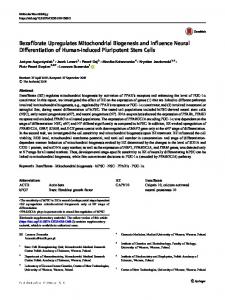
Fig. I c. Figure 1. Abscissa: T i m e after subcultivation (days); Ordinate: Cell n u m b e r per 25 c m 2 flask. Proliferation o f early passage WI-38 cells in t h e presence of different antibiotics: CAP, CHI a n d EB. A n t i b i o t i c t r e a t m e n t b e g a n o n d a y one. (a) Effect of c h l o r a m p h e n i c o l c o n c e n t r a t i o n s o n g r o w t h o n WLB8 cells: 9 O, 0; 9 -', 0.2; 9 9 0.5; a n d 4 #, 1.0 m g / m l . (b) Effect of c y c l o h e x i m i d e c o n c e n t r a t i o n s o n g r o w t h o f WI-S8 cells: o o, 0 ; 9 ~, 0.1; s s , 0.5; a n d 9 9 1.0/lg/ml. (c) Effect of e t h i d i u m b r o m i d e c o n c e n t r a t i o n s o n g r o w t h of WI-38 cells: o o, 0; -" 9 ;9 9 1.0; 9 9 5; and $ #, 10 ktg/ml.
Electron Microscopy Cells growing in plastic 25 cm 2 flasks were fixed with 2% buffered glutaraldehyde after four days treatment with antibiotics. The cells were subsequently post-fixed with 1% buffered osmium tetroxide~ stained with 0.5% aqueous uranyl acetate, dehydrated in a graded series of ethanol concentrations, and embedded in Epon using a modified technique of Brinkley et al. [4]. Thin sections were obtained using a
90
L. PACKER, J. S. NOLAN, S. KATYARE AND J. SMITH
MT-2 ultramicrotome, and stained with 0.5% uranyl acetate and lead citrate. Sections were examined with a Siemens IA electron microscope. For determination of mitochondrial area, photographs were Xeroxed, mitochondrial areas, cut out and weighed. Mitochondrial areas could be calculated by this procedure.
Cytochrome Oxidase For determination of cytochrome oxidase activity, the cells were gently scraped from 75 cm 2 flasks and washed once in 0.25 M sucrose containing 2 mM EDTA and 0.1% ethanol, pH 7.5. The cells were suspended in the same medium, homogenized- and sonicated for 2 sec using the microprobe of Biosonik III sonicator and salt-ice water bath. Cytochrome oxidase activity was measured according to a microprocedure developed by Sun and Poole [5]. The assay medium consisted of 100 mM Tris-HCL, pH 7.5, 1 mM EDTA, 0.05 mM reduced cytochrome c and 0.1% emasol. Cytochrome c was reduced with sodium dithionite just before use. The assay was carried out at 37~ Oxidation of cytochrome c was followed at 550 nm using a Cary 14 recording spectrophotometer. The activities are expressed as nmoles cytochrome c oxidized/min/mg protein. Protein was determined by a modification of the method of Lowry et al. [6] using crystalline bovine serum albumin as a standard.
Results Cell Proliferation-Acute Treatment with Antibiotics Typical growth curves and the effects of CAP, CHI, and EB on proliferation of human diploid fibroblasts is shown in Fig. la, b, and c respectively. Each antibiotic, at the concentrations used in this series of experiments, manifests inhibition of cell proliferation. The threshold concentrations of antibiotics, i.e., which arrest growth, may cause a slight enlargement of cell size as judged by mean cell volume registered in a Coulter Counter or qualitatively by light microscopy.
Gross Morphology and Mitochondrial Content Morphological examination was carried o u t on cells grown at the antibiotic concentration for the threshold level for inhibition of cell proliferation. Antibiotic treatments at the threshold of growth inhibition have marked effects on cellular and mitochondrial structure. Results are summarized below:
MITOCHONDRIAL BIOGENESIS IN HUMAN FIBROBLASTS
91
Figure 2. High magnification electron micrograph of untreated early passage WI-38 cells, showing distribution of mitochondria in peripheral region. Control
Electron micrographs of peripheral regions show normal mitochondria (Fig. 2). The inner and outer membranes and the cristae are evident. Ethidium bromide
In cells treated with 2 ug/ml (Fig. 3) and 5 Ug/ml of EB (Fig. 4) marked changes in mitochondrial structure are observed. Increasing the concentration of EB progressively causes the appearance of smaller mitochondria which seem to be fragmented. At higher concentrations of EB, mitochondrial structure is not well defined; the inner membrane and cristae are disorganized (Fig. 4). The effect of EB on cell structure is
92
L. PACKER,J. S. NOLAN, S. KATYAREAND J. SMITH
Figure 3. High magnification electron micrograph of early passage WI-38 cells treated with 2//g/ml of ethidium bromide. An area peripheral to the nuclear region is shown. pleomorphic. There seems to be an enhanced appearance of microfilamentous structures. The loss of m i t o c h o n d r i a m a y reveal these structures more d e a r l y , or there could be a real increase in their occurrence. Changes in the endoplasmic reticulum following EB t r e a t m e n t were also noted. The cisternal diameter is larger (Fig. 4) and the occurrence of Golgi regions is n o t very evident after higher EB t r e a t m e n t .
Chlorarnphenicol T r e a t m e n t with CAP at 200/~g/ml has the remarkable effect of increasing the n u m b e r of m i t o c h o n d r i a and mitochondria-like structures.
MITOCHONDRIAL BIOGENESIS IN HUMAN FIBROBLASTS
93
Figure 4. High magnification electron micrograph of early passage WI-38 cells treated with 5/lg/ml of ethidium bromide. An area peripheral to the nuclear region is shown. Figure 5 shows the a p p e a r a n c e of n u m e r o u s m i t o c h o n d r i a , which retain their n o r m a l a p p e a r a n c e in some instances, b u t in others, are closely associated with myelin-like structures which a p p e a r m o r p h o l o g i c a l l y to be derived f r o m m i t o c h o n d r i a . There is a larg e increase in the p r o p o r t i o n of m i t o c h o n d r i a to o t h e r organelles in CAP t r e a t e d cells. O t h e r cellular structures following CAP t r e a t m e n t a p p e a r normal.
Cycloheximide C y c l o h e x i m i d e t r e a t m e n t at 1 / l g / m l does n o t m a r k e d l y affect gross m o r p h o l o g y (Fig, 6) or the n u m b e r of m i t o c h o n d r i a as c o m p a r e d to u n t r e a t e d cultures. The m o s t r e m a r k a b l e effect appears on the s m o o t h
94
L. PACKER, J. S. NOLAN, S. KATYARE AND J. SMITH
Figure 5. High magnification electron micrograph of a peripheral region of early passage WI-38 cells treated with 200~g/ml of chloramphenicol. Myelinated mitochondrial structures are evident.
surfaced elements of the endoplasmic reticulum which are more numerous. A more elaborate development of Golgi vesicles is also observed; these vesicles are not filled with electron dense material. These effects may be related to the known inhibition of' cytoplasmic protein synthesis by cycloheximide. Mitochondrial Content The effect of the various antibiotic treatments on the occurrence of mitochondria is summarized in Table I. The mitochondrial content was evaluated by determining the relative areas they occupied in photographs (cf. Methods) of regions of WI-38 cells peripheral to the nucleus.
MITOCHONDRIAL BIOGENESIS IN HUMAN FIBROBLASTS
95
Figure 6. High magnification electron rnicrograph of a peripheral region of early passage WI-cellstreated with 1/lg]ml of cycloheximide. Mitochondria decreased both in number and size with EB; with CAP, at the concentration employed, the number of mitochondria increased, even apart from the occurrence of myelin figures which may also be attributed to mitochondria. A small decrease in the number of mitochondria occurred with cycloheximide.
Effect of Ethidium Bromide, Chloramphenicol and Cycloheximide on long-term growth potential and cytochrome oxidase activity of fibroblasts In order to answer the question as to whether mitochondria per se are important to the limited in vitro lifespan in human fibroblasts,
96
L. PACKER, J. S. NOLAN, S. KATYARE AND J. SMITH
TABLE I. Effect o f ethidium b r o m i d e , chloramphenicol, and cycloheximide on biogenesis o f m i t o c h o n d r i a in h u m a n diploid (WI-38) cells Cell area (/.t2)
Number of mitochondria
66.67
Conditions
Control Ethidium b r o m i d e (2/sg/ml) (5 ktg/ml) Chloramphenicol (200/ag/ml) Cycloheximide (1 /~g/ml)
Mitochondrial area #2
% of t o t a l
14.5
3.10
4.65
68.38 61.30
9.7 9.0
2.53 0.96
3.71 1.57
68.44
19.6 3.3 a
6.21 0.88 a
9.08 1.29 a
64.62
13.3
2.13
3.30
a Proportion of mitochondrial "myelin figures" present.
antibiotics that interfere with the normal biogenesis of mitochondria were tested under chronic conditions of treatment (cf. Methods). Concentrations considerably lower than threshold levels had to be used for chronic experiments. As shown in Fig. 7, when the antibiotics were present in the growth medium at sufficiently low levels, there was no effect on the cell yield after eight days of growth nor on the in vitro lifespan. However, when the antibiotic concentration was increased by a ETHIDIUM BROMIDE (p.g/ml) [] Control 0.005 9 0.015
t0.06
'0 6 X
eaE 5 o
E
.
to
r~ 4 tiff
~3 z
2
J .J
0
t
30 Fig. 7a.
32
i
34
316
i
38
i
40
POPULATION
i
42
414
DOUBLING
4'6
48
LEVEL
510
5=2
CYCLOHEXIMIDE (Fg/ml)
I0 -x
O
~o
6-
[] ,& A 0
0 (Control) 0.015 0.06
0
1.0
97
0.25
C
E 5-
E
u
r~ ~ 4w
z
.J .J u.I o
.2 ~g '7, 2-
f
I
22
I
I
26
30
34
3J8
42
4]6
50
5=4
POPULATION DOUBLING LEVEL
Fig. 7b.
CHLORAMPHENICOL (/~g/ml) [] 0 Control A3 z~ 12 0 50
$ ' 0- 6
*C
0
NE 5 ~
~E
"4 W
~
53
_J hi U I 0
22 Fig. 7c.
2'6
3'0
34 ' 3 8 42. . 46. . 50 54 POPULATION DOUBLING LEVEL
Figure 7. Abscissa: p o p u l a t i o n doubling level; ordinate: cell n u m b e r / 7 5 cm 2 (x 10 -6 ). Effect of prolonged t r e a t m e n t of antibiotics, EB, CHI and CAP on the g r o w t h of WI-38 cells. (a) Concentration of ethidium b r o m i d e (/~g/ml): ~ D, 0; A A, 0.005; o - - - - - o , 0.015; and = e,0.06. (b) Concentration of cycloheximide (~g/ml): []G, 0; ~ - - - - A , 0.015; • A, 0.06; o o, 0.25; and 9 -~, 1.0. (c) Concentration of chloramphenicol (/2g/ml): z] D, 0; AA, 3; z~ -~, 12; o o, 50; and 9 -', 200.
98
L. PACKER,J. $. NOLAN, S. KATYAREAND J. SMITH
factor of four there was an immediate effect on growth rate and the cultures were lost after only a few subcultivations. Cultures treated with CHI behaved s o m e w h a t differently from those treated with EB or CAP. When treated with 0.06/2g/ml CHI (four times the c o n c e n t r a t i o n which had n o influence on growth) the cultures showed a nearly n o r m a l growth rate for several generation doublings and the culture was able to survive for ten p o p u l a t i o n doublings. A s u m m a r y of the effect of various c o n c e n t r a t i o n s of the antibiotics on lifespan is given in Table II. In addition to the n u m b e r of p o p u I a t i o n doublings accrued during t r e a t m e n t , we also d e t e r m i n e d the total p r o t e i n as an alternative measure of the a m o u n t of growth achieved during t r e a t m e n t . F r o m Table II it is seen that the total p r o t e i n is roughly p r o p o r t i o n a l to the n u m b e r of p o p u l a t i o n doublings. At each s u b c u l t i v a t i o n during the antibiotic t r e a t m e n t c y t o c h r o m e oxidase activity was d e t e r m i n e d in cultures parallel to the s u b c u l t i v a t i o n series. The results of these experiments are shown in Fig. 8. Cultures treated with a c o n c e n t r a t i o n of CAP which did n o t i n h i b i t growth or decrease lifespan (3.0/2g/ml) possessed only 50% of the c y t o c h r o m e
TABLE II. Effect of chronic antibiotic treatment on lifespan, cellular proteins and cytochrome c oxidase activity of WI-38 cells Medium concentration (/~g/ml)
Population doublings accrued
Total protein (mg)
Average cytochrome oxidase (% of control)
Cycloheximide control 0.015 0.06 0.25
30 27 10 4
7.6 7.3 2.2 1.1
100 98 103 92
1.0
2
0.5
77
19 20 3 1
4.9 3.7 0.5 -
100 89 30 -
30 28
7.6 6.5
5 3
0.9 0.6
2
0.2
100 51 42 48 32
Ethidium bromide control 0.005 0.015 0.06 Chloramphenicol control 3.O 12.0
50.0 200.0
The cells were grown on different antibiotic concentrations as described in Materials and Methods section and as shown in Figs. 7 and 8. Cytochrome c oxidase activity is expressed as % of control. As indicated in Fig. 8a, the activity in control remained more or less constant at different passage levels.
MITOCHONDRIAL BIOGENESIS IN HUMAN FIBROBLASTS
99
oxidase activity of the u n t r e a t e d cultures (Fig. 8c). A t higher concentrations the activity was s o m e w h a t lower, b u t even at c o n c e n t r a t i o n s which i n h i b i t e d g r o w t h very q u i c k l y the c y t o c h r o m e oxidase activity was still a b o u t 30% o f the control. By contrast, c o n c e n t r a t i o n s of CHI (Fig. 8b) which did n o t allow cell g r o w t h h a d very little effect on the c y t o c h r o m e oxidase activity. T r e a t m e n t with EB (Fig. 8a) at a c o n c e n t r a t i o n which did n o t inhibit growth a p p e a r e d to cause an initial decrease in c y t o c h r o m e oxidase activity. A f t e r a few p o p u l a t i o n doublings, c y t o c h r o m e oxidase activity was regained and r e m a i n e d at a b o u t the c o n t r o l level until the culture neared the end of its in vitro lifespan when it a p p a r e n t l y decreased again. Also shown in Fig. 8a is the specific activity of u n t r e a t e d cultures as a f u n c t i o n of p o p u l a t i o n d o u b l i n g level. There was no decrease in activity as the culture a p p r o a c h e d the end of its in vitro lifespan. T h e average c y t o c h r o m e oxidase activity during t r e a t m e n t with various concentrations of antibiotics is given in Table II.
Recovery of Cytochrome Oxidase Activity and Growth Potential after Antibiotic Treatment Table III shows the effect of treating cultures with antibiotics for two or f o u r subcultivation periods on the lifespan of the cultures. In the case of CHI and EB, g r o w t h for two s u b c u l t i v a t i o n periods in c o n c e n t r a t i o n s t h a t d~astically inhibit g r o w t h does n o t decrease the lifespan of the cultures. S o o n after the antibiotics were r e m o v e d the g r o w t h rate r e t u r n e d to t h a t of cultures which h a d n o t b e e n treated. However, when cultures were t r e a t e d for two s u b c u l t i v a t i o n periods TABLE III. Effect of removal of the antibiotics on the growth potentia! of WI-38 cells Medium concentration (//g/ml) Cycloheximide
1.0 0.25 0.06 0.015 Chloramphenicol 3.0 3.0 12.0 Ethidium bromide 0.015 Control 0
Number of population Length of antibiotic doublings accrueda treatment (transfers) b 29 34 30 31 30 26 19 27 30
a Sum of population doublings during and after antibiotic treatment. b 4 x 10³ cells/cm2 inoculated at eight day intervals. The details are as described in the Materials and Methods section.
2 2 4 4 2 4 4 2 0
100
L. PACKER, J. S. NOLAN, S. KATYARE AND J. SMITH
ETHIDIUM
BROMIDE
Z [xl I-O [K O.
(Fg/ml) [] 0 Control 0.005 0 0.015
0 rr-
~z 1 4 0 o
.E
u. 1200
hi (1/)
o
la,l N
-I00>_
100
I,,-
-80 >
80-
1.1,,1
L)
o
0
- 6 0 (j
60"
E
hi
~0 4 0 ~
-40
%
(E -r
(J 0 F0
-20
20-
F--
~
_1 0
IE c
0
30 Fig. 8a.
!
i
34
38
/
42
POPULATION
!
i
i
46
50
54
DOUBLING LEVEL
0 Z 0 Ii0
120-
IO0c'~ X
o
80-
[] O,Control 9 0.015 A 0.06 00.25
600 nr 4 0 ~ -r 0 0
@1.0
F-20).. L)
0 22 Fig. 8b.
i6
,
i
i
30
34
38
POPULATION
!
42
i
i
46
50
DOUBLING
LEVEL
i
54
101
M I T O C H O N D R I A L B I O G E N E S I S IN H U M A N F I B R O B L A S T S
0 ].Z 0 0 ~IO0 [iJ (t) '~ 8 0 -
CHLORAMPHENICOL (/=g/ml)
X 0 u
[] 0 Control
A3
60-
A 12 9 50 9 200
uJ 400 n"1-
c) 200 F>o
Fig. 8c.
O 22
2'6
~0 3'4 38 4'2 4~6 ,50 ,54 POPULATION DOUBLING LEVEL
Figure 8. Abscissa: population doubling level; ordinate (left): cytochrome c oxidase activity (% of control), (right in Fig. 8a): cytochrome c oxidase activity of control, n moles cytochrome c oxidized/min/mgprotein; m~ ". (a) Concentration of ethidium bromide (/lg/ml): D - - [ ] , 0; ~ ,6, 0.005; o--------o, 0.015. (b) Concentration of cycloheximide (/lg/ml): [] D, 0; A A, 0.015; /~ ~, 0.06; o o, 0.25; and o e, 1.0. (c) Concentration of chloramp.henicol (#g/ml): [] [],0;A A, 3;A ZX,12 ; O------O, 50; and eo, 200.
with high concentrations of CAP (50 or 200/Jg/ml) they were unable to recover after removal of the antibiotic. Cultures treated during four subcultivation periods with 12 tzg/ml of CAP recovered from the antibiotic treatment slowly and the growth rate never became as high as that of untreated cultures. Furthermore, these cultures were unable to undergo as m a n y population doublings as untreated cultures. Treatment with CAP (12 #g/ml) and EB (0.015 pg/ml) resulted in c y t o c h r o m e oxidase activities 40~ and 25% respectively of that in untreated cultures (see Table IV). When the antibiotics were removed from the culture medium the c y t o c h r o m e oxidase activity returned to that of untreated cultures within one or two sucultivation periods. The c y t o c h r o m e oxidase activity remained high t h r o u g h o u t the lifespan of these cultures.
Discussion Biogenesis of Mitochondria in Human Cells Earlier studies with EB and CAP in yeast [7], regenerating rat liver [8] and L-cells [9] have shown that these antibiotics inhibit the
102
L. PACKER,J. S. NOLAN, S. KATYAP,E AND J. SMITH
TABLE IV. Recovery of cytochrome c oxidase activity in WI-38 cells after removal of the antibiotics Medium concentration (pg/ml)
Cytochrome oxidase activity (% of control)
Chloramphenicol 12.0 (4)a
40 110
Ethidium bromide 0.015 (2)a
80 90 25 114 123 105 90
No. of transfers after antibiotic removal 1
2 3 -2 6 9 10
a Number of transfers in presence of antibiotic.
synthesis of functional cytochromes a, a3, b and cl. The effects of EB and CAP on growth, cytochrome oxidase and morphology in this investigation indicate that qualitatively similar effects occur in human diploid fibroblasts. In particular, the morphological effects of these antibiotics reagents are interesting because they indicate the selectivity of their effects. Ethidium bromide treated fibroblasts show the presence of fragmented mitochondria, similar to changes brought about by EB in Tetrahymena [10]. The effect of CAP on mitochondrial structure in human fibroblasts and other systems also seems similar. Prolonged CAP treatment caused the appearance of fewer cristae in He La cells [11, 12], Polytomella [13] and regenerating rat liver [8]. In Ochromonas, CAP treatment leads to the presence of prolamellar bodies and of abnormal thylakoid membranes [14]. The presence of myelin-like bodies observed in the present study have also been seen in other systems [15] ; they may represent degenerating structures or non-functional mitochondria. The occurrence of myelin-like figures is usually associated with the formation of lamellar membranous structures which are low in protein content. They are characteristic of myelin itself and the myelin-like membranes found in the surfactin producing type II lung alveolar cells, and perhaps other abnormal conditions where protein synthesis has been inhibited. These results suggest "decoupling" of the membrane protein synthesis from membrane lipid synthesis leading to the production of multi-layered myelin-like structures by CAP.
Lifespan of Human Fibroblasts Normal human cells manifest a finite lifespan when serially subcultivated in vitro. The factors which limit the in vitro lifespan of
MITOCHONDRIAL BIOGENESIS IN HUMAN FIBROBLASTS
103
such ceils are presently unknown. Limitation of lifespan may relate to the state of differentiation [2] or alternatively, environmental conditions (e.g. the constraints of diffusion-limited in vitro culture conditions may impose a critical dependence on mitochondrial oxidative metabolism for cell proliferation). In this investigation, we have studied the effect on lifespan of mitochondrial oxidative metabolism. The results dearly show- that cells treated with EB and CAP manifest an inhibition of synthesis of normal mitochondria as shown by decrease in cytochrome oxidase and morphological changes; while CHI, an inhibitor of cytoplasmic protein synthesis, had no effect on cytochrome oxidase activity even at levels which greatly inhibited growth. Treatment of WI-38 cultures with 0.005/~g/ml of EB caused an initial decrease in cytochrome oxidase activity, but on continued subcultivation in its presence the activity returned to the control value. When the EB treated cultures entered Phase III the cytochrome oxidase activity again decreased. It may be that during continuous growth in EB the mitochondrial population became resistant to its action, but that resistance was lost when the culture reached the end of its in vitro lifespan. EB resistance has been reported in SV-40 transformed WI-38 celIs [16] and CAP resistance has been reported in He La cells [17]. Also irreversible morphological changes in mitochondria of He La cells have been obtained b y growth in EB [18]. Although EB and CAP, at low concentrations, cause a decrease in cytochrome oxidase activity they exert no effect on the proliferative capacity. This is especially apparent in cultures treated with 3,/zg/ml CAP. During the last half of their in vitro life span the cytochrome oxidase activity was maintained at 50% of the normal level but there was no decrease in the number of population doublings achieved in culture
(Table II). EB has been shown to cause the disappearance of the circular form of mitochondrial DNA in several mammalian ceil lines [191. This change was reversible by subsequent growth in EB-free medium. This is similar to the behavior of cytochrome oxidase activity in our cultures. EB when present at 0.015 #g/ml caused a 75~ decrease in cytochrome oxidase activity and inhibited cell division. However, the effects of this antibiotic were reversible and b o t h the cytochrome oxidase activity and proliferative capacity returned to normal when the drug was removed. Similar results have been reported after periods of anaerobic incubation of TC 12266 human diploid fibroblasts by Hakami and Pious [20]. In contrast, CAP when present in the culture medium for four subcultivation periods at 12/lg/ml did cause a decrease in the in vitro lifespan. However, the inhibition of cytochrome oxidase activity was not as great with CAP as with EB and the activity returned to the control level within one subcultivation period after removal of CAP. We also found that the specific activity of cytochrome oxidase did not change
104
L. PACKER,J. S. NOLAN, 8. KATYAREAND J. SMITH
during the last half of the in vitro lifespan of u n t r e a t e d cultures (Fig 8a). This is in agreement with the findings of Hakami and Pious [ 2 1 ] . It appears that the p o t e n t i a l for oxidative m e t a b o l i s m is more t h a n sufficient to p r o m o t e cell proliferation t h r o u g h o u t the lifespan of h u m a n diploid cell cultures. Since decreasing the c y t o c h r o m e oxidase activity b y 50% during the last half of the i n vitro lifespan has no effect o n the lifespan, we conclude that older cultures are n o t d e p e n d e n t o n oxidative m e t a b o l i s m to a n y greater e x t e n t t h a n y o u n g cells. It is possible, however, that m i t o c h o n d r i a m a y be indirectly involved in limiting the in vitro lifespan. Cytoplasmic damage m a y arise as a result of t h e c o n t i n u a l t u r n o v e r of m i t o c h o n d r i a since the highest c o n c e n t r a t i o n s of free radical generating reactions and u n s a t u r a t e d f a t t y acids which are susceptible to lipid peroxidation, and calcium activated phospholipase, occur in m i t o c h o n d r i a l m e m b r a n e s . In damaged m e m b r a n e s , phospholipase causes the further release of u n s a t u r a t e d fatty acids, which themselves are damaging to membranes. These reactions are n o r m a l l y latent in m i t o c h o n d r i a u n t i l m e m b r a n e damage occurs during mitochondrial turnover [22].
References 1. L. Hayflick and P. S. Moorhead, Exptl. Cell Res., 25 (1961) 585. 2. L. Hayflick, Exptl. Cell Res., 37 (1965) 614. 3. J. S. McHale and L. Packer, Methods in Enzymology, Biomembranes (1973) Vol. I, in press. 4. B.R. Brinkley, P. Murphy and L. C. Richardson, J. Cell. Biol., 35 (1967) 279. 5. A. S. K. Sun and B. Poole, Fractionation of mitochondria, lysosomes, plasma membrane and peroxisomes from rat embryo fibroblasts (in preparation). 6. O. H. Lowry, N. J. Rosebrough, A. L. Fan" and R. J. Randall, ]. Biol. Chem., 193 (1951) 265. 7. G. M. Kellerman, D. R. Biggs and A. W. Linnane,J. CellBiol., 42 (1969) 378. 8. F.C. Firkin and A. W. Linnane, Exptl. Cell Res., 55 (1969) 68. 9. M. E. King, G. C. Godman and D. W. King, J. CellBiol., 53 (1972) 127. 10. D.C. Rein and R. R. Meyer, J. CellBiol., abstracts (1971) 478. 11. R. Lenk and S. Penman, J. CellBiol., 49 (1971) 541. 12. N. Kislev, C. M. Spolsky andJ. M. Eisenstadt, J. CelIBiol., 57 (1973) 571. 13. D. A. Evans and D. Lloyd, BiochemJ., 103 (1967) 22P. 14. H. Smith-Johannsen and S. P. Gibbs, J. Cell Biol., 52 (1972) 598. 15. A. Adoutte, M. Balmefrezol, J. Beisson and J. Andre,.J. Cell Biol., 54 (1972) 8. 16. W. Klietmann, K. Kato, N. Sato and H. Koprowski, Fed. Proc., 31 (1972) 620 (abst.). 17. W. Klietmann, K. Kato, B. Gabara, H. Koprowski and N. Sato, Exptl. CellRes., 78 (1973) 47. 18. C. M. Spolsky andJ. M. Eisenstadt, FEBS Lett., 25 (1972) 319. 19. M. M. K. Nass, ExptL CellRes, 72 (1972) 211. 20. N. Hakami and D. A. Pious, Nature, 216 (1967) 1087. 21. N. Hakami and D. A. Pious, Exptl. Cell Res., 53 (1968) 135. 22. A. Scarpa andJ. G. Lindsay, Eur. J. Biochem., 27 (1972) 401.
MITOCHONDRIAL BIOGENESIS IN HUMAN FIBROBLASTS
].05
Acknowledgements T h e a u t h o r s are grateful to Pauline Sallata for technical assistance with e x p e r i m e n t s , to K a t h y D o n o v a n for assistance with cell culture, a n d to S u s a n L. Tinsley and L o u W o r t h i n g t o n for assisting w i t h electron m i c r o s c o p y . This research was s u p p o r t e d b y USPHS G r a n t HD 0 5 8 7 5 a n d the Veterans A d m i n i s t r a t i o n .