Molecular cloning of a cDNA encoding a human macrophage migration inhibitory factor. (lymphokine/expression cloning/DNA sequence analysis/insertional ...
Proc. Natl. Acad. Sci. USA Vol. 86, pp. 7522-7526, October 1989 Immunology
Molecular cloning of a cDNA encoding a human macrophage migration inhibitory factor (lymphokine/expression cloning/DNA sequence analysis/insertional mutational analysis)
WEISHUI Y. WEISER*, PATRICIA A. TEMPLEt, JOANN S. WITEK-GIANNOTTIt, HEINZ G. REMOLD*, STEVEN C. CLARKt, AND JOHN R. DAVID* *Department of Medicine, Harvard Medical School, and the Department of Rheumatology and Immunology, Brigham and Women's Hospital, Boston, MA 02115; and tThe Genetics Institute, Cambridge, MA 02140
Communicated by Barry R. Bloom, June 5, 1989 (received for review March 17, 1989)
We have employed functional expression cloning in mammalian cells to identify novel lymphokines with MIF activity. Here we report the isolation of a cDNA encoding a protein that inhibits the migration of macrophages in vitro. The availability of recombinant MIF will facilitate the analysis of the role of this lymphokine in host defense and in inflammation.t
A cDNA encoding a human macrophage miABSTRACT gration inhibitory factor (MIF) was isolated, through functional expression cloning in COS-1 cells, from a cDNA library prepared from a lectin-stimulated T-cell hybridoma, T-CEMB. The 115-amino acid polypeptide encoded by the MIF cDNA (p7-1) was effectively released from the transfected COS-1 cells and yielded readily detectable MIF activity in the culture supernatant despite the apparent lack of a classical protein secretory sequence. Insertional mutational analysis and elution of MIF activity from polyacrylamide gel slices demonstrated that the Mr 12,000 protein with MIF activity released by the COS-1 cells is encoded by p7-1. The p7-1 cDNA hybridized with a 700-base mRNA expressed by Con-A-stimulated lymphocytes but not unstimulated lymphocytes. The availability of the MIF cDNA clone and recombinant MIF will facilitate the analysis of the role of this lymphokine in cell-mediated immunity, immunoregulation, and inflammation.
MATERIALS AND METHODS Cell Line. The T-CEMB cell line is a human T-cell hybridoma line generated by fusion of a hypoxanthine/aminopterin/thymidine-sensitive T-lymphoblastoid line (CEMWH4) with Con A-stimulated human peripheral blood T cells (14). MIF Assay. The MIF assay (14, 15) employed human peripheral blood monocytes as indicator cells in an agarosedroplet assay system. The area of migration was calculated by the following formula: migration = (diameter of total area/diameter of agarose droplet)2 - 1. The percentage of inhibition of each sample was derived as follows: % inhibition = 100 - (average migration of test samples/average migration of control samples) x 100. Inhibition of 20% or greater was considered to be significant (11). Isolation of mRNA and Construction of cDNA Library. Total RNA was extracted (16) from T-CEMB cells that had been stimulated with phytohemagglutinin (PHA, 1%) and phorbol 12-myristate 13-acetate (PMA, 10 ng/ml) for 18 hr. Five micrograms of mRNA prepared by oligo(dT)-cellulose chromatography (17) was used to synthesize double-stranded cDNA as described (18). The double-stranded DNA was blunted and ligated to a 5-fold excess of synthetic semi-Xho adapters (19). The semi-Xho-adapted cDNA was sizefractionated by agarose gel electrophoresis. Gel containing DNA of .500 base pairs was excised and the cDNA fragments were isolated from the gel slice by adherence to glass powder (20). The COS-1 cell expression vector pXM was linearized and ligated to equimolar amounts of the semiXho-adapted cDNA. The ligation reaction was used to transform Escherichia coli strain HB101, thus generating a library of -60,000 ampicillin-resistant colonies. DNA Preparation and COS-1 Cell Transfection. Bacterial colonies from the library were replica-plated onto nitrocellulose filters. Colonies from each filter were scraped into Luria broth and plasmid DNA was isolated. Each primary DNA sample was prepared from a pool of 200-500 colonies. Five micrograms of each plasmid DNA was used to transfect COS-1 cells (simian virus 40-transformed monkey kidney cell line) as described (21, 22). Culture supernatant fluid from
Lymphocytes secrete a large number of protein mediators, known as lymphokines, in response to antigenic or mitogenic stimulation. These lymphokines play an important role in immunoregulation, inflammation, and cellular immunity (1, 2). Migration inhibitory factor (MIF) for guinea pig macrophages was the first lymphokine to be discovered (3, 4). This factor originally was identified by its ability to prevent the migration of guinea pig macrophages out of capillary tubes in vitro (3, 4). Subsequently, the expression of MIF activity was found to correlate well with delayed hypersensitivity and cellular immunity in animal models and in humans (3-6). MIF activity has been detected in leukocyte culture supernatants of mice during allograft rejection (7, 8), in the synovia of patients with rheumatoid polyarthritis (9), and in a variety of chronic inflammatory loci (10). The expression MIF at sites of inflammation suggests a role for the mediator in regulating the function of macrophages in host defense. The biochemical characterization of MIF activity has proved to be difficult largely because of the low levels of activity expressed by natural sources of the mediator. Our studies with human MIF, which also functions with guinea pig target cells, have shown that human MIF obtained from lectin-stimulated T cells or T-cell lines is heterogeneous, with at least three different species distinguishable by gel chromatography, isoelectric focusing, or sensitivity to enzymatic treatments (11). In addition, several other cytokines, including interferon y (IFN-'y) and interleukin 4 (IL-4) can inhibit macrophage migration (12, 13). Thus the MIF activity present in activated T-cell supernatants is likely to result from the presence of multiple cytokines capable of influencing macrophage migration.
Abbreviations: MIF, migration inhibitory factor; IFN, interferon; IL, interleukin; PHA, phytohemagglutinin; PMA, phorbol 12myristate 13-acetate. MThe sequence reported in this paper has been deposited in the GenBank data base (accession no. M25639).
The publication costs of this article were defrayed in part by page charge payment. This article must therefore be hereby marked "advertisement" in accordance with 18 U.S.C. §1734 solely to indicate this fact. 7522
Immunology: Weiser et al. each dish oftransfected COS-1 cells was harvested 72 hr after transfection and examined for MIF activity. Protein Analysis. COS-1 cells transfected with recombinant DNA of MIF-positive clones were incubated with 0.5 mCi of [35S]methionine (1 mCi = 37 MBq) for 4 hr at 370C. Radiolabeled supernatant was subjected to NaDodSO4/PAGE (15%). After electrophoresis, the gel was immersed in EN3HANCE (New England Nuclear) dried, and exposed to x-ray film. To investigate whether MIF activity in COS supernatant could be recovered after NaDodSO4/PAGE, the polypeptide of interest was excised from the polyacrylamide gel and electroeluted at 6 W for 2 hr in elution buffer containing 50 mM NH4HCO3 and 200 ng of human serum albumin per ml. The latter was added to prevent nonspecific sticking. In parallel, polypeptides with the same apparent molecular weight from supernatant of mock-transfected COS cells were also excised and subjected to electroelution. The eluant was reconstituted with medium and examined for MIF activity. DNA Sequence Analysis. The nucleotide sequence of the cDNA insert of p7-1 was determined as described (18, 19) by generating ordered sets of overlapping fragments by BAL-31 nuclease digestion and subcloning into M13 vectors. Singlestranded DNA was prepared, and the nucleotide sequence was determined by the dideoxynucleotide chain-termination procedure (23). Generation of Insertional Mutants of the MIF Clone. Although the MIF cDNA clone p7-1 contained a single Pst I site, the Pst I sites in the adapters flanking the insert rendered this site unsuitable for mutational analysis of the MIF cDNA. Therefore, we removed the two flanking Pst I sites by treating the p7-1 insert (isolated after partial digestion of the plasmid with Pst I) with T4 DNA polymerase, then ligating EcoRI adapters to the resulting flush ends. This adapted fragment was subcloned into the single EcoRI site of a derivative of pXM designated pXMT4, which has no Pst I sites. A clone, p7-1-24, with the cDNA in the correct orientation was selected for mutational analysis. Two insertional mutants of MIF were constructed. The first mutant was generated by inserting a 14-base oligodeoxynucleotide (5'-TGTAATTACATGCA-3') at the single Pst I site of p7-1-24. This sequence was designed such that the MIF coding region could be interrupted by a termination codon (TAA) regardless of the orientation of insertion. The second insertional mutant was constructed by inserting a 99-base oligodeoxynucleotide into the Pst I site of p7-1-24. The sequence was designed to add 33 amino acids to the MIF
Proc. Natl. Acad. Sci. USA 86 (1989)
7523
coding region when inserted in either orientation into the Pst I site of p7-1-24. Clones containing the 14-base oligonucleotide or the 99-base oligonucleotide were identified by hybridization with 32P-labeled 14-base oligomer or 99-base oligomer.
RNA Analysis. Twenty micrograms of total cellular RNA from PHA/PMA-stimulated or unstimulated T-CEMB cells, Con A-stimulated or unstimulated human peripheral blood lymphocytes, or CEM cells was electrophoresed in a 1.2% agarose gel containing 2.2 M formaldehyde (24). The formaldehyde-denatured RNA was transferred to a nylon filter (Zetabind; Cuno). cDNA probe was prepared by cleaving the cDNA inserts from the vector with restriction endonuclease Xho I. The inserts were labeled with 32P by random priming (25). The nylon filter was prehybridized (4 hr at 43°C) and then hybridized (16 hr at 43°C) with 32P-labeled cDNA probe in 6x SSC/0.5% NaDodSO4/0.1% Ficoll/0.1% polyvinylpyrrolidone/0.1% bovine serum albumin containing denatured salmon sperm DNA at 100 ug/ml. (SSC is 0.15 M NaCl/0.015 M sodium citrate, pH 7.) After hybridization, the filter was washed twice with 10 mM sodium phosphate/0. 1% NaDodSO4/1 mM EDTA/lx SSC for 30 min at room temperature and twice with 10 mM sodium phosphate/0. 1% NaDodSO4/1 mM EDTA/0.2x SSC for 15 min at 68°C, dried, and exposed to x-ray film.
RESULTS Identification and Isolation of a Human MIF cDNA Clone by Expression. mRNA from PHA/PMA-stimulated T-CEMB cells (14), which produce MIF without concomitant production of significant levels of IFN-y, was used to construct a cDNA library comprising -60,000 clones with the COS cell expression vector pXM. This library was screened by transfecting pools of plasmid DNAs, each containing 200-500 clones, into COS-1 cells as described (19); the resulting supernatants were tested for MIF activity. Our initial screen of 100 pools yielded several samples with various levels of MIF activity (Table 1). Each of these samples was tested five times, using cells from different donors to minimize the chance of obtaining falsely positive samples. Pool 44, which yielded the highest reproducible level of MIF activity, was selected and subdivided into smaller pools. These were retested for their ability to induce MIF expression when introduced into COS cells. This process was repeated until a single clone (p7-1) was identified that consistently yielded the highest level of MIF activity when transfected into COS cells.
Table 1. MIF activity in supernatants from COS-1 cells transfected with DNAs from the T-CEMB cDNA expression library MIF activity, % inhibition Transfected DNA Exp. 1 Exp. 2 Exp. 3 Exp. 4 Exp. 5 Sample 2 17 200-500 clones, -9 31 -3 28 undiluted 10 -1 10 33 7 10 23 -23 47 18 -3 -22 34 39 59 69 50 34 43 23 21 4 27 40 44 52 71 37 40 36 -7 -11 -4 -21 Mock 3 5 20-43 clones, 39-32b 16 26 -6 17 10-fold dilution 44-11a 28 11 -7 5 0 34 44-lib 30 15 26 36 2 2 Mock 3 -5 -9 24 37 4411b7-1* 39 26 39 Single clone, 5 4 3 -7 -11 20-fold dilution Mock Of the 100 supernatants from the COS-1 cell transfections of the primary pools, sample 44 showed the best overall MIF activity. The samples were then subdivided to contain fewer clones until individual clones were isolated. *Plasmid p7-1.
7524
Proc. Natl. Acad. Sci. USA 86 (1989)
Immunology: Weiser et al.
A 5-fold dilution of the conditioned medium from this transfection also inhibited the migration of guinea pig and mouse peritoneal macrophages by 38% and 31%, respectively, consistent with the previous finding of non-species specificity of MIF (5, 6). Identification of the MIF Polypeptide. The polypeptide encoded by the cDNA of p7-1 was identified in metabolic labeling experiments. NaDodSO4/PAGE of polypeptides secreted by COS-1 cells transfected with p7-1 DNA revealed a polypeptide of Mr 412,000 (Fig. 1, lane 2) which was absent from the mock-transfected control (lane 1). The region of the gel containing the Mr 12,000 polypeptide was excised and electroeluted. Strong MIF activity was found in the eluant (Fig. 2). The molecular weight of the MIF-specific polypeptide is in agreement with the molecular weight of MIF produced by several lymphoid cell lines (26-28). No MIF activity was detected from the same molecular weight region of gel lanes containing proteins of mock-transfected cells
/00
801-
60F 400
co
:z 2
-2 0 -4
(Fig. 2).
Sequence Analysis of the cDNA Insert of Plasmid p7-1. The sequence of the p7-1 cDNA insert contains an open reading frame of 345 nucleotides, beginning with the ATG codon at nucleotides 51-53 (Fig. 3). The ATG is followed by 114 codons downstream by a TAA termination triplet at nucleotides 396-398. The 345 nucleotides encode a 115-amino acid polypeptide with a calculated molecular weight of 12,650, which is in agreement with the apparent molecular weight of the MIF-specific protein band observed in metabolic labeling experiments (Fig. 1, lane 2). Although MIF is thought to be a secreted protein, the predicted sequence does not contain a stretch of hydrophobic amino acids that resembles conventional secretory leader sequences (29), either at the N terminus or internally. A very hydrophobic sequence, characteristic of a protein signal peptide, is also lacking in IL-la and IL-1,B (30), acidic and basic fibroblast growth factors (31, 32), endothelial cell growth factor (33), and MIF-related proteins (9). The apparent absence of a leader sequence suggests that the mechanism of secretion of this MIF is distinct from that of typical secretory proteins, which involves passage through the Golgi apparatus and endoplasmic reticulum (34). Cells may export 1
2 3 4
43.0-
25.7-
18.4-
1436 0FIG. 1. Polypeptides secreted by transfected COS-1 cells. [35S]Methionine-labeled supernatants (20 ,l per lane) were analyzed by NaDodSO4/PAGE and the labeled proteins were visualized by fluorography. Shown are supernatants from mock-transfected cells; (lane 1), from cells transfected with DNA of p7-1 (lane 2), from cells transfected with DNA of p7-1-24B2 (its cDNA insert contains a stop codon at the Pst I site) (lane 3), and from cells transfected with DNA of p7-1-24232 (its cDNA insert contains a 99-base oligomer at the Pst I site) (lane 4). The molecular weight (Mr x 10-3) of standard proteins in the same gel is indicated at left. Arrows indicate the Mr 12,000 polypeptide present only in lane 2 (p7-1-transfected) and the Mr 15,500 polypeptide present only in lane 4 (p7-1-24232-transfected).
20
-610
I
F 1:7
I
114
1:28
1:56
DILUTION FIG. 2. MIF activity of proteins obtained by electroelution. Supernatants (300 ,ul) from COS-1 cells transfected with p7-1 DNA or mock-transfected were loaded onto the same polyacrylamide gel and were subjected to NaDodSO4/PAGE. Regions corresponding to Mr -12,000 were excised from both. MIF activity of the Mr 12,000 regions (o, p7-1; o, mock) was examined after electroelution. As an additional control, elution buffer (o) was included in the same assay.
MIF by a mechanism similar to that of IL-la, which at present remains unclear. However, it has been reported that IL-1,B is not anchored on the plasma membrane and that its secretion occurs by a novel mechanism that does not use a secretory leader sequence or the classical secretory pathway
(34).
Analysis of the DNA sequence revealed several other interesting features. The cDNA encodes two potential asparagine-linked glycosylation sites at amino acids 73-75 (AsnArg-Ser) and 100-112 (Asn-Asn-Ser) (35). The presence of two potential asparagine-glycosylation sites has been reported in numerous cytokines (18, 19, 36-38). There are three cysteine residues, located at positions 57, 60, and 81. We have often observed that MIF loses its biological activity upon storage. The presence of three cysteine residues may account at least in part, for this observation. Indeed, IFN-,8 Ser-17 (39), which was obtained through site-specific mutagenesis and differed from IFN-,B Cys-17 (40) only by the substitution of serine for one of the three cysteine residues, was found to be far more stable to long-term storage than IFN-p8 Cys-17 (39). We compared the nucleotide sequence of p7-1 cDNA with the nucleotide sequences recorded in GenBank (May 25, 1988) and found that the p7-1 cDNA shares no sequence homology with IFN-y or IL-4, both of which have been reported (12, 13) to exhibit MIF-like activity. It also shares no sequence homology with other cytokines or with the two cDNAs encoding two MIF-related proteins, MRP-8 and MRP-14 (9). Insertional Mutational Analysis of the MIF cDNA. The relatively efficient secretion of the Mr 12,000 protein with MIF activity from p7-1-transfected COS cells, despite the lack of a clear signal peptide in the coding sequence, raised the possibility that the molecularly cloned protein was not MIF but an inducer of endogenous MIF expression by the COS cells. To test this possibility, we constructed two insertional mutations of the coding region of the p7-1 cDNA.
Immunology: Weiser et al.
Proc. Natl. Acad. Sci. USA 86 (1989)
7525
30
10
CTCGAGCTGCAGAGCTGCCTCTGCGCGGGTCTCCTGGTCCTTCTGCCATCATGCCGATGT M P M F 70
90
110
TCATCGTAAACACCAACGTGCCCCGCGCCTCCGTGCCGGACGGGTTCCTCTCCGAGCTCA I
V
N
T
N
V
P
R
A
130
S
V
P
D
G
F
150
L
S
E
L
T
170
CCCAGCAGCTGGCGCAGGCCACCGGCAAGCCCCCCCAGTACATCGCGGTGCACGTGGTCC Q Q L A Q A T G K P P 0 Y I A V H V V P 190
210
co
230
CGGACCAGCTCATGGCCTTCGGCGGCTCCAGCGAGCCGTGCGCGCTCTGCAGCCTGCACA D Q L M A F G G S S E P C A L C S L H S 250
270
I-
21.-*
290
GCATCGGCAAGATCGGCGGCGCGCAGAACCGCTCCTACAGCAAGCTGCTGTGCGGCCTGC I G K I G G A Q N R S Y S K L L C G L L 310
330
350
TGGCCGAGCGCCTGCGCATCAGCCCGGACAGGGTCTACATCAACTATTACGACATGAACG A
E
R
L
R
I
S
P
D
370
R
V
Y
I
N
390
Y
Y
D
M
N
410
CGGCCAGTGTGGGCTGGAACAACTCCACCTTCGCCTAAGAGCCGCAGGGACCCACGCTGT A
S
V
430
G
W
N
N
S
T
F
A
*
450
470
CTGCGCTGGCTCCACCCGGGAACCCGCCGCACGCTGTGTTCTAGGCCCGCCCACCCCAAC 490
0
A
510
CTTCTGGTGGGGAGAAATAAACGGTTTAGAGACAGCTCTGCAG
FIG. 3. Nucleotide sequence and predicted amino acid sequence of human MIF cDNA clone p7-1. The consensus sequences for asparagine-linked glycosylation sites are underlined. Amino acids are designated by the single-letter code, and an asterisk indicates the termination codon.
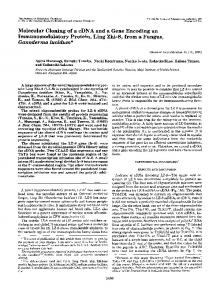
The first mutant, p7-1-24B2, was generated by inserting a 14-mer into the unique Pst I site so that a termination codon disrupted the open reading frame. The second mutant, p71-24232, was created by insertion of a 99-mer, at the same site, that extended the open reading frame by 33 amino acids regardless of the orientation of the insertion. Each of these plasmids was tested for ability to induce secretion of the Mr 12,000 protein observed with the original p7-1 plasmid as well as MIF activity when transfected into COS cells. Neither the Mr 12,000 protein (Fig. 1, lane 3) nor MIF activity (Fig. 4) was detected in the supernatants from the COS cells transfected with the truncated form of MIF (p7-1-24B2). Transfection of COS cells with the mutant having the extended coding region also failed to yield detectable levels of MIF activity (Fig. 4) or Mr 12,000 protein. However, the COS cell supernatant contained a novel species of apparent Mr -15,500, consistent with the expected size of the extended coding region of the insertional mutant p7-1-24232 (Fig. 1, lane 4). The results from these experiments further support the conclusion that the induced Mr 12,000 species from p7-1-transfected COS cells is encoded by the cDNA insert of p7-1 plasmid and that this protein has MIF activity. Expression of MIF mRNA. We examined mRNA from several cell sources for ability to hybridize with the cDNA insert of p7-1. Blot analysis revealed that the T-cell line CEM, the T-cell hybridoma line T-CEMB, and lectin-stimulated human peripheral blood lymphocytes synthesized readily detectable levels of mRNA that hybridized with the MIF clone (Fig. 5). However, the messenger was not detected in RNA samples from unstimulated lymphocytes despite prolonged autoradiographic exposure (Fig. 5, lane 1). The presence of the RNA transcript in activated human lymphocytes
1:5
1: /
1:100
DILUTION FIG. 4. MIF activity of supernatants from COS-1 cells transfected with p7-1 (e), p7-1-24 (o), p7-1-24B2 (A), or p7-1-24232 ( o) or mock-transfected (o). p7-1-24B2 and p7-1-24232 were constructed as insertional mutants of p7-1 containing 14-mer or 99-mer, respec-
tively.
(Fig. 5, lane 4) suggests that the human MIF gene is expressed and that MIF is a product of activated lymphocytes.
DISCUSSION We have identified and isolated a cDNA clone that encodes a biologically active human MIF by detection of the functional polypeptide produced by mammalian cells. The protein encoded by the MIF cDNA has a molecular weight of :12,000 and its bioactivity is recovered even after NaDodSO4/PAGE. Insertion of a termination codon into the coding region of the MIF cDNA disrupted the production of the Mr 12,000 protein and MIF activity by transfected COS cells. Extension of the open reading frame of MIF-cDNA by 33 amino acids produced a polypeptide of Mr =15,500, consistent with the expected size of the extended coding region of the insertional mutant. The results of insertional mutational analysis of MIF cDNA further support the finding that the Mr 12,000 MIF is encoded by the p7-1 cDNA. The definitive proof will require the purification and the determination of the amino acid sequence of the Mr 12,000 MIF protein. The apparent molecular weight of the MIF-specific polypeptide obtained by NaDodSO4/PAGE corresponds well with the number of amino acids encoded by the p7-1 cDNA 1 23 4 5
7,5-.. 4z4-
2 2.44- ::: 4.40-
*. -
0.3- .
FIG. 5. RNA blot analysis of MIF mRNA prepared from human peripheral blood lymphocytes and human T-cell lines. Twenty micrograms of total RNA from unstimulated lymphocytes (lane 1), unstimulated CEM cells (lane 2), unstimulated TCEMB cells (lane 3), Con A-stimulated lymphocytes (lane 4), or PHA/PMA-stimulated T-CEMB cells (lane 5) was electrophoresed in a 1.2% agarose gel containing 2.2 M formaldehyde. MIF-encoding sequences were identified by blot hybridization using the p7-1 cDNA as probe. Sizes of standards (RNA ladder; Bethesda Research Laboratories) run in the same gel are indicated in kilobases.
7526
Immunology: Weiser et al.
and is in agreement with the molecular weight of MIF produced by several lymphoid cell lines (26-28). However, the undenatured MIF species elaborated by lectin-stimulated lymphocytes have molecular weights of 25,000-68,000 (11, 41, 42). It is possible that the native MIF exists as a dimer or multimer of individual subunits. Indeed, evidence for subunit structure in human MIF was reported by Possanza et al. (26). Furthermore, glycosylation may also affect molecular weight estimates. Consistent with this explanation is the presence of two potential asparagine-glycosylation sites in the predicted protein sequence, at amino acids 73-75 and 110-112. In addition, human MIF elaborated by mitogen-stimulated peripheral blood lymphocytes has been found to be heterogeneous, comprising at least three molecular species (11). The MIF encoded by p7-1 cDNA is an independent molecular entity and shares no sequence homology with other cytokines, including IFN-y and IL-4, both of which are capable of inhibiting migration of macrophages (12, 13). In this regard, it is of note that tumor necrosis factor and bacterial lipopolysaccharide have also been observed to inhibit the migration of macrophages (data not shown), supporting the notion that MIF activity in culture supernatant of mononuclear cells or at the site of inflammation may result from the inhibitory activity of multiple cytokines or from the combined effect of cytokines and bacterial products. It also suggests independently evolving genes whose dissimilar protein products share some similar biological activities. In preliminary experiments, we have observed that supernatant derived from p7-1-DNA-transfected, but not mocktransfected, COS-1 cells induced human monocyte-derived macrophages to express IL-1,8 and to up-regulate HLA-DR gene expression (data not shown), suggesting that the p7-1 MIF is involved in the activation of macrophages. The availability of recombinant MIF will enable us to analyze its role in immunoregulation, delayed hypersensitivity and cellular immunity, and to delineate its involvement in chronic inflammatory processes. We thank Lu-Ann Pozzi for excellent technical assistance. This work was supported by Grants A122801, A122530, and A122532 from the National Institutes of Health. 1. Oppenheim, J. J. & Cohen, S., eds. (1983) in Interleukins, Lymphokines and Cytokines, Proceedings of the Third Internatinal Lymphokine Workshop (Academic, New York), pp.
511-576. 2. Miyajima, A., Miyatake, S., Schreurs, J., De Vries, J., Arai,
N., Yokota, T. & Arai, K. (1988) FASEB J. 38, 2462-2473. 3. Bloom, B. R. & Bennett, B. (1966) Science 153, 80-82. 4. David, J. R. (1966) Proc. Natl. Acad. Sci. USA 56, 72-77. 5. David, J. R. & David, R. A. (1972) Prog. Allergy 16, 300-449. 6. Rocklin, R. E., Rosen, F. & David, J. R. (1970) N. Engl. J. Med. 282, 1340-1343. 7. Al-Askari, S., David, J. R., Lawrence, H. S. & Thomas, L. (1965) Nature (London) 205, 916-917. 8. Harrington, J. T. (1977) Cell. Immunol. 30, 261-271. 9. Odink, K., Cerletti, N., Bruggen, J., Clerc, R. G., Tarcsay, L., Zwadlo, G., Gerhards, G., Schlegel, R. & Sorg, C. (1987) Nature (London) 330, 80-82. 10. Burmeister, G., Zwadlo, G., Michels, E., Brocker, E. & Sorg, C. (1984) Lymphokine Res. 3, 236 (abstr.). 11. Weiser, W. Y., Greineder, D. K., Remold, H. G. & David, J. R. (1981) J. Immunol. 126, 1958-1962. 12. Thurman, G. B., Braude, I. A., Gray, P. W., Oldham, R. K. & Stevenson, H. C. (1985) J. Immunol. 134, 305-309. 13. McInnes, A. & Rennick, D. M. (1988) J. Exp. Med. 167, 598-611. 14. Weiser, W. Y., Remold, H. G. & David, J. R. (1985) Cell.
Proc. Natl. Acad. Sci. USA 86 (1989) Immunol. 90, 167-178. 15. Harrington, J. T. & Stastny, P. (1973) J. Immunol. 110, 752759. 16. Chirgwin, J. M., Przybyla, A. E., MacDonald, R. J. & Rutter, W. J. (1979) Biochemistry 18, 5294-5299. 17. Aviv, H. & Leder, P. (1972) Proc. Natl. Acad. Sci. USA 69, 1408-1412. 18. Wong, G. G., Witek, J. S., Temple, P. A., Wilkens, K. M., Leary, A. C., Luxenberg, D. P., Jones, S. S., Brown, E. L., Kay, R. M., Orr, E. C., Shoemaker, C., Golde, D. W., Kaufman, R. J., Hewick, R. M., Wang, E. A. & Clark, S. C. (1985) Science 228, 810-815. 19. Yang, Y. C., Ciarletta, A. B., Temple, P. A., Chung, M. P., Kovacic, S., Witek-Giannotti, J. A., Leary, A. C., Kriz, R., Donahue, R. E., Wong, G. G. & Clark, S. C. (1986) Cell 47, 3-10. 20. Vogelstein, B. & Gillespie, D. (1979) Proc. Natl. Acad. Sci. USA 76, 615-619. 21. Sompayrac, L. M. & Danna, K. J. (1981) Proc. Natl. Acad. Sci. USA 78, 7575-7578. 22. Luthman, H. & Magnusson, G. (1983) Nucleic Acids Res. 11, 1295-1308. 23. Sanger, F., Nicklen, S. & Coulson, A. R. (1977) Proc. Natl. Acad. Sci. USA 74, 5463-5467. 24. Lehrach, H., Diamond, D., Wozney, J. M. & Boedtker, H. (1977) Biochemistry 16, 4743-4750. 25. Feinberg, A. P. & Vogelstein, B. (1983) Anal. Biochem. 132, 6-13. 26. Possanza, G., Cohen, M. C., Yoshida, T. & Cohen, S. (1979) Science 205, 300-301. 27. Papageorgiou, P. S., Henley, W. L. & Glade, P. R. (1972) J. Immunol. 108, 494-504. 28. Yoshida, T., Kuratsuji, T., Takada, A., Minowada, J. & Cohen, S. (1976) J. Immunol. 117, 548-554. 29. Perlman, D. & Halvorson, H. 0. (1983) J. Mol. Biol. 167, 391-409. 30. March, C. J., Mosley, B., Larsen, A., Cerretti, D. P., Braedt, G., Price, V., Gillis, S., Henney, C. S., Kronheim, S. R., Grabstein, K., Conlon, P. J., Hopp, T. P. & Cosman, D. (1985) Nature (London) 315, 641-647. 31. Giminez-Gallego, G., Rodkey, J., Bennet, C., Rios-Candelore, M., Di Salvo, J. & Thomas, K. (1985) Science 230, 1385-1388. 32. Abraham, J. A., Mergia, A., Whang, J. L., Tumolo, A., Friedman, J., Hjerrild, K. A., Gospodarowicz, D. & Fiddes, J. C. (1986) Science 233, 545-548. 33. Jaye, M., Howk, R., Burgess, W., Ricca, A., Chiu, I. M., Ravera, M. W., O'Brien, S. J., Modi, W. S., Maciag, T. & Drohan, W. N. (1986) Science 233, 541-545. 34. Singer, I. I., Scott, S., Hall, G. I., Limjuco, G., Chin, J. & Schmidt, J. A. (1988) J. Exp. Med. 167, 389-407. 35. Winzler, R. J. (1973) in Hormonal Proteins and Peptides, ed. Li, C. H. (Academic, New York), Vol. 1, p. 1. 36. Gray, P. W., Leung, D. W., Pennica, D., Yelverton, E., Najarian, R., Simonsen, C. C., Derynck, R., Sherwood, P. J., Wallace, D. M., Berger, S. L., Levinson, A. D. & Goeddel, D. V. (1982) Nature (London) 295, 503-508. 37. Kawasaki, E. S., Ladner, M. B., Wang, A. M., Van Arsdell, J., Warren, M. K., Coyne, M. Y., Schweickart, V. L., Lee, M., Wilson, K. J., Boosman, A., Stanley, E. R., Ralph, P. & Mark, D. F. (1985) Science 230, 291-296. 38. Namen, A. E., Lupton, S., Hjerrild, K., Wignall, J., Mochizuki, D. Y., Schmierer, A., Mosley, B., March, C. J., Urdal, D., Gillis, S., Cosman, D. & Goodwin, R. G. (1988) Nature (London) 333, 571-573. 39. Mark, D. F., Lu, S. D., Creasey, A. A., Yamamoto, R. & Lin, L. S. (1984) Proc. Natl. Acad. Sci. USA 81, 5662-5666. 40. Taniguchi, T., Ohno, S., Fuji-Kuriyama, Y. & Muramatsu, M. (1980) Gene 10, 11-15. 41. Rocklin, R. E., Remold, H. G. & David, J. R. (1972) Cell. Immunol. 5, 435-441. 42. Block, L. H., Jakshe, H., Bamberger, S. & Ruhentroth-Bauer, G. (1978) J. Exp. Med. 147, 541-553.