Cell-free translation in a rabbit reticulocyte lysate (Promega; nuclease-treated) .... 6360. Biochemistry: Zarucki-Schulz et aL. A. B. Isoelectric focusing. --. -. pH 3.5. Mr ..... discussions and suggestions; Dr. B. S. Dunbar and Donnie Bund- man for ...
Proc. NatI. Acad. Sci. USA Vol. 81, pp. 6358-6362, October 1984 Biochemistry
Molecular cloning of a cDNA for the chicken progesterone receptor B antigen (cDNA library/monoclonal antibody screening/hybrid-selected translation/RNA gel analysis/steroid hormone regulation)
TANYA ZARUCKI-SCHULZ*, MARKKU S. KULOMAA*t, DENIS R. HEADON*t, NANCY L. WEIGEL*, MELVYN BAEZ*, DEAN P. EDWARDS§, WILLIAM L. MCGUIRE§, WILLIAM T. SCHRADER*, AND BERT W. O'MALLEY* *Department of Cell Biology, Baylor College of Medicine, Houston, TX 77030; and §Department of Medicine, University of Texas Health Sciences Center, San Antonio, TX 78284
Communicated by Theodore T. Puck, July 5, 1984
ty exclusively with the subunit B antigen (15). To identify the corresponding cDNA(s) for the protein(s), immunological screening of a cDNA expression library prepared from sizeselected total poly(A)+ RNA of estrogen-treated chicken oviduct was performed (16-20). In this communication, we report the isolation and characterization of a cDNA coding for a steroid receptor antigen. Using this cDNA probe, we found that the mRNA levels for this protein in oviduct tissue are dependent on the estrogen level.
A cDNA for the chicken progesterone recepABSTRACT tor B subunit antigen (Mr, 108,000) has been isolated from a cDNA library prepared from size-selected chicken oviduct poly(A)+ RNA. A specific monoclonal antibody raised against hen progesterone receptor B subunit (aPR-B) was used to screen the library. Recombinant clones reacting with the antibody by virtue of antigen expression were used in hybrid-selected translation. A single clone, pPRB-1, hybridized specifically to a mRNA that yielded a Mr 108,000 protein when translated in vitro and which was immunoprecipitable by the aPR-B antibody. This cDNA represents a 470-base-pair portion of the PR-B nucleotide sequence. Additional clones have been subsequently isolated from the recombinant library using the insert from pPRB-1 as a specific probe. A mRNA size of approximately 3000 nucleotides was determined for the chicken progesterone receptor B subunit by formaldehyde/agarose gel electrophoresis and blot hybridization using pPRB-1 as a probe. Preliminary studies show that withdrawal of hormone from chickens treated chronically with estrogen leads to a dramatic decrease in the cellular RNA concentration of receptor B, indicating that target tissue levels of receptor B RNA are under hormonal control.
EXPERIMENTAL PROCEDURES Poly(A)+ RNA Isolation and cDNA Synthesis. Total cellular RNA was extracted from oviducts of 5-week-old White Leghorn chickens treated with diethylstilbestol (DES) for 3 weeks or from White Leghorn laying hens by phenol/NaDodSO4 extraction (21) or by the guanidine thiocyanate method (22). Poly(A)+ RNA was isolated by two cycles of oligo(dT)-cellulose chromatography (23). Double-stranded cDNA synthesis, homopolymeric (dC) addition, chimeric plasmid formation with poly(dG)-tailed Pst I-cut pBR322, and transformation of Escherichia coli RRI followed previously described methods (24, 25). Screening the cDNA Library. Antibody screening of the oviduct cDNA library with a monoclonal aPR-B antibody, 9G10 (15), was carried out by a modification of the method of Kemp and Cowman (26) This antibody is specific for the PR-B antigen, but binds only the "inactive" form, which does not bind hormone. Nitrocellulose filters containing lysed bacterial colonies were washed to remove bacterial debris and incubated in 50 mM Tris-HCI, pH 7.5/150 mM NaCl/3% bovine serum albumin for 2 hr to block nonspecific protein binding. Overnight incubation with aPR-B was followed by overnight incubation in the presence of rabbit anti(rat)IgG. Bound rabbit IgG was detected by a 4-hr incubation with 125I-labeled protein A (1-2 x 10⁶ cpm/ml) prepared by the Iodobead procedure (27). The washing/blocking protocol was repeated between all steps. The washed and dried filters were exposed overnight to x-ray film at -80°C. Groups of potential positive colonies were then screened by translation of hybrid-selected mRNA. Cell-Free Translation and Immunoprecipitation of PR-B. Cell-free translation in a rabbit reticulocyte lysate (Promega; nuclease-treated) containing [35S]methionine (-1200 Ci/ mmol; 1 Ci = 37 GBq; Amersham) was carried out at 30°C for 90 min and the mixture was centrifuged (110,000 x g, +4°C, 1 hr). The supernatant containing released polypep-
Steroid hormone receptors appear to function as gene regulatory elements, possessing a high degree of specificity for affecting defined genes in hormone target cells (1-8). The chicken oviduct progesterone receptor has been shown to consist of two subunits, A and B, that contain functional progesterone binding sites (9). The receptor subunit A (Mr, 79,000) contains a DNA-binding site that recognizes specific sequences in the 5'-flanking region of target genes, such as ovalbumin, and presumably acts as a gene "activator" protein (10). The receptor subunit B (Mr, 108,000 ± 5000) is structurally similar to subunit A, but has a distinctly different amino acid sequence and binds to chromatin at physiological ionic strength in preference to DNA. Its role may be as a "specifier protein" that directs the activator (A subunit) to the site of action or modifies chromatin proteins. In addition to the apparent role of the progesterone receptor as a regulatory protein, the progesterone receptor gene itself may be a target gene for steroid hormone action; levels of receptor are regulated as much as one order of magnitude by the level of estrogen present in the animal (11-14). These interesting properties of the progesterone receptor make it a unique candidate for studies on the mechanism of hormone
action. The molecular cloning of the chicken oviduct progesterone receptor gene(s) has been undertaken using a monoclonal antibody, 9G10 (a rat IgG2a), that reacts with high affini-
Abbreviations: PR-B, progesterone receptor B subunit; aPR-B, progesterone receptor B subunit monoclonal antibody (9G10). tPermanent address: Department of Biomedical Sciences, University of Tampere, Tampere 10, Finland. tPermanent address: Cell and Molecular Biology Group, Department of Biochemistry, University College, Galway, Ireland.
The publication costs of this article were defrayed in part by page charge payment. This article must therefore be hereby marked "advertisement" in accordance with 18 U.S.C. §1734 solely to indicate this fact.
6358
Proc. NatL Acad Sci. USA 81 (1984)
Biochemistry: Zarucki-Schulz et aL tides was diluted (50 mM Tris HCl, pH 7.5/0.5 M NaCl/1% Triton X-100/0.05% NaDodSO4/0.5%, bovine serum albumin/0.02% NaN3) and then incubated first with aPR-B overnight at 230C in the absence of additional unlabeled antigen and then at 40C for 4 hr with an affinity-purified rabbit anti(rat)IgG. Immunocomplexes were precipitated by addition a 10% Staphylococcus aureus ceU suspension (+40C, 1 hr) og and then centrifuged through a sucrose step gradient (0.5 and 1.0 M sicrose in 10 mM Na phosphate buffer, pH 7.2/0.5 M NaCl/10 mM EDTA/1% Triton X-100) at 5000 x g, +40C, for 7-8 min. The frozen -pellet was cut out, dissolved in sample buffer, and analyzed on a NaDodSO4/polyacrylamide gel (28). Protein bands were detected by fluorography at
-80°CC.
To isolate the [.35S]methionine-labeled antigen for characterization in two-dimensional gels (29) or by partial digestion with S. aureus V8 protease (30, 31), goat anti-(rat)IgG-agarose was used to separate immunocomplexes. Bound antigens were eluted with 0.1 M glycineHCl buffer (pH 2.5), neutralized, dialyzed against 50 mM. NH4HCO3, and lyophilized. The pellet was dissolved directly in a sample buffer. hybrid-Selection of mRNA. Plasrtid DNA from the clones identified by antibody screening was prepared by an alkaline cleared-lysate procedure (32, 33) afid denatured by incubation at room temperature for 1 hr in 1 M NaOH. An equal volume of 3 M NaCl was added and 20 jig of DNA was absorbed onto nitrocellulose filters (9 x 9 mm). Filters were washed, and DNA was immobilized as described by Parnes et al. (34), and prehybridized at 50'C for 2 hr in a hybridization solution [65% (vol/vol) deionized formamide/0.4 M NaCI/20 mM Pipes, pH 6.4/2 mM EDTA/0.2% NaDodSO4/ E. coli tRNA, 100 ,ug/mlJ containing poly(A) (1 ttg/ml). Hybridization (10-15 filters per ml) was carried out in the presepce of chicken oviduct poly(A)+ RNA (150-250 ,Ag/ml) at 500C for 3 hr with gentle shaking. Filters were washed extensively, and bound RNA was eluted and ethanol precipitated (34). The precipitates were washed twice with 70% (vol/vol) ethanol, dried, and then suspended in 5 td of sterile water for translation in vitro. Agarose Gel Electrophoresis and Blot Hybridization of RNA. RNA was fractionated by electrophoresis in agarose gels containing 2.2 M formaldehyde, then transferred to nitrocellulose and hybridized to 32P-labeled cDNA insert using established procedures (35, 36). The specific activity of the nick-translated probes was routinely 1-4 x 10⁸ cpm/,tg.
RESULTS In Vitro Translation, Immunoprecipitation, and Characterization of the Chicken Progesterone Receptor B Subunit A monoclonal antibody against the chicken progesterone receptor subunit B, aPR-B, was used to identify the PR-B antigen synthesized in vitro. In addition, it was employed as a specific probe in the selection of recombinant clones expressing the PR-B antigen. The antibody, a rat IgG2a, reacts specifically with the antigen in both crude cytosol and purified B-subunit preparations as determined by protein-blotting analysis, which yields a single protein band of the correct molecular weight (15)-. It does not react with the native (hormone binding) form of the receptor B subunit. Cell-free translation products were analyzed either directly or after immunoprecipitation with aPR-B by NaDodSO4/ polyacrylamide gel electrophoresis (Fig. 1). Translation of chicken oviduct total poly(A)+ RNA yielded a large number of distinct products whereas immunoprecipitation by qPR-B gave a major band at a molecular weight of '108,000. An endogenous protein of Mr -110,000 was frequently observed in the rabbit reticulocyte lysate system. However, a band of this size was not immunoprecipitated by either aPR-B or by a monoclonal antibody against chicken ovalbumin. The Mr
1
6359
2 3 4 5 6 7 89 Mr
Mr X 1o03
X
lo-3
-200
-116 -97
108-
-67 -43
40-
_ -30
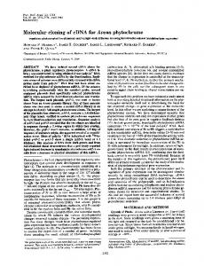
FIG. 1. Immunoprecipitation of cell-free translation products by aPR-B. One microgram of chicken oviduct total poly(A)+ RNA was translated in vitro in the presence of [35S]methionine. Aliquots of the translation products were either used directly or immunoprecipitated with specific antibodies and electrophoresed in NaDodSO4/ polyacrylamide (10%) gels. Lanes: 1, no RNA translation, product immunoprecipitated by aPPR-B; 2, total translation products from poly(A)+ RNA; 3-7, translation of poly(A)+ RNA followed by immunoprecipitation by antiovalbumin (lane 3), by aPR-B (lhne 4), by rabbit anti-(rat)IgG and S. aureus (lane 5), by S. aureus alone (lane 6), or by aPR-B in the presence of 45 /xg of authentic hen PR-B (lane 7); 8 and 9, immunoprecipitation by aPR-B of translation products using poly(A)+ RNA from hen thigh (lane 8) or heart muscle (lane 9). Mr standards are indicated on the right. X-ray film was exposed at -80C for 3 days.
108,000 band in the immunoprecipitate was dependent on the addition of the antibody to PR-B and did not require addition of carrier antigen. Moreover, addition of excess purified hen PR-B was competitive. The Mr 108,000 protein band was undetectable, at this level of sensitivity, when hen thigh or heart muscle poly(A)+ RNA was translated in vitro. The immunoprecipitable Mr 108,000 peptide synthesized in vitro was further examined by two-dimensional gel electrophoresis. The isolated 35S-labeled antigen, to which authentic hen PR-B was added, was run in an isoelectric focusing gel (pH 3.5-8) and then electrophoresed through a linear 10-20o polyacrylamide gel in the presence of NaDodSO4 (29). The 35S-labeled antigen comigrated with authentic hen PR-B (Fig. 2). The existence of multiple spots observed by Coomassie staining is indicative of differential phosphorylation of the hen PR-B (9). Since the reticulocyte lysate does not result in phosphorylated translation products, the antigen acted essentially as a single molecular species at the most basic pI observed for authentic hen PR-B (Fig. 2). Mild proteolytic digestion of the hen progesterone receptor subunits under denaturing conditions has been shown to result in characteristic peptide patterns on analysis by NaDodSO4/polyacrylamide gel electrophoresis (31). Consequently, a mixture of authentic hen PR-B and previously immunoprecipitated [35S]methionine-labeled antigen was electrophoresed in a 7.5% NaDodSO4/polyacrylamide gel. The Mr 108,000 band, identified by Coomassie staining and fluorography, was cut from the gel and partially digested with S. aureus V8 protease as described by Cleveland et al. (30). The presence of purified hen PR-B was necessary, because the digestion pattern is dependent on the concentration ratio
6360
Proc. NatL Acad ScL USA .81
Biochemistry: Zarucki-Schulz et aL
(1984)
B
A
Isoelectric focusing
--
pH 8
pH 3.5 Mr o ix
10
3 '9
CL)
-o U) 0
0a 67 -
z
43-
30 --
2014-
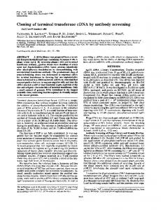
FIG. 2. Two-dimensional gel analysis of authentic hen PR-B and the in vitro-transiated Mr 108,000 protein immunoprecipitated with aPR-B. Chicken oviduct total poly(A)+ RNA was translated in vitro and immunoprecipitated with aPR-B. The 35S-labeled antigen (20,000 cpm) was electrophoresed (first dimension) in an isoelectric focusing gel (pH 3.5-8) in the presence of 5 Ug of authentic hen tSR-B and then (second dimiension) in a linear 10-20% NaDodSO4/polyacrylamide gel. (A) Coomassie blue stain; the arrow indicates the position at authehtic hen PRB. (B) Fluorograph of the same gel; the arrow indicates the position of the 35S-labeled immunoprecipitated Mr 108,000 protein.
of total protein to enzyme (30). Comparison of the proteolytic products from hen PR-B (Coomassie stain) and 35S-labeled antigen (fluorograph) showed similar digestion patterns in a 15% denaturing polyacrylamide gel (Fig. 3), presenting addition'al evidence for identity between the in vitro-synthesized Mr 108,000 peptide and authentic hen PR-B. Only one 35S_ labeled band was detected that was more abundant than the corresponding stained homolog. It is not clear whether this 35S-labeled band was prominent because of different rates of enzyme digestion of the in vitro-synthesized protein compared with added receptor protein, uneven distribution of methionine in the small fragments, or modified amino acid residues. Isolation and Identification of a Partial cDNA Clone for the Chicken Progesterone Receptor B-Subunit Gene. Because of the predicted low abundance of PR-B based on ligand-binding analysis (16), poly(A)+ RNA isolated from diethylstilbestrol-stimulated chicken oviduct was enriched for PR-B-specific mRNA by size selection (37, 38). Approximately 50,000 recombinants were screened using aPR-B by a method found to be sensitive enough to recognize -0.1 ng of PR-B. Plasmid DNA prepared from recombinant clones reacting with aPR-B was bound to nitrocellulose filters and used in hybrid-selection of chicken oviduct total poly(A)+ RNA. Typical endogenous bands from the rabbit reticulocyte lysate were observed after gel analysis of the total products from translation of RNA hybrid-selected by pBR322 (Fig. 4). They were however not immunoprecipitable by aPR-B. A plasmid containing the cDNA of ovalbumin mRNA, pOV230 (25), was used as a positive control for hybrid-selection. This resulted in the detection of a major band at MAr 40,000, corresponding in size to ovalbumin (the spreading to other lanes is due to sample overloading). One of the antibodyidentified clones, pPRB-1, contained a sequence that hybridized to and specifically selected mRNA for PR-B. This was evident from both the size (Mr, 108,000) and specific immunoprecipitation with aPR-B of the subsequent translation products as determined by polyacrylamide gel electrophoresis (Fig. 4, lanes 5 and 15). Minor bands at Mr 90,000 and -80,000 were also synthesized and were recognized by aPRB. These could be either related peptides or the result of nonspecific precipitation. No specific bands were detected when total translation products of mRNA after hybrid-selection by other clones identified by aPR-B were examined,
suggesting that they were false positives obtained during antibody screening (Fig. 4, lanes 7-10). Plasmid DNA prepared from pPRB-1 was found to contain an insert of 470 base pairs when digested by Pst I (data not shown). This fragment was nick-translated to high specific activity and used as a probe to rescreen the entire cDNA
library by colony hybridization (39). Examination of one of the identified clones, pPRB-2, by hybrid-selected translation and immunoprecipitation showed bands identical to those obtained with pPRB-1 (Fig. 4). pPRB-2 containednan insert of 820 nucleotides. By restriction -mapping, pPRB-1 was found to be entirely contained with pPRB-2. 2
1 fMr,
'-.v
10 3
43-
31 &IO. 21 - a,ip-*~
A
_W
14.4 WIW
_-
i.TI FIG. 3. Comparison by partial proteolysis of authentic hen PR-B and the in vitro-translated Mr 108,000 antigen. Authentic hen PR-B (5 ug) was mixed with 50,000 cpm of immunoprecipitated [35S]ihethionine-labeled peptide synthesized in vitro from oviduct total poly(A)+ RNA. The sample was electrophoresed on a 7.5% NaDodSO4/polyacrylamide gel and the Mr 108,000 band was cut out for proteolytic digestion. The gel slice was applied to a 15% polyacrylamide gel, overlayed with 20 IAI of S. aureus V8 protease at 5 jug/ml, and digested for 30 min, and the peptides were separated by electrophoresis. Lanes: 1, Coomassie blue stain showing the digestion pattern of authentic hen PR-B; 2, fhiorograph of the 35S-labeled antigen. The x-ray film was exposed for 1 week at -800C.
Biochemistry: Zarucki-Schulz et aL A
1 2
3 4
Proc. Natl. Acad. Sci USA 81 (1984)
5 6 7 8 9 10
x 10
11 12 13 14 15 16
B
Mr
Mr
M.
X
3
6361
10
200
\ 10
3
3
_
116
08
--
97
-_-108
_
67 43
30
-¢--
20
_
FIG. 4. Immunoprecipitation of products from hybrid-selected mRNA translation. Plasmid DNA from antibody-screened clones was bound to nitrocellulose filters and mRNA complementary to the inserts was hybrid-selected from oviduct total poly(A)+ RNA and translated in vitro. Translation products were analyzed on NaDodDSO4/polyacrylamide (10%) gels directly (A) or following immunoprecipitation (B). Lanes: 1-10, total translation products synthesized in the absence of RNA (lane 1) and in the presence of oviduct total poly(A)+ RNA (lane 2); mRNA hybrid-selected by pBR322 (lane 3), by pOV230 (lane 4), by pPRB-1 (lane 5), by pPRB-2 (lane 6), and by clones pA, pB, pC, and pD (negative in antibody screening) (lanes 7-10, respectively); 11, immunoprecipitation by a-ovalbumin of translation products encoded in mRNA hybridselected by pOV230; 12 and 13, immunoprecipitation by aPR-B of translation products prepared in the absence of RNA (lang 12) or in the presence of chicken oviduct total poly(A)+ RNA (lane 13); 14-16, immunoprecipitation by aPR-B of translation products from mRNA hybridselected by pBR322 (lane 14), by pPRB-1 (lane 15), or by pPRB-2 (lane 16). M, standards are between the gels. X-ray film was exposed for 3 days at -800C.
Size Analysis and Hornional Regulation of Progesterone Receptor Subunit B RNA Levels in Chicken Ovidlqct Tissue. The presence and size of a mRNA recognized by pPRB-1 was assessed by agarose gel electrophoresis using denaturing conditions followed by blot hybridization (Fig, 5). When the 32P-labeled iDNA insert from pPRB-1 was used as a hybridization probe, a single prominent band corresponding to a mRNA of approximately 3Q00 nucleotides' (± 150) was detected. Estrogen regulation of PR-B mRNA levels was apparent because the message concentration in hormone-withdrawn chicken oviduct (lape 1) was greatly reduced in comparison with that of chickens chronically stimulated by diethylstilbestrol (lane 2)*
results is that the antigen protein differs from the hormone binder by a post-translational modification. The identification of a clone by hybrid-selected translation is dependent on successful translation of the mRNA and immunpprecipitation of the [35S]methionine-labeled protein of interest. Chicken oviduct total poly(A)+ RNA was translated in a rabbit reticulocyte lysate system and a protein was produced that could be specifically immunoprecipitated by aPR-B and Fomrigrated with authentic hen PR-B in NaDodbp
5450
DISCUSSION A monoclonal antibody directed against the B-subunit antigen of the chicken oviduct progesterone receptor was used to identify subclones fronm a gDNA library that were capable of expressing the antigenic sequence. The antibody had previously been shown by immunoblotting to recognize only one band (at Mr 108,0Q0) following denaturing gel electrophoresis of either chicken oviduct' cytosol or purified authentic hen B subunit (15). This antibody does not react with native receptor B-subunit protein having hormone-binding activity. Thus, the receptor B antigen recognized by antibody 9G10 appears to be modified in some way from the hormope-binding form. The two forms (antigen and hormone binder) are indistinguishable by tryptic peptide mapping. We have used the nomenclature "receptor B drntigen" in the present communication to define unequivocally the reactive protein. We cannot exclude at this time the possibility that the B antigen and receptor B hormone binder are alleles or products of related genes. Preliminary studies (unpublished) of genomic DNA blots by the method of Southern (40) have failed to give evidence for a gene family; the most likely interpretation of our
1700
1 2
FIG. 5. RNA gel analysis of PR-B antigen in total chicken oviduct RNA. :fen micrograms of total cellular RNA isolated from chicken oviduct was electrophoresed in a 1% agarose gel in the presence of 2.2 M formaldehyde. The RNA was transferred to a nitrocellulose filter and hybridized to 2 x 10⁷ cpm of nick-translated pPRB-1 insert. Molecular size standards were determined using an ovomucoid-specific nucleic acid probe. The variability in this gel system is ±300 base pairs (bp). X-ray film was exposed at -80'C for 3 days. Lanes: 1, RNA from estrogen-deprived chicken oviduct; 2, RNA from chronically diethylstilbestrol-stimulated chicken oviduct.
Biochemistry: Zarucki-Schulz et aL
6362
S04/polyacrylamide gel electrophoresis. The Mr 108,000 protein from in vitro translation also comigrated with purified hen PR-B in two-dimensional gels, indicating similar charge distribution. Furthermore, the peptide pattern obtained from the [35Slmethionine-labeled antigen by S. aureus V8 protease digestion was similar to the characteristic set of peptide fragments obtained from authentic hen PR-B (31). These studies suggest strongly that the Mr 108,000 protein synthesized by the cell-free translation system and identified by specific immunoprecipitation with aPR-B is indeed the chicken progesterone receptor B subunit. By antibody screening and hybrid-selected translation of combined groups of clones, a single clone, pPRB-1, was identified from 50,000 recombinants from the chicken oviduct cDNA library. The clone hybridized to a mRNA that directed the synthesis in vitro of'a Mr 108,000 protein that comigrated with authentic hen PR-B in NaDodSO4/polyacrylamide gels and could be immunoprecipitated by aPR-B. Unequivocal identification of a progesterone receptor subunit requires reintroduction of a cloned full-length cDNA into an expression system that produces sufficient mature protein to detect by specific high-affinity binding of radiolabeled progesterone. In addition to coding for a regulatory protein, the progesterone receptor gene itself appears to be a target for hormone action. It has been shown that hormone-binding capacity can be regulated by estrogen and progesterone in various species (11-13). However, a detailed study of this phenomenon at the mRNA level has yet to be performed. In preliminary experiments, mRNA for PR-B was easily detectable in the oviduct of estrogen-treated chickens by RNA gel analysis and hybridization to a 32 P-labeled insert whereas it was barely found in hormone-deprived oviduct. These results are consistent with inductive regulation by estrogen of the gene coding for the chicken progesterone receptor B subunit. Precise studies on the mechanisms of hormonal regulation of the chicken PR-B antigen at the molecular level can now be carried out. Note Added in Proof. Additional longer cDNA clones have now been isolated that contain nucleotide sequences corresponding to three separate peptide sequences determined by amino acid sequence analysis of the hen PR-B antigen, thus confirming the conclusions of this manuscript.
We thank Drs. T. Chandra and Savio L. C. Woo for many helpful discussions and suggestions; Dr. B. S. Dunbar and Donnie Bundman for two-dimensional isoelectric focusing analysis; Esther M. Presente, Margaret E. Rickaby, and Theresa Wedrychowicz for superb technical assistance; and Cheryl McCarthy for excellent secretarial help. This work was supported in part by National Institutes of Health Grant HD-07857. D.R.H. is the recipient of Fogarty International Center National Institutes of Health Fellowship 1 F05 TW 03360-01. M.S.K. is a recipient of a Special Postdoctoral Fellowship from the Rockefeller Foundation (New York). D.P.E. is the recipient of American Cancer Society Grant BC-319A. 1. Compton, J. G., Schrader, W. T. & O'Malley, B. W. (1983) Proc. Nati. Acad. Sci. USA 80, 16-20. 2. Payvar, F., Wrange, O., Carlstedt-Duke, J., Okret, S., Gustafsson, J.-K. & Yamamoto, K. R. (1981) Proc. NatI. Acad. Sci. USA 78, 6628-6632. 3. Mulvihill, E. R., LePennec, J.-P. & Chambon, P. (1982) Cell 24, 621-632. 4. Chandler, V. L. , Maler, B. A. & Yamamoto, K. R. (1983) Cell
33, 489-499.
5. Dean, D.
C., Knoll, B. J., Riser, M. E. & O'Malley, 13. W.
(1983) Nature (London) 305, 551-554. 6. Gronemeyer, H. & Pongs, 0. (1980) Proc. NatI. Acad. Sci. USA 77, 2108-2112. 7. Sibley, C. H. & Tomkins, G. M. (1974) Cell 2, 221-227.
Proc. NatL Acad Sci. USA 81
(1984)
8. Bourgeois, S., Newly, R. F. & Huet, M. (1978) Cancer Res. 38, 4279-4284. 9. Weigel, N. L., Tash, J. S., Means, A. R., Schrader, W. T. & O'Malley, B. W. (1981) Biochem. Biophys. Res. Commun. 102, 513-519. 10. Schrader, W. T., Birnbaumer, M. E., Hughes, M. R., Weigel, N. L., Grody, W. W. & O'Malley, B. W. (1981) Recent Prog. Harm. Res. 37, 583-633. 11. Frenette, G., Dube, J. Y. & Tremblay, R. R. (1982) J. Steroid Biochem. 17, 271-276. 12. Pageaux, J. F., Laugier, C., Pal, D. & Pacheco, H. (1983) J. Steroid Biochem. 18, 209-214. 13. Milgrom, E., Thi, L., Atger, M. & Baulieu, E. E. (1973) J. Biol. Chem. 248, 6366-6374. 14. Toft, D. D. & O'Malley, B. W. (1972) Endocrinology 90, 10411045. 15. Edwards, D. P., Weigel, N. L., Schrader, W. T., O'Malley, B. W. & McGuire, W. L. (1984) Biochemistry, in press. 16. Boyd-Leinen, P. A., Fournier, D. & Spelsberg, T. C. (1982) Endocrinology 111, 30-36. 17. Helfman, D. M., Feramisco, J. R., Fiddes, J. C., Thomas, G. P. & Hughes, S. H. (1983) Proc. Nat4.Acad. Sci. USA 80, 31-35. 18. Yodng, R. A. & Davis, R. W. (1983) Proc. Nati. Acad. Sci. USA 80, 1194-1198. 19. O'Malley, K. L., Mauron, A., Raese, J., Barchas, J. D. & Kedes, L. (1983) Proc. Natl. Acad. Sci. USA 80, 2161-2165. 20. Whittle, D. J., Kilburn, D. G., Warren, R. A. J. & Miller, R. C. (1982) Gene 17, 139-145. 21. Rosen, J. H., Woo, S. L. C., Holder, J. W., Means, A. R. & O'Malley, B. W. (1975) Biochemistry 14, 69-78. 22. Ulrich, A., Shine, J., Chirgwin, J., Pictet, R. E., Tischer, E., Butler, W. J. & Goodman, H. M. (1977) Science 196, 13131316. 23. Aviv, M. & Leder, P. (1972) Proc. Natl. Aead. Sci. USA 69,
1408-1412. 24. Monahah, J. J., Harris, S. E., Woo, S. L. C., Robberson, D. L. & O'Malley, B. W. (1976) Biochemistry 15, 223-233. 25. McReynolds, L. A., Catteral, J. F. & O'Malley, B. W. (1977) Gene 2, 217-231. 26. Kemp, D. J. & Cowman, A. F. (1981) Proc. Natl. Acad. Sci. USA 78, 4520-4524. 27. Markwell, M. A. K. (1982) Anal. Biochem. 125, 427-432. 28. Porzio, M. A. & Pearson, A. M. (1977) Biochim. Biophys. Acta 490, 27-34. 29. Dunbar, 13. S. (1984) in Laboratory Methods Manualfor Hormone Action and Molecular Endocrinology, eds. Schrader, W. T. & O'Malley, B. W. (Baylor College of Medicine, Houston, TX), 8th Ed., Chapt. 15, pp. 1-31. 30. Cleveland, D. W., Fischer, S. G., Kirschner, M. W. & Laemmli, U.. K. (1976) J. Biol. Chem. 252, 1102-1106. 31. Weigel, N. L., Minghetti, P. P., Stevens, B., Schrader, W. T. & O'Malley, B. W. (1984) in Steroid Hormone Receptors: Structure and Function, Nobel Symposium No. 57, eds. Eriksson, M. & Gustafsson, J.-A. (Elsevier, Amsterdam), pp. 2544. 32. Birnhoim, H. D. & Doly, J. (1979) Nucleic Acids Res. 7, 15131523. 33. Katz, L., Kingsbury, D. J. & Helinski, D. R. (1973) J. Bacteriol. 114, 577-591. 34. Parnes, J. R., Velan, B., Felsenfeld, A., Ramanathan, L.,, Ferrini, U.,,Appella, E. & Sidman, J. G. (1981) Proc. NatI. Acad. Sci. USA 78, 2253-2257. 35. Lehrach, M., Diamond, D., Wozney, J. M. & Boedtker, H. (1977) Biochemistry 16, 4743-4751. 36. Maniatis, T., JeffreyJ A'. & Kleid, D. G. (1975) Proc. Nat/. Acad. Sci. USA 71, 1184-1188. 37. Bailey, J. M. & Davidson, N. (1976) Anal. Biochem. 70, 7585. 38. Scherrer, K. (1969) in Fundamental Techniques in Virology, eds. Habet, K. & Solyman, N. P. (Academic, New York), pp. 413-432. 39. Gergen, J. P., Stern, R. H. & Wensink, P. C. (1979) Nucleic Acids Res. 7, 2115-2136. 40. Southern, E. M. (1972) J. Mol. Biol. 93, 503-517.