Revista Latinoamericana de Microbiología Volumen Volume
47
Número Number
3-4
Julio-Diciembre July-December
2005
Artículo:
Molecular detection of dengue viruses in field caught Aedes aegypti mosquitoes from northeastern Argentina Derechos reservados, Copyright © 2005: Asociación Mexicana de Microbiología, AC
Otras secciones de este sitio:
Others sections in this web site:
! Índice de este número ! Más revistas ! Búsqueda
! Contents of this number ! More journals ! Search
edigraphic.com
Revista Latinoamericana de
MICROBIOLOGÍA
Vol. 47, Nos. 3-4 July - September. 2005 October - December. 2005 pp. 82 - 87
ORIGINAL ARTICLE
Molecular detection of dengue viruses in field caught Aedes aegypti mosquitoes from northeastern Argentina Domingo Javier Liotta,* Gustavo Cabanne,* Rodolfo Campos,** Sergio Andrés Tonon*
ABSTRACT. Most molecular protocols for Dengue virus detection described so far are time consuming and cumbersome with mosquito samples. In order to count with a sensitive and specific molecular detection system for monitoring possible Dengue outbreaks and circulating viral serotypes in field-caught Aedes aegypti populations from Northeastern Argentina, a RT-PCR and RFLP assay was developed. The original RT-PCR assay proposed by Sudiro et al. for human serum was optimized for mosquito samples. Modifications were done at the RNA extraction-purification and at the thermal profile steps. The generic 230 bp amplicon was validated by RFLP assay and cycle sequencing. Results showed that, due to the generic characteristic of the primers used, certain mosquito genome regions could be co-amplified, making confirmation of the Dengue specific amplicon by RFLP assay a required step. Under these conditions, the proposed method can be employed as a Dengue viral generic screening procedure in Aedes aegypti mosquito samples, giving in our hands an estimated 99.52% of confirmed negatives (207/208 tested samples).
RESUMEN. Numerosos protocolos propuestos para la detección del virus del Dengue requieren de un tiempo prolongado de ejecución o bien presentan dificultades metodológicas con muestras de mosquitos. Por este motivo, se desarrolló un ensayo de RT-PCR y RFLP con el objeto de disponer de un sistema molecular sensible y específico, capaz de detectar potenciales brotes epidémicos de Dengue y serotipos virales circulantes en mosquitos Aedes aegypti capturados a campo en la región nordeste de Argentina. El protocolo original de RT-PCR propuesto por Sudiro et al para la detección viral en suero humano fue optimizado para muestras de mosquitos. Las modificaciones se realizaron a nivel de las etapas de extracciónpurificación y ciclado térmico. El producto genérico de 230 bp fue autenticado por RFLP y secuenciación cíclica. Los resultados mostraron que debido a la naturaleza genérica de los cebadores empleados, ciertas regiones genómicas del vector podrían ser co-amplificadas, considerando por ello necesario la validación del producto mediante RFLP. Bajo estas condiciones de trabajo, el método propuesto puede ser empleado en procedimientos de tamizaje viral genérico con muestras de vector Aedes aegypti, generando un 99.52% de negativos confirmados (207/208 muestras analizadas).
Key words: Dengue, Aedes aegypti, RNA, RT-PCR, RFLP.
Palabras clave: Dengue, Aedes aegypti, ARN, RT-PCR, RFLP.
INTRODUCTION Dengue Fever (DF) and Dengue Haemorrhagic Fever (DHF) are caused by infection with any of the four Dengue virus serotypes (DV1-4). These pathologies have special sanitary interest due to their social and economic impact during epidemics.18 Dengue viruses are mainly transmitted by Aedes aegypti, a mosquito that grows up in breeding containers maintained by rain or human activity. Its expansion to different areas of the American continent is determined by ecological changes and human behavior that enhance the ocurrence of breeding places.17 The 1981 and 1997 outbreaks suffered in Cuba showed that Dengue does
occur when no precautions are taken regarding vector surveillance.11 Thus, prevention and control of epidemic outbreaks are sustained by vector surveillance and by the identification of declared human cases. A permanent surveillance system allows the implementation of direct actions, like the application of insecticides and destruction of larvae population habitat.17 By 1955, when Aedes aegypti eradication campaign began in Argentina, the infested area included all the northern provinces with template and subtropical weather. In 1963, the program considered Argentina free of Aedes aegypti,19 but unfortunately, in 1986 the reinfestation was officially declared, being the aforementioned regions regained by the mosquito.4 In accordance with this data, between October 1998 and December 2000 we have continuously detected, at predetermined monitoring stations, the presence of Aedes aegypti in Posadas, Northeastern Argentina. Although no epidemic has ocurred at Posadas yet, the present situation is of concern due to the regional epidemiological history. More than 25,000 cases have ocurred in nearby Paraguay from December 1999 to March
edigraphic.com
* Laboratorio de Biología Molecular Aplicada, Facultad de Ciencias Exactas, Químicas y Naturales, Universidad Nacional de Misiones, Argentina. ** Cátedra de Virología, Facultad de Farmacia y Bioquímica, Universidad de Buenos Aires, Argentina. Received November 17, 2003; received in revised form February 3, 2005; accepted May 19, 2005.
Liotta et al
Molecular detection of dengue viruses in field caught Aedes aegypti mosquitoes
83
Rev Latinoam Microbiol 2005; 47 (3-4): 82-87
2000, being 9 of them Dengue haemorragic fever cases.3,14 Adding up to this perspective, the frontier between Posadas (Argentina) and Encarnación (Paraguay) is under a relaxed control system, and is not properly prepared to detect imported Dengue human cases. This situation is similar to that occurred in Salta, a northwest province from Argentina at the borderline with Bolivia, before the Dengue outbreak of 1997.2 When adult mosquito density is low, direct entomological monitoring is not a sensitive indicator for outbreaks prevention. It is in this particular situation that detection of Dengue viruses in vector populations becomes an important element as part of an early alert system. Moreover, this kind of analysis, allows to position the vector as the primary and necessary element in the transmission cycle during epidemiological evaluations,6,23 MATERIAL AND METHODS Dengue virus controls for RT-PCR set-up Four different types of controls were employed: I) 1x10⁷ PFU/ml of DV2 viral particles, resuspended in PBS; II) 1x10⁷ PFU/ml of DV2 viral particles, resuspended in human serum; III) 1x10⁷ PFU/ml of DV2 viral particles resuspended in Aedes aegypti mosquito heads. 16 IV) Uninfected mosquito heads used as negative control. The DV2 strain was kindly provided by the Flavivirus Laboratory, Oswaldo Cruz Institute, RJ, Brazil. Although transovarial transmission is a rarely occurring event in Aedes aegypti, in order to avoid this phenomena all mosquitoes employed as negative control came from pools grown up in laboratory conditions. RT-PCR protocol tested with mosquito samples from natural populations Two hundred and eight adult female Aedes aegypti mosquitoes were captured from five monitoring stations placed at Posadas, Province of Misiones, Argentina. The captures were performed with traps developed by the Center for Disease Control (U.S.A.) and/or a hand nest6, 10 on a weekly basis during the last two hours of the evening. Trapped mosquitoes were put into a 1.5 ml microcentrifuge tube and placed on ice, transported to the laboratory and frozen at -20°C until being analyzed. Taxonomic classification of captured mosquitoes was done in collaboration with the Ecology Department, National University of Misiones, in accordance with Consoli & de Oliveira7 guidelines.
Total RNA extraction and purification from mosquitoes Mosquito heads were separated from their bodies using a pair of sterile needles and put into a 1.5 ml microcentrifuge tube. One hundred microliters from each Dengue RT-PCR control was added to individual laboratory grown mosquitoes heads, and mixed with 100 µl of lysis buffer (6M guanidine isothiocianate, 50 mM sodium citrate, 1% Sarkosil, 20 mg/ml E. coli tRNA, 100 mM β-mercaptoethanol).9 Alternatively, 100 µl of phosphate buffered saline (PBS) was added to single mosquito heads coming from field samples (natural populations), and mixed with lysis buffer. All heads were macerated in a glass micromortar. Finally, they were clarified by centrifugation for 1 minute at 1,000 rpm. Originally, the supernatant was purified twice with 100 µl of phenol/chloroform/isoamyl alcohol mixture (25:24:1). In order to optimize the retention of RNA molecules in the aqueous phase,22 this step was later modified by using acid phenol instead of phenol pH 8. Five microliters of acid silica solution were added to each sample for RNA concentration purposes.5 The mixture was incubated for 5 minutes at room temperature and pelleted by centrifugation. Each sediment was treated twice with 200 µl of washing buffer (50% ethanol, 10 mM Tris pH 7.4, 1 mM EDTA, 50 mM NaCl).9 Finally, samples were resuspended and incubated for 5 minutes at 55°C in 15 µl of nuclease free water, containing 5 mM DTT, and 1U/µl RNAseOUT (GIBCO BRL). After centrifugation, the supernatant was transferred to a new tube and frozen until amplification. Reverse Transcription and generic PCR The RT-PCR reaction was performed with the primers originally described by Sudiro et al., that target the regulatory 3’UTR genome region of all four DV and giving a single product of 230 bp. 20 In this work, the AMV retrotranscriptase was replaced by the rTth pol enzyme (Promega) for its property of being a dual enzyme, and the thermal profile included: 10 minutes at 50°C, 10 minutes at 55°C and 20 minutes at 60°C, followed by 10 cycles of 45 seconds at 92°C, 45 seconds at 56°C, 1 minute at 72°C and lastly by 30 cycles of 45 seconds at 92°C, 45 seconds at 53°C, 1 minute at 72°C with a final extension step of 10 minutes at 72°C. The reaction mixture for RT was: 10 mM Tris-HCl pH 8.3, 90 mM KCl, 1 mM MnCl 2, 200 µM of each dNTP, 5 mM DTT, 2 U/µl RNAseOUT (GIBCO BRL), 1 pmol/µl antisense primer, and 0,25 U/µl rTth pol enzyme, in a final volume of 10 µl. The PCR reaction mixture contained quelate buffer (10 mM Tris-HCl pH 8.3, 0.1
edigraphic.com
84
Liotta et al
Molecular detection of dengue viruses in field caught Aedes aegypti mosquitoes
Rev Latinoam Microbiol 2005; 47 (3-4): 82-87 MG
M KCl, 0.75 mM EGTA, 0.05% Tween20™ 5 % glycerol), 2 mM MgCl 2, 200 µM of each dNTP, and 1 pmol/µl sense primer, in a final volume of 25 µl.
Terminator Cycle Sequencing Kit, Amersham Pharmacia Biotech). The obtained sequences were compared against those from Genbank.
RFLP Assay
RESULTS
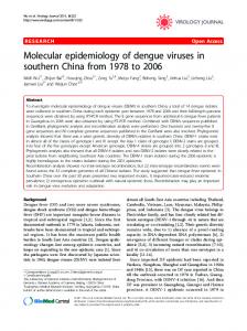
Data from DV 3’UTR region was obtained from Genbank (DV1 S275/90 strain, M87512; DV2 Jamaica strain, M20558; DV3 H87 strain, M93130; and DV4 Caribbean 814669 strain, M14931). Multiple sequence alignment was done with the Clustal W 1.74 computer program and a conserved RsaI restriction site was found for all four serotypes. Besides, an additional conserved RsaI site was located on DV 3 and 4 (Fig. 1a). Controls named as PTA-D1.1 (DV1), PTA-D2.5 (DV2), PTA-D3.5 (DV3) and PTA-D4.2 (DV4) were amplified and analyzed by RFLP. For DV1 and 2, the RFLP pattern produced two fragments of approximately 160 bp and 70 bp, while for DV3 and 4 three fragments lighter than 100 bp, as detected by agarose gel electrophoresis (Fig. 1b).
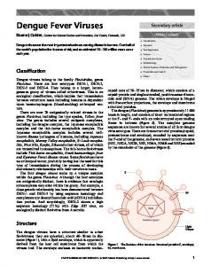
In order to develop a sensitive and specific molecular method for DV monitoring in field caught Aedes aegypti mosquitoes, standard assays proposed for human serum samples were employed as a starting point. The generic method proposed by Sudiro et al20 was selected in first instance, although its application to mosquito samples and controls produced several unexpected electrophoretic bands (Fig. 2). Nevertheless, in this first approximation 10 field caught mosquito samples also showed a 230 bp amplification product as the expected DV generic band (Fig. 2, lanes 8-12). Repurification of mosquito samples with acid phenol and the introduction of modifications in RT-PCR conditions eliminated the nonspecific band pattern observed, being now the 230 bp product present only in one mosquito sample (A1-2) (Fig. 3). This putative positive amplification fragment was further checked by RFLP, giving two fragments of, approximately, 160 bp and 70 bp as analyzed by agarose gel electrophoresis (data not shown). When the digestion mixture was separated by polyacrilamide gel electrophoresis and compared to those coming from a DV2 control, the increased gel resolution allowed to observe differences in the migration band pattern (Fig. 4a). Complete sequencing of the aforementioned amplification product was compared against the four DV genomes. The alignment showed only 31.4% sequence homology. Therefore, although having a proper size and a RsaI restriction site, this amplicon did not belong to any
RT-PCR amplicon cloning and cycle sequencing RT-PCR products (controls and samples) were subcloned into the pGEM-T easy (Promega) cloning vector, and transformed into E. coli DH5aF’ strain. The recombinant plasmids were recovered by the alkaline lysis method. The 230 bp cloned fragments were recovered by Eco RI enzymatic digestion, and separated by LMP agarose gel electrophoresis. Final fragment purification was done by spin silica columns method (Wizard SV Gel and PCR Clean-up System, Promega Corp.). One hundred and fifty nanograms from each purified fragment was employed for cycle sequencing, following manufacturer specifications (Thermo Sequenase Radiolabeled
edigraphic.com
Figure 1a. Multiple sequence alignment of DV 3’UTR partial region. The first nucleotide corresponds to genome position 10406 (DV1, M87512), 10408 (DV2, M20558), 10387 (DV3, M93130) and 10322 (DV4, M14931). Complete sequence homology is highlighted in grey. RsaI restriction sites (GTAC) are remarked in black.
Liotta et al
85
Molecular detection of dengue viruses in field caught Aedes aegypti mosquitoes Rev Latinoam Microbiol 2005; 47 (3-4): 82-87
1
2
3
5
4
6
7
8
9
1
10 11
2
3
4
5
6
7
8
9
230 bp 100 bp
160 bp
Figure 1b. RFLP analysis of DV controls. Lanes 1-2: DV1, 3-4: DV2, 5-6: DV3, 7-8: DV4, 9: 100 bp molecular weight ladder, 10-11: PCR controls (+ and -). Odd lanes, uncut PCR products. Even lanes, RsaI digested PCR products.
Putative (-) mosquito samples
Putative (+) mosquito samples
Figure 3. Standard vs modified protocol conditions. Lanes 1-2: sample A1-2; lanes 3-4: uninfected mosquito head, spiked with DV2 viral particles; lanes 5-6: uninfected mosquito head (negative control). Odd lanes: standard protocol, even lanes: modified protocol. Lane 7: 100 bp Molecular weight ladder; lanes 8-9: RT-PCR controls (+ and -).
MW C(+) 1
2
3
4
5
6
7
8
9
10
11
12
230 bp
Figure 2. RT-PCR DV human serum generic protocol applied to mosquito samples. Lanes 1 to 5 and 8 to 12: mosquito samples; lane 6: 100 bp Molecular weight ladder; lane 7: DV2 positive control. The arrow points to the 230 bp diagnostic generic RT-PCR product.
DV genome (Fig. 4b), making RFLP analysis by polyacrilamide gel electrophoresis a required step for this proposed methodology. Taking into account all these considerations, the present method showed a good negative prediction value, established as 99.52% of confirmed negatives in our hands (207/208 tested samples).
detection method for infected mosquitoes would be very useful in screening field caught mosquito pools in Dengue prone or vulnerable areas. This approach could be applied in an early warning strategy, helping public health policy design. Ideally, a general screening strategy would be benefited with a single round RT-PCR amplification method having the capability of detecting all DV serotypes in mosquitoes. Several RT-PCR protocols for DV detection in human serum samples have been proposed during recent years. 8,12,13,21 They vary, mainly, in target regions and sensitivity levels. In this work, we have adapted a generic detection method for DV in human serum samples21 to the more complex Aedes aegypti mosquito samples. In order to increase method specificity and sensitivity, the following changes were introduced to the original protocol: I) RNA extraction was optimized by the incorporation of an acid phenol-chloroform-isoamyl alcohol purification step. This modification selectively retains RNA in the aqueous phase, eliminating non desirable genomic DNA.22 II) Replacement of AMV by rTth polymerase enzyme, for increasing stringency of the RT reaction by setting the elongation temperature at 60°C. III) Utilization of a touchdown approach to the annealing temperature profile, starting at 56°C (first 10 cycles) and reaching 53°C (last 30 cycles). Before the introduction of these modifications to the original protocol, 10 samples out of 208 analyzed (4.8%)
edigraphic.com
DISCUSSION Since Aedes aegypti is the main transmission vector for Dengue viruses,1,15,17,23 an affordable and rapid molecular
86
Liotta et al
Molecular detection of dengue viruses in field caught Aedes aegypti mosquitoes
Rev Latinoam Microbiol 2005; 47 (3-4): 82-87 MG
showed a putative positive amplicon and heavy banding sustraídode-m.e.d.i.g.r.a.p.h.i.c pattern (Fig. 2). The new conditions allowed the dissacihpargidemedodabor pearance of unspecific bands and the retention of only one sample (A1-2) with the putative diagnostic amplicon (Fig. 3). These preliminary results, prompted us to check the specificity of the methodology and to authenticate the
DV2
1
257
2
A1-2
3
4
MW
249 195
171
86
54
Figure 4a. RFLP analysis of field sample A1-2. Differences observed in fragments size at the polyacrilamide resolution level. Lane 1-2: uncut and digested DV2 control RT-PCR fragment. Lane 3-4: uncut and digested A1-2 sample RT-PCR fragment; lane 5: 100 bp Molecular weight ladder.
primarily assigned positive sample.:rop Theodarobale generic nature FDP of the primers originally proposed by Sudiro et al allowed the posibilityVC of ed a complementarity AS, cidemihparG with some regions of the vector genome. To authenticate the 230 bp obtained amplicon, a RFLP assay was designed emarap ploying the restriction enzyme RsaI (Fig. 1). This extra methodological step adds:cihpargideM sensitivity without inacidémoiB arutaretiL creasing complexity to the system. When applyed to the putative positive product, the analysis by polyacsustraídode-m.e.d.i.g.r.a.p.h.i.c rilamide gel electrophoresis showed a readily visible difference in migration patterns against a DV2 control. This discrepancy was, initally, attributed to a unspecific amplification product or to the presence of other flavivirus genome. Complete sequencing showed no significant homology to any DV serotype or any other known flavivirus, confirming that product A1-2 was not Dengue related. This result established that a unspecific amplification product with a very close molecular weight to that expected was obtained, possible from interfering mosquito RNA. Therefore, in this context RFLP patterns must be analyzed by polyacrilamide gel eletrophoresis. Even though, molecular detection techniques are nowadays widespread primary tools in large screenings during DV epidemics and in the continuous monitoring of endemic areas; extreme care should be employed regarding sample complexity and analysis of results. In our experience, molecular techniques should be used as a first step and not as the only analytical tool for detection of infected Aedes aegypti. Other methods must be applyed as confirmatory stages, preferably viral particle isolation from putative positive mosquito pools and/or sequencing of diagnostic amplicons, taking extreme care before reaching any conclusion.
edigraphic.com
Figure 4b. Multiple sequence alignment of DV and sample A1-2 3’UTR region amplified fragment. Homology between DV and sample A1-2 is highlighted by asterisks. The RsaI sites are remarked in black: 1) DV RsaI conserved site, 2) sample A1-2 RsaI site (detected by nucleotide sequencing).
Liotta et al
Molecular detection of dengue viruses in field caught Aedes aegypti mosquitoes
87
Rev Latinoam Microbiol 2005; 47 (3-4): 82-87
ACKNOWLEDGMENTS The authors are indebted to Dr. H. Schatzmayr from the Oswaldo Cruz Institut, Rio Janeiro, Brazil and Dr. A. Rothman from the University of Massachusetts Medical Center, U. S. A. for providing Dengue controls; Flavia Kristicevic, Nelsi Pascual and Prof. Aida Tricio from Ecology Department, National University of Misiones, Argentina, for mosquito taxonomy.
REFERENCES 1.
Acha, P. and Szyfres, B., 1997. In: Zoonosis y enfermedades transmisibles comunes al hombre y a los animales. Pan American Health Organization (Editor), Washington D.C.; Scientific publication No. 503, pp 302-305. 2. Avilés, G., Rangeón, G., Vorndam, V., Briones, A., Baroni, P., Enria, D., Sabattini, M.S., 1999. Dengue Reemergence in Argentina. Emerg Infect Dis 5(4): 575-578. 3. Avilés, G., Paz, V., Rangeón, G., Ranaivoarisoa, M. Verzeri, N., Roginski, S., Baroni, P., Enria, 2003. Laboratory Surveillance of Dengue in Argentina, 1995-2001. Emerg Infect Dis 9(6): 738-742. 4. Boffi, R. 1998. Programa de prevención del Dengue y control del Aedes aegypti. In: Temas de Zoonosis y enfermedades emergentes. II Congreso Argentino de Zoonosis, I Congreso Argentino y Latinoamericano de Enfermedades Emergentes y Asociación Argentina de Zoonosis (Editors), pp 413-419. 5. Boom, R., Sol, C.J., Salimans, M.M., Jansen, C.L., Wertheim-van Dillen, P.M., van der Noorda, J., 1990. Rapid and simple method for purification of nucleic acids. J Clin Microbiol 28: 495-503. 6. Chow, V., Chan, Y., Yong, R., Lee, M., Lim, L., Chung, Y., LamPhua, S., Tan, B., 1998. Monitoring of Dengue Viruses in FieldCaught Aedes aegypti and Aedes albopictus Mosquitoes by a Typespecific Polimerase Chain Reaction and Cycle Sequencing. Am J Trop Med Hyg 58(5): 578-586. 7. Consoli, R. and de Oliveira, R., 1994. Principais Mosquitos de Importancia Sanitaria no Brasil. Fiocruz Editor, Brazil, pp 104-117. 8. Deubel, V., Laille, V., Hugnot, J., Chunge, E., Guesdon, J., Drouet, M., Bassot, S., Chevrier, D., 1990. Identification of Dengue sequences by genomic amplification: rapid diagnosis of Dengue Virus serotypes in peripheral blood. J Virol Methods 30: 41-54. 9. Harris, E., Roberts, G., Smith, L., Selle, J., Kramer, L., Valle, S., Sandoval, E., Balmaseda, A., 1998. Typing of Dengue viruses in clinical specimens and mosquitoes by single-tube multiplex Reverse Transcriptase PCR. J Clin Microbiol 36: 2634-2639. 10. Jensen, T., 1994. Comparison of Bi-directional Fay, Omnidirectional, CDC, and duplex cone traps for sampling adult Aedes albopictus and Aedes aegypti in north Florida. J Am Mosq Control Assoc 10(1): 74-78.
11. Kourí, G., Guzmán, M.G., Valdés, L., Carbonel, I., del Rosario, D., Vazquez, S., Laferté, J., Delgado, J., Cabrera, M., 1998. Reemergence of Dengue in Cuba: A 1997 Epidemic in Santiago de Cuba. Emerg Infect Dis 4(1): 89-92. 12. Kuno, G., 1998. Universal diagnostic RT-PCR protocol for Arboviruses. J Virol Methods 72: 27-41. 13. Lanciotti, R.S., Calisher, C., Gubler, D., Chang, G., Vorndam, V., 1992. Rapid detection and typing of Dengue Viruses from clinical samples by using Reverse Transcriptase-Polymerase Chain Reaction. J Clin Virol 30: 545-551. 14. León, T., 2001. Epidemiology Chief, National Ministery of Health, Republic of Paraguay. Personal communication. 15. Martínez Torres, E., 1998. Dengue y Dengue Hemorrágico. Universidad Nacional de Quilmes & Elea Laboratorio (Editors), pp 43-56. 16. Miagostovich, M.P., Nogueira, R.M., Schatzmayr, H., Lanciotti, R., 1998. Molecular Epidemiology of Den-2 Virus in Brazil. Mem Inst Oswaldo Cruz 93(5): 625-626. 17. Pan American Health Organization, 1994. Dengue and Dengue Haemorrhagic Fever: guidelines for prevention and control. Washington D.C. Scientific publication No. 548. 18. Peters, C.J., 1997. Viral Hemorrhagic Fevers. In: Viral Pathogenesis. Nathanson N. (Editor). Lippincott-Raven Publishers, Philadelphia, pp 779-799. 19. Sabattini, M.S., Avilés, G., Monath, T.P., 1998. Historical, epidemiological, and ecological aspects of arboviruses in Argentina: Flaviviridae, Bunyaviridae and Rhabdoviridae. In: An overview of arbovirology in Brazil and neighboring countries. Travassos da Rosa, Vasconcelos, Travassos da Rosa (Editors). Belem, Brazil. Instituto Evandro Chagas, pp 113-134. 20. Sudiro, T., Ishiko, H., Green, S., Vaughn, D., Nisalak, A., Kalayanarooj, S., Rothman, A., 1997. Rapid diagnosis of Dengue viremia by Reverse Transcriptase-Polymerase Chain Reaction using 3´-noncoding region universal primers. Am J Trop Med Hyg 56: 424-429. 21. Tanaka, M., 1993. Rapid identification of Flavivirus using the Polymerase Chain Reaction. J Virol Methods 41: 311-322. 22. Wallace, D.M., 1987. In: Methods in Enzymology. Berger and Kimmar (Editors), Academic Press. Molecular Cloning Techniques Series 152, pp 33-41. 23. World Health Organization, 1997. Dengue Haemorrhagic Fever: Diagnosis, treatment, prevention and control. 2nd Edition, Geneva, pp 60-66. Correspondence to: Lic. D. J. Liotta. Laboratorio de Biología Molecular Aplicada, Facultad de Ciencias Exactas Químicas y Naturales, Universidad Nacional de Misiones, Avenida Mariano Moreno 1375, Posadas, Argentina. 3300; E-mail:
[email protected] Phone: +54 3752 427687 Fax: +54 3752 435102
edigraphic.com