VECTOR/PATHOGEN/HOST INTERACTION, TRANSMISSION
Molecular Identification of Bloodmeals From Sand Flies and Mosquitoes Collected in Israel LEA VALINSKY,1 GONEN ETTINGER,1 GILA KAHILA BAR-GAL,2
AND
LAOR ORSHAN1,3
J. Med. Entomol. 51(3): 678Ð685 (2014); DOI: http://dx.doi.org/10.1603/ME13125
ABSTRACT In Israel, sand ßies are the vectors of Leishmania Ross and mosquitoes are the vectors of West Nile Virus. In the Judean Desert and Tiberias, the sand ßy Phlebotomus sergenti Parrot is the vector of Leishmania tropica (Wright) and the rock hyrax (Procavia capensis Pallas) is considered the main reservoir animal. The main vectors of West Nile Virus are Culex pipiens L. and Culex perexiguus Theobald. Bloodmeals of engorged Þeld-caught female sand ßies and mosquitoes are an important source for deÞning host preferences. Recent progress in DNA molecular techniques has enabled the accurate identiÞcation of blood sources within the arthropod gut. In this study, we applied molecular approach for species-speciÞc identiÞcation based on polymerase chain reaction and nucleotide sequence analysis of polymorphic regions along two mitochondrial genes, 12S and 16S rRNA. The research was carried out on 261 engorged female sand ßies collected in the Judean Desert and Tiberias and 50 engorged female mosquitoes collected in Tel-Aviv and Arava. Species identiÞcation of bloodmeals was successful in 92% of the samples. Rock hyrax was the most abundant host in bloodmeals of P. sergenti, while human blood was found in only seven (3%) females. L. tropica DNA was detected in three P. sergenti females from Tiberias that contained rock hyrax blood. Avian sequences were detected in 67% (10 of 15) of the identiÞed bloodmeals from Cx. perexiguus and in 10% (3 of 29) of the identiÞed meals from Cx. pipiens. Human sequences were found in 14% of the identiÞed bloodmeals from Cx. pipiens. The successful analysis of the majority of the bloodmeals performed on wild sand ßies and mosquitoes suggests that bloodmeal identiÞcation can be applied as one of the routine procedures in vector surveillance programs. KEY WORDS bloodmeal, host, Phlebotomus sergenti, Culex, Leishmania tropica
Cutaneous leishmaniases (CL) are endemic zoonotic diseases in Israel (Jaffe et al. 2004). In recent years, Leishmania tropica (Wright) has extended its geographical distribution in Israel to the western margins of the Judean Desert (Singer et al. 2008) and the western slopes of the Samaria Mountains (unpublished data). Phlebotomus (Paraphlebotomus) sergenti Parrot is the sole vector of L. tropica in most foci in Israel. In a few foci in the north of the country, Phlebotomus (Adlerius) arabicus Theodor is an additional vector (Jacobson et al. 2003, Schnur et al. 2004, Svobodova´ et al. 2006). The nature of leishmaniasis as a result of L. tropica in Israel is zoonotic, unlike its anthroponotic characteristics in many other foci (Jacobson 2011). In 2005, a leishmaniasis surveillance and research program was launched in Israel to study L. tropica infections in reservoir animals and sand ßy vectors. At the time, evidence incriminating rock hyraxes (Procavia capensis Pallas) as the only signiÞcant reservoir 1
Central Laboratories, Ministry of Health, Jerusalem, Israel. Koret School of Veterinary Medicine, The Robert H. Smith Faculty of Agriculture, Food and Environment, The Hebrew University of Jerusalem, Rehovot, Israel. 3 Corresponding author, e-mail:
[email protected]. 2
of L. tropica in Israel was partial and circumstantial (Jacobson et al. 2003). Citizens and local municipalities in foci of the disease demanded urgent action against the probable reservoir. Intervention to control protected wildlife species such as the rock hyrax from the vicinity of inhabited areas required stronger evidence of its signiÞcant role as the reservoir host for Leishmania. Therefore, the study progressed in two parallel directions: 1) sampling wild and domestic animals for Leishmania infections (Talmi-Frank et al. 2010), and 2) determining the reservoir by identiÞcation of sand ßy bloodmeals and detection of Leishmania infections in engorged female sand ßies. Culex perexiguus and Culex pipiens are the main vectors of West Nile Virus (WNV) in Israel (Orshan et al. 2008). WNV infection rates in Cx. perexiguus are higher than those found in Cx. pipiens, but the latter is more abundant. Data on bloodmeal origin could help clarify the relative role of the two species in the endemic transmission cycle of the virus in Israel. Traditional serological methods for bloodmeal identiÞcation require production of speciÞc antibodies to all potential hosts. The major limitations of these methods are the lack of shelf products for exotic animals, low sensitivity, and cross-reactivity between
0022-2585/14/0678Ð0685$04.00/0 䉷 2014 Entomological Society of America
May 2014
VALINSKY ET AL.: BLOODMEAL IDENTIFICATION OF SAND FLIES IN ISRAEL
related species. In recent years, speciÞc and sensitive molecular methods based on DNA polymorphisms have been used by surveillance laboratories (Kent 2009). Targets used for identifying the host source of vector bloodmeals to the species level include mitochondrial genes cytochrome b, cytochrome c oxidase, rRNA genes (12S, 16S, and 18S), and a nuclear gene (prepronociceptine). Techniques used include groupspeciÞc polymerase chain reaction (PCR), PCR followed by DNA sequencing, PCRÐrestriction fragment length polymorphism, PCR followed by reverse line blot hybridization (RLB), real-time PCR, and heteroduplex analysis (Kent 2009). In this study, we used PCR ampliÞcation of the mitochondrial 12S and 16S ribosomal genes followed by direct DNA sequencing. The advantage of this approach is that species identiÞcation of the host from bloodmeals of various arthropods can be determined even in the lack of previous knowledge of the fauna in the studied region. The long-term goals of this study included determining associations between vectorÐreservoir(s) and vectorÐ humans as well as establishing baseline data to estimate transmission risk levels. The speciÞc goals were: 1) to develop a universal molecular method for identifying arthropod bloodmeals, 2) to identify the origin of bloodmeals in Israeli exophilic P. sergenti sand ßies from the Judean Desert and Tiberias, 3) to coidentify vertebrate hosts and Leishmania infections in engorged female sand ßies, and 4) to identify bloodmealsÕ origin in the mosquito vectors of WNV. Materials and Methods Field Specimens. Sand ßies were collected outdoors from CL foci due to L. tropica in the Judean Desert and Tiberias during 2005Ð2006. ModiÞed Centers for Disease Control and Prevention (CDC) light traps baited with dry ice (Orshan et al. 2010) were placed overnight with their openings ⬇5 cm above the ground. The freshly collected live catches were transferred to ⫺20⬚C and kept in this temperature until processing. Engorged females were picked out from the catch and kept individually in disposable tubes at ⫺80⬚C for further analysis. For each sand ßy specimen, the bloodmeal condition was recorded. Bloodmeal size and color were observable through the light-colored, half-transparent sand ßy integument. Sand ßy bloodmeals were classiÞed into four categories of size and two categories of color based on their visible characteristics (modiÞed from Svobodova´ et al. 2003). Abdomens Þlled with blood were classiÞed as large meals. Half-Þlled abdomens were medium and one-fourthÞlled abdomens were designated as small. Very small bloodmeals were assigned when only a trace of blood could be seen. The two color categories were red blood and brown blood. Heads and abdomens tips were mounted for species identiÞcation (Lewis 1982, Lewis and Buttiker 1982). Mosquitoes were collected from Arava, Jerusalem, and Tel-Aviv in the frame of the mosquito and WNV surveillance program. ModiÞed CO2-baited CDC light traps were placed overnight with their openings ⬇1 m above the ground.
679
Engorged Cx. pipiens and Cx. perexiguus females were separated from mosquito collections. Live specimens were identiÞed on laboratory chill tables (1,431; BioQuip Rancho, Dominguez, CA) under a dissecting microscope (Orshan et al. 2008). The bloodmeal size and condition were difÞcult to distinguish in the intact dark mosquitoes and were not recorded. Intact identiÞed specimens were kept individually at ⫺80⬚C until processed. DNA Extraction. DNA was extracted from the thorax and abdomen of sand ßies and from intact mosquitoes, using the DNeasy Blood & Tissue Kit (QIAGEN, Valencia, CA). Each specimen was crushed with the needle side of a disposable inoculation loop (Cat. 8177CS20H, Copan Diagnostics Inc., Murrieta, CA) in a 0.2-ml disposable microcentrifuge tube and incubated in the lysis buffer for 18 h, after which the DNA extraction was carried out according to the manufacturerÕs protocol. DNA was eluted in 50 l of elution buffer. Bloodmeal Primer Sets. Bioinformatics analysis was carried out before the selection of a Þnal primer set and the design of an additional set. Published sequences representing the major vertebrate taxon (Murphy et al. 2001) of four mitochondrial genes (cytochrome b and 12S, 16S, and 18S rRNA) were aligned by ClustalW software (http://www.ebi.ac.uk/ Tools/msa/clustalw2). Potential primers were tested bioinformatically against DNA sequences of hematophagus arthropods including sand ßies, mosquitoes, ßeas, and ticks. Based on the alignments, the number of taxa represented and sequence polymorphism, we used a published primer set from the 12S mitochondrial rRNA gene (12S3F [5⬘-GGGATTAGATACCCCACTATGC3⬘] and 12S5R [5⬘-TGCTTACCATGTTACGACTT-3⬘]; Roca et al. 2004). To retest negative results and validate identiÞcations, we searched for an additional primer set. None of the published primers Þt; thus, we designed an additional primer set based on the alignments described. The new primers target the 12S and 16S mitochondrial rRNA gene delineated polymorphic regions among a large diversity of species (12Ð16SF [5⬘-ACACCGCCCGTCACCCTCC-3⬘] and 12Ð16SR [5⬘-AACCAGCTATCACCAGGCTCG-3⬘]). The two primer sets ampliÞed amplicons of ⬇500 bp. PCR for Host Identification. All DNA extracts were ampliÞed with the previously described 12S rRNA primer set using a Ti thermocycler (Biometra, Gottingen, Germany). Selected samples were ampliÞed for further validation with the 12Ð16S primer set. All PCRs were performed using a Þnal volume of 15 l, containing 0.7 M of each primer, 7.5 l of preprepared PCR reaction mix (200 M each dNTP, 1.5 mM MgCl2, and 0.8U Taq polymerase; Reddymix ABgene, Epsom, The United Kingdom), and 3 l of DNA. The cycling parameters were 94⬚C for 2 min; followed by 35 cycles of 40 s at 94⬚C, 40 s at 56⬚C, and 40 s at 72⬚C; and 72⬚C for 4 min. PCR products were puriÞed for direct sequencing with either the QIAquick PCR puriÞcation kit (Qiagen, Valencia, CA) or ExoSAP-IT (USB, Cleveland, OH). In each PCR, negative and positive (100 ng of human DNA) controls were included. To prevent
680 Table 1.
JOURNAL OF MEDICAL ENTOMOLOGY
Vol. 51, no. 3
Number of sand flies and mosquitoes tested, listed by species and location
Location
P. sergenti
P. syriacus
P. papatasi
Kfar Adumim MaÕale Adumim Tiberias Arava Jerusalem Tel Aviv Total
70 132 32
7
20
Cx. perexiguus
Cx. pipiens
Total
5 13 16 34
70 159 32 19 13 18 311
14 234
7
contamination, the different processes (DNA extraction, PCR setup, and sample addition) were carried out in separate rooms in ultraviolet hoods, in a designed oneway workßow. Sequence Analysis. DNA extracted from known animal species and laboratory Cx. pipiens females fed on known hosts was used for validation of the method. All PCR fragments were direct sequenced with the 12S-3 F sense primer at the Center for Genome Technology at the Hebrew University of Jerusalem. Positive ampliÞcations of 50 samples were further sequenced with both sets of primers (sense and antisense). DNA sequences were compared using the BLAST algorithm and the GenBank database (http://blast.ncbi.nlm. nih.gov/Blast.cgi). Species-level identiÞcation was determined when sequences exhibited ⬎98% identity spanning at least 300 bp. Lower similarities of highquality sequences were used to characterize the sample to the family and/or order level, assuming that they originated from animal species for which sequences were not available in GenBank. Detection of Leishmania by PCR. All sand ßy females were coanalyzed for Leishmania infections by PCR. The ribosomal internal transcribed spacer 1 region was ampliÞed using the primer set L5.8S (5⬘TGATACCACTTATCGCACTT-3⬘) and LITSR (5⬘CTGGATCATTTTCCGATG-3⬘; El Tai et al. 2000). The reactions were performed in a Þnal volume of 25 l using a PCR preprepared mix (200 M each dNTP, 1.5 mM MgCl2, and 0.8U Taq polymerase; PCR-Ready High SpeciÞcity, Syntezza Bioscience, Jerusalem, Israel), 0.5 M of each primer, and 3 l DNA. The cycling parameters were 95⬚C for 4 min; 40 cycles of 95⬚C for 45 s, 54⬚C for 45 s, and 72⬚C for 50 s; and then 72⬚C for 5 min using Ti thermocycler (Biometra, Gottingen, Germany). The successful ampliÞcation was Sanger-sequenced to validate the identiÞcation of Leishmania. Statistical Analysis. Correlations between detectability by PCR and bloodmeal condition and size were tested using general linear model, SPSS 17.02 (SPSS, Chicago, IL).
2 16
20
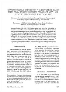
of three Phlebotomus species (P. sergenti, Phlebotomus papatasi (Scopoli), and Phlebotomus syriacus Adler & Theodore) and 50 mosquito females of two Culex species (Cx. pipiens and Cx. perexiguus) were analyzed during 2006 and 2007 (Table 1). Bloodmeal Condition. Sand ßies were screened for bloodmeal freshness and size (Table 2). The screening detected a higher proportion of large and medium bloodmeals (67%) than small (27%) and very small bloodmeals (6%). The majority of the large and medium bloodmeals were red in color, while the majority of the smaller bloodmeals were brown (Table 2). The size and color of the bloodmeal had a combined signiÞcant effect (F ⫽ 3.35; df ⫽ 3; P ⫽ 0.016) on PCR detectability. Detectability was not affected by the color of the medium and large bloodmeals. Detectability of small red bloodmeals was similar to that of larger bloodmeals but was reduced in small brown bloodmeals. Further decreases were noted in very small brown bloodmeals. The signiÞcance of the separate effects was F ⫽ 4.8; df ⫽ 1; and P ⫽ 0.029 for freshness and F ⫽ 6.08; df ⫽ 3; and P ⫽ 0.001 for size. IdentiÞcation of bloodmeals could not be accomplished in 26 samples because of failure of PCR ampliÞcation and/or low sequence quality, even after several ampliÞcation attempts (Fig. 1). Host Identification. Validation of the method was carried out on DNA extracted from blood samples of known vertebrate species (hyrax, dog, cat, sheep, partridge, and human), and laboratory mosquitoes fed on humans, quails, mice, and hamsters. The samples were ampliÞed and sequenced using both primer sets. DNA polymorphism was detected among all studied sam-
Table 2. Correlation between sand fly bloodmeal size and freshness and successful PCR analysis Bloodmeal characteristics Size
Color
Very small
Brown Red Total Brown Red Total Brown Red Total Brown Red Total
Small
Results Field Specimens. Using CDC light traps, blood-fed female sand ßies were collected from three different known CL foci: MaÕale Adumim (159), Kfar Adumim (70), and Tiberias (32). The blood-fed mosquitoes were collected from Arava (19), Tel-Aviv (18), and Jerusalem (13; Table 1). Overall, 261 sand ßy females
Medium Large Total
No. analyzed
No. analyzed successfully (%)
11 4 15 36 35 71 39 75 114 15 46 61 261
6 (55%) 3 (75%) 9 (60%) 26 (72%) 34 (97%) 60 (85%) 36 (92%) 73 (97%) 109 (96%) 15 (100%) 43 (93%) 58 (95%) 236 (90%)
May 2014
VALINSKY ET AL.: BLOODMEAL IDENTIFICATION OF SAND FLIES IN ISRAEL M 1
2
3
4
5
6
7
8
681
9 10 11 12 13 14 15
1000pb
A
600bp 500bp
1000bp
B
600bp 500bp
Fig. 1. PCR ampliÞcation of selected specimens using the 12S (A) and the 12Ð16S (B) primers.
ples for both gene regions (Supp Table 1 [online only]: A, 12S; B, 12Ð16S). The validity was further tested on 50 bloodmeals using the two primer sets. Species identiÞcation based on both primer sets was obtained in the majority of the samples (37, 74%). Ten bloodmeals (20%) were identiÞed only by primer pair 12S and two (4%) were identiÞed only by primer pair 12Ð16S. Inconsistent species identiÞcation using two primer sets was observed for only one sample. Successful identiÞcation of the host from bloodmeals was achieved in 285 (92%) of all samples (Table 3): comprising sand ßies (P. sergenti [221, 94%], P. papatasi [15, 75%], and P. syriacus [5, 71%]) and mosquitoes (Cx. perexiguus [15, 94%] and Cx. pipiens [29, 85%]). The identiÞed bloodmeals were of 22 domestic and wild vertebrate species, comprising 13 mammals and 9 birds. Similarity with ⬍2% sequence divergence was achieved for 12 mammalian (93%) and 4 (44%) avian hosts. Lower similarities (90 Ð96%) of high-quality sequences enabled identiÞcation to the genus level in the case of Gazella (Mammalia: Cervidae) and to the family level in the case of the Phasmidae (Aves: Table 3.
Host DNA identification in field-collected sand flies and mosquitoes determined by 12S–16S rRNA gene analysis
Host species
Total
Pr. capensis syriaca (hyrax) Felis silvestris catus (domestic cat) Gazella sp. (gazelle) A. chukar (partridge) Canis familiaris (domestic dog) Homo sapiens Phasianidae (two species)a Vulpes vulpes (red fox) Columbidae (pigeons and doves) Passer domesticus (sparrow) Ovis aries (domestic sheep) Equus caballus (domestic horse) Bos taurus (domestic cow) Sylvia atricapilla (blackcap) Acomys cahirinus (spiny mouse) Capra hircus (domestic goat) Gallus gallus (chicken) Hystrix indica (porcupine) Turdus philomelos (song thrush) Equus asinus (domestic donkey) Aves unidentiÞed (bird)b NDc Total
96 68 21 18 17 13 11 10 10 5 3 2 2 2 1 1 1 1 1 1 1 26 311
a b c
Galliformes). Among the Phasmidae, 18 samples belonging to one group were identical to the local partridge (Alectoris chukar), common in the study areas. The other 11 Phasmidae sequences, as yet, were not identiÞed to species level. An additional group of 10 similar avian samples with sequence divergence of 6 Ð 8% from GenBank sequences were identiÞed as pigeons (Aves: Columbiformes: Columbidae). One avian sequence with similarity of ⬍90% to the closest GenBank sequences was identiÞed to the class Aves. Species identiÞcation via DNA sequencing determined that there was almost always one host per bloodmeal. Only a few chromatograms had double peaks, interpreted as representing multiple host origin. Sequences of DNA mixtures prepared from two known vertebrate species returned inconsistent chromatogram results. Thus, we were not able to attribute multiple blood origin to chromatograms with double peaks. Twenty-one different host taxa were identiÞed in the bloodmeals extracted from P. sergenti sand ßy females collected in the three CL foci (Table 3). The
Cx. perexiguus
Cx. pipiens
P. papatasi
P. sergenti
P. syriacus
2
13 2
3 1 6
1 1
2
7 4
8 1
1 1
92 51 13 17 5 7 10 10 1 3 2 2 1
1 1
1 16
2 2 1
1 1
1
1
5 34
5 20
Sequences split into two distinct groups with 100% similarity within each group, not identiÞed to species. Similarity of ⬍90% to the closest GenBank sequences. PCR product or acceptable sequence could not be obtained.
1 1 1 1 1 1 1 13 234
2 7
682
JOURNAL OF MEDICAL ENTOMOLOGY
Vol. 51, no. 3
Table 4. The number of times and frequency (%) in which host species were identified in bloodmeals of P. sergenti collected at three different locations Location Kfar Adumim MaÕale Adumim Tiberia
Hyrax
Domestic Cat
70
33 47% 32 24% 27 84% 92 39%
3 4% 47 36% 1 3% 51 22%
132 32
Total
a
Total
234
Gazelle 9 13% 4 3% 13 6%
Human
Partridge
Pheasanta
Other birds
Other mammals
10 14%
6 5% 1 3% 7 3%
2 3% 13 10% 2 6% 17 7%
1 1% 6 5%
10 4%
7 3%
8 11% 15 11% 1 3% 24 10%
ND 4 6% 9 7% 13 6%
Sequences split into two distinct groups with 100% similarity within each group, not identiÞed to species.
majority (85%) were from 13 different mammalian hosts. Three species of ground-nesting and grounddwelling pheasants comprised 80% (27/34) of the bloodmeals of avian origin. Of the 221 identiÞed bloodmeals, rock hyrax DNA was found in 42% of the females, and domestic cat DNA in 23%. The proportion of these two most common hosts was different in the three study sites (Table 4), reßecting host availability. Hyrax DNA was found in 84% of the females collected in Tiberias, in 47% of the females from Kfar Adumim, and in only 24% of the females from MaÕale Adumim. About one-third (36%) of the females from MaÕale Adumim contained DNA of domestic cat, while only few bloodmeals from domestic cats were detected in sand ßies collected in Tiberias and in Kfar Adumim. Six host species were identiÞed in 15 bloodmeals from P. papatasi and Þve host species in 5 bloodmeals from P. syriacus. The blood of rock hyrax, domestic cat, and domestic dog was found in both species. Gazelle DNA was identiÞed in six P. papatasi females (Table 3). The majority of the identiÞed Cx. pipiens bloodmeals were from mammals (26 of 29, 90%), while a high proportion of identiÞed Cx. perexiguus bloodmeals were of avian origin (10 of 15, 67%; Table 3). Human DNA was identiÞed in 13 bloodmeals sampled from Cx. pipiens (4 of 29, 11%), P. papatasi (2 of 15, 13%), and P. sergenti (7 of 221, 3%). Leishmania Detection. All 261 sand ßy samples were screened for Leishmania DNA by PCR using a M 1 2 3 4
5
previously published primer set (El Tai et al. 2000). Leishmania DNA was found in three P. sergenti sand ßy females (3 of 27 [11%]) from Tiberias that contained hyrax blood, and it was not detected in any of the females fed on other hosts or in the other sand ßy species (Fig. 2). Discussion The current study focused on the blood feeding behavior of P. sergentiÑthe vector of L. tropica in Israel (Jacobson et al. 2003, Schnur et al. 2004, Svobodova´ et al. 2006). The study also includes the results of bloodmeal identiÞcation from the two other Phlebotomus species (P. papatasi and P. syriacus) caught in the same traps with P. sergenti (Table 1). Bloodmeal analysis from Cx. pipiens and Cx. perexiguus mosquito females provides initial data on the feeding habits of the two important WNV vectors in Israel (Orshan et al. 2008). Successful identiÞcation of bloodmeal source was obtained in 92% (285 of 311) of the engorged females tested (Table 3), using two primer sets for the mitochondrial rRNA genes, designed to amplify vertebrate species but not arthropod DNA. The majority of the hosts were mammalian species, both wild and domestic. In addition, several avian species were identiÞed as hosts, with partridge (A. chukar) as the main species. The common species found as hostsÑrock hyrax, domestic cat, and partridgeÑ correlate with the fre-
6 7 8 9 10 11 12 13 M 14 15 16 17 18 19 20 21 22 23 24 25 26 M
300bp
Fig. 2. PCR ampliÞcation of Leishmania DNA from engorged P. sergenti females collected in Tiberias.
May 2014
VALINSKY ET AL.: BLOODMEAL IDENTIFICATION OF SAND FLIES IN ISRAEL
quency of these animals in the vicinity of the villages (L. O., unpublished data). The successful host identiÞcation and the process described demonstrate the versatility and efÞcacy of the sequencing method for identiÞcation of hosts from bloodmeals, especially in diverse environments when the knowledge of potential hosts is partial or lacking. The exponentially growing number of sequences deposited in public databases will continue to facilitate the direct identiÞcation of unknown hosts to the species level. The results of this study further demonstrate the possibility of conducting retrospective, targeted searches to identify hosts to the species level. Overall, the detection sensitivity of bloodmeal hosts in Þeld-collected sand ßies in this study was high (92%) compared with other published studies (Haouas et al. 2007, SantÕAnna et al. 2008, Abbasi et al. 2009, Svobodova´ et al. 2009, Garlapati et al. 2012). We believe that several factors contributed jointly to the high detection levels observed in this study: high copy number of the target mitochondrial 12SÐ16S rRNA gene, direct transfer of live sand ßies to ⫺20⬚C, fast desiccation of sand ßies in these conditions, and the use of two primer sets. The direct sequencing approach cannot identify bloodmeals from multiple hosts. Determining the host origin of multiple bloodmeals requires cloning of the PCR fragments and sequence analysis of many clones. Based on laboratory studies (Haouas et al. 2007), red color bloodmeals indicated a fresh meal taken during the night. Females caught in the trap containing brown meals indicated older meals that had gone through a level of digestion. Red (fresh) and brown (older) meals were found in female sand ßies in all four size categories. The percentage of red blood among very small and small meals probably points to disrupted feeding in those individuals, a phenomenon of great signiÞcance regarding pathogen transmission (Davies 1990). The size and color of sand ßy bloodmeals had a combined effect on PCR detectability (Table 2). The high (⬇95%) detection sensitivities of large and medium bloodmeals regardless of the freshness of the blood indicate enough DNA template even in older meals, while DNA degradation in brown small and very small bloodmeals is probably the reason for the reduced detection success (72 and 55%, respectively; Svobodova´ et al. 2003, Haouas et al. 2007, SantÕAnna et al. 2008, Abbasi et al. 2009). The observed lower detection sensitivity in mosquitoes (88%) in comparison with sand ßies is surprising given the smaller volume of sand ßy bloodmeals (0.2Ð 0.3 l in Burniston et al. (2010) and 0.5Ð1 l in Daba et al. (2004)) in comparison with that of mosquitoes (2Ð 6 l; Clements 1992). Nonetheless, the sample size of the mosquitoes tested was too small to distinguish between a signiÞcant or random result. All Þve Diptera species examined in the current study fed from various hosts (Table 3) and can be regarded as nonselective opportunistic blood feeders on mammals and birds. The high variability of hosts, the different feeding rates on the same host in the different collection sites, and the high proportions of avian blood from ground-dwelling birds (Tables 3 and
683
4) indicate a nonselective blood feeding behavior of P. sergenti. Opportunistic feeding of P. sergenti has been reported from Syria (Maroli et al. 2009). In the endemic area of CL in Sanliurfa, Turkey, a high proportion of chicken blood was found in P. sergenti females that were abundant in cellars where poultry was kept (Svobodova´ et al. 2003). The results of this study provide preliminary information related to opportunistic feeding behavior of P. papatasi and P. syriacus. In an earlier study conducted in Israel (Abbasi et al. 2009), almost all of the P. papatasi females collected inside houses contained human blood, while the majority of females that were collected near a small farm contained blood of farm animals. An opportunistic feeding habit has also been observed in females of P. neglectusÑa species closely related to P. syriacus (Velo et al. 2005). Human genes were detected in 3 and 14% of the identiÞed P. sergenti and P. papatasi bloodmeals, respectively. Similar results were reported for the two species in the endemic area of CL in Sanliurfa, Turkey (Svobodova´ et al. 2003). Larger proportions of bloodmeals of human origin were found in Allepo, Syria. The highly anthropophilic blood-feeding behavior of P. papatasi, the vector of Leishmania major in indoor environments, was reported from Israel (Abbasi et al. 2009) and from various other locations (Pandya 1985, el Sawaf et al. 1989, Srinivasan & Panicker 1992, Yaghoobi-Ershadi et al. 1995, Palit et al. 2005, Burniston et al. 2010). Thus, in Israel, high transmission risk of CL as a result of L. major by the highly endophilic anthropophagic vector P. papatasi exists even in situations of low sand ßy densities and low Leishmania infection rates in the zoonotic cycle. This is in contrast to CL as a result of L. tropica. Exposure of people to the exophilic exophagic vector P. sergenti is less frequent, and transmission of Leishmania parasites to people requires high sand ßy densities and high Leishmania infection rates in the zoonotic cycle. All of the sand ßy females were also screened for Leishmania DNA. Three P. sergenti females from Tiberias, which contained hyrax blood, were positive for L. tropica (11%). In addition, L. tropica infection rates in pools of unfed sand ßy females were in correlation with the proportion of rock hyrax bloodmeals from the same location: highest in Tiberias and lowest in MaÕale Adumim (L. O., unpublished data). The vectorÐreservoir contact results strengthen the evidence (TalmiFrank et al. 2010) pointing to the rock hyrax as the relevant reservoir animal of L. tropica in the Judean Desert and Tiberias and probably in other leishmaniasis foci because of L. tropica in Israel. Avian hosts were detected in 67% of the bloodmeals analyzed from Cx. perexiguus in comparison with only 7% of the bloodmeals from Cx. pipiens. Human genes were present in 14% of Cx. pipiens bloodmeals, but they were not found in Cx. perexiguus. Because birds are considered the main reservoir of WNV (Kramer et al. 2008), these initial results indicate Cx. perexiguus as the main vector of the sylvatic transmission cycle of WNV in Israel. Cx. pipiens probably plays a major role in the transmission of the virus to people. A larger
684
JOURNAL OF MEDICAL ENTOMOLOGY
sample of blood-fed females of the two species from many locations and collection dates is needed to further elucidate the vectorial role of the two species in Israel and to calculate WNV transmission risks to people. Mosquitoes and sand ßies of public health importance are monitored in Israel. The data collected are used to plan and evaluate intervention activities and to study the effects of climate, demographic, and landuse changes on pathogen transmission. Adding bloodmeal analyses and host identiÞcations to the variety of tests performed in Israel today contributes valuable data to vector-borne pathogen transmission cycles and risks in Israel. CoidentiÞcation of species, pathogens, and hosts of engorged vector females by metagenomics together with reduction in costs and increase in open-access sequences will enable the analysis of large samples and establish comprehensive databases. Evidence-based information enables better understanding of the eco-epidemiology of vector-borne diseases and assists in reducing transmission risks to people. Acknowledgments We thank Abed Naseraddin and Prof. Charles Jaffe for their help in establishing the molecular method of Leishmania detection and providing Leishmania cultures. Noteworthy is the special contribution of Prof. Avi Israeli to the establishment of the Anti-Leishmaniasis program in 2005 in Israel and his continued interest and support. We acknowledge the involvement and encouragement of Ruth Yishai, and Emanuel Gazit. We would like to warmly thank Fouad Akad for his assistance and Heather Schnur for her help.
References Cited Abbasi, I., R. Cunio, and A. Warburg. 2009. IdentiÞcation of blood meals imbibed by Phlebotomine sand ßies using cytochrome b PCR and reverse line blotting. Vector Borne Zoonotic Dis. 9: 79 Ð 86. DOI: 10.1089/vbz.2008.0064. Burniston, I., L. Roy, A. Picado, M. Das, S. Rijal, M. Rogers, M. Coosemans, M. Boelaert, C. Davies, and M. Cameron. 2010. Development of an enzyme-linked immunosorbent assay to identify host-feeding preferences of Phlebotomus species (Diptera: Psychodidae) in endemic foci of visceral leishmaniasis in Nepal. J. Med. Entomol. 47: 902Ð906. Clements, A. 1992. The biology of mosquitoes, vol 1: Development Structure and Reproduction. Chapman & Hall, London, United Kingdom. Daba, S., A. Daba, M. G. Shehata, and B. M. el-Sawaf. 2004. A simple micro-assay method for estimating blood meal size of the sand ßy, Phlebotomus langeroni (Diptera: Psychodidae). J. Egypt. Soc. Parasitol. 34: 173Ð182. Davies, C. R. 1990. Interrupted feeding of blood-sucking insects: causes and effects. Parasitol. Today 6: 19 Ð22. El Tai N. O., O. F. Osman, M. El Fari, W. EI Presber, and G. Scho¨ nian. 2000. Genetic heterogeneity of ribosomal internal transcribed spacer in clinical samples of Leishmania donovani spotted on Þlter paper as revealed by singlestrand conformation polymorphisms and sequencing. Trans. R. Soc. Trop. Med. Hyg. 94: 575Ð579. Garlapati, R. B., I. Abbasi, A. Warburg, D. Poche´, and R. Poche´. 2012. IdentiÞcation of bloodmeals in wild caught blood fed Phlebotomus argentipes (Diptera: Psychodidae)
Vol. 51, no. 3
using Cytochrome b PCR and reverse line blotting in Bihar, India. J. Med. Entomol. 49: 515Ð521. (doi: http:// dx.doi.org/10.1603/ME11115). Haouas, N., B. Pesson, R. Boudabous, J.-P. Dedet, H. Babba, and C. Ravel. 2007. Development of a molecular tool for the identiÞcation of Leishmania reservoir hosts by blood meal analysis in the Insect Vector. Am. J. Trop. Med. Hyg. 77: 1054 Ð1059. Jacobson, R. L. 2011. Leishmaniasis in an era of conßict in the Middle East. Vector-Borne Zoonotic Dis. 11: 247Ð258. doi:10.1089/vbz.2010.0068. Jacobson, R. L., C. L. Eisenberger, M. Svobodova´ , G. Baneth, J. Sztern, J. Cavalho, A. Nasereddin, M. El Fari, U. Shalom, P. Volf, et al. 2003. Outbreak of cutaneous leishmaniasis in Northern Israel. J. Infect. Dis. 188: 1065Ð1073. Jaffe, C. L., G. Baneth, Z. A. Abdeen, Y. Schlein, and A. Warburg. 2004. Leishmaniasis in Israel and the Palestinian Authority. Trends Parasitol. 20: 328 Ð332. Kent, R. J. 2009. Molecular methods for arthropod bloodmeal identiÞcation and applications to ecological and vector-borne disease studies. Mol. Ecol. Resour. 9: 4 Ð18. doi: http://dx.doi.org/10.1111/j.1755Ð 0998.2008.02469.x. Lewis, D. J. 1982. A taxonomic review of the genus Phlebotomus (Diptera: Psychodidae). Bull. Br. Mus. Nat. Hist. (Ent). 45: 121Ð209. Lewis, D., and W. Buttiker. 1982. Insects of Saudi Arabia: the taxonomy and distribution of Saudi Arabian Phlebotomus sandßies (Diptera: Psychodidae). Fauna Saudi Arabia 4: 353Ð383. Kramer, L. D., L. M. Styer, and G. D. Ebel. 2008. A global perspective on the epidemiology of West Nile virus. Annu. Rev. Entomol. 53: 61Ð81. (doi:10.1146/annurev.ento.53. 103106.093258). Maroli, M., L. Jalouk, M. Al Ahmed, R. Bianchi, G. Bongiorno, C. Khoury, and L. Gradoni. 2009. Aspects of the bionomics of Phlebotomus sergenti sandßies from an endemic area of anthroponotic cutaneous leishmaniasis in Aleppo Governorate, Syria. Med. Vet. Entomol. 23: 148 Ð 154. DOI: 10.1111/j.1365Ð2915.2009.00808.x. Murphy, W. J., E. Eizirik, W. E. Johnson, Y. P. Zhang, O. A. Ryder, and S. J. O’Brien. 2001. Molecular phylogenetics and the origins of placental mammals. Nature. 409: 614 Ð 618. Orshan, L., H. Bin, H. Schnur, A. Kaufman, A. Valinsky, L. Shulman, L. Weiss, E. Mendelson, and H. Pener. 2008. Mosquito vectors of West Nile fever in Israel. J. Med. Entomol. 45: 939 Ð947. Orshan, L., D. Szekely, Z. Khalfa, and S. Bitton. 2010. Distribution and seasonality of phlebotomus sand ßies in cutaneous leishmaniasis foci, Judean Desert, Israel. J. Med. Entomol. 47: 319 Ð328. Palit, A., S. K. Bhattacharyaa, and S. N. Kundu. 2005. Host preference of Phlebotomus argentipes and Phlebotomus papatasi in different biotopes of West Bengal, India. Int. J. Environ. Health Res. 15: 449 Ð 454. (doi:10.1080/ 09603120500392525). Pandya, A. P. 1985. Bloodmeals of phlebotomine sandßies of Surat district (Gujarat state) India. Indian J. Med. Res. 81: 46 Ð 48. Roca, A. L., G. Kahila Bar-Gal, E. Eizirik, K. M. Helgen, R. Maria, M. S. Springer, S. J. O’Brien, and W. J. Murphy. 2004. Mesozoic origin for West Indian insectivores. Nature 429: 649 Ð 651. Sant’Anna, M.R.V., N. G. Jones, J. A. Hindley, A. F. MendesSousa, R. J. Dillon, R. R. Cavalcante, B. Alexander, and P. A. Bates. 2008. Blood meal identiÞcation and parasite detection in laboratory-fed and Þeld-captured Lutzomyia longipalpis by PCR using FTA databasing paper. Acta Trop.
May 2014
VALINSKY ET AL.: BLOODMEAL IDENTIFICATION OF SAND FLIES IN ISRAEL
107: 230Ð237. (http://dx.doi.org/10.1016/j.actatropica.2008. 06.003). el Sawaf, B. M., N. S. Mansour, S. M. el Said, S. Daba, F. G. Youssef, M. A. Kenawy, and J. C. Beier. 1989. Feeding Patterns of Phlebotomus papatasi and Phlebotomus langeroni (Diptera: Psychodidae) in El Agamy, Egypt. J. Med. Entomol. 26: 497Ð 498. Schnur, L. F., A. Nasereddin, C. L. Eisenberger, C. L. Jaffe, M. El Fari, K. Azmi, G. Anders, M. Killick-Kendrick, R. Killick-Kendrick, J.-P. Dedet, et al. 2004. Multifarious characterizations of Leishmania tropica from a Judean Desert focus, exposing intraspeciÞc diversity and incriminating Phlebotomus sergenti as its vector. Am. J. Trop. Med. Hyg. 70: 364 Ð372. Singer, S. R., N. Abramson, H. Shoob, O. Zaken, G. Zentner, and C. Stein-Zamir. 2008. Ecoepidemiology of cutaneous leishmaniasis outbreak, Israel. Emerg. Infect. Dis. 14: 1424 Ð1426. Srivinasan, R., and K. N. Panicker. 1992. IdentiÞcation of bloodmeals of phlebotomine sandßies using the agarose gel diffusion method. The Southeast Asian Journal of Tropical Medicine and Public Health 23(3): 486 Ð 488. Svobodova´ , M., J. Sadlova, K. P. Chang, and P. Volf. 2003. Distribution and feeding preference of the sandßies Phlebotomus sergenti and P. papatasi in a cutaneous leishmaniasis focus in Sanliurfa, Turkey. Am. J. Trop. Med. Hyg. 68: 6 Ð9. Svobodova´ , M., J. Voty´pka, J. Peckova, V. Dvorˇ a´ k, A. Nasereddin, G. Baneth, J. Sztern, V. Kravchenko, A. Orr, D.
685
Meir, L. F. Schnur, P. Volf, and A. Warburg. 2006. Distinct transmission cycles of Leishmania tropica in 2 adjacent foci, Northern Israel. Emerg. Infect. Dis. 12: 1860 Ð 1868. Svobodova´ , M., B. Alten, L. Zı´dkova´ , V. Dvorˇ a´ k, J. Hlavacˇ kova´ , J. Mysˇkova´ , V. Sˇ eblova´ , O. E. Kasap, A. Belen, J. Voty´pka, et al. 2009. Cutaneous leishmaniasis caused by Leishmania infantum transmitted by Phlebotomus tobbi. Int. J. Parasitol. 39 251Ð256. (http://dx.doi.org/ 10.1016/j.ijpara.2008.06.016). Talmi-Frank, D., C. L. Jaffe, A. Nasereddin, A. Warburg, R. King, M. Svobodova´ , O. Peleg, and G. Baneth. 2010. Leishmania tropica in rock hyraxes (Procavia capensis) in a focus of human cutaneous leishmaniasis. Am. J. Trop. Med. Hyg. 82: 814 Ð 818. (doi:10.4269/ajtmh.2010.090513). Velo, E., A. Paparisto, G. Bongiorno, T. Di Muccio, C. Khoury, S. Bino, M. Gramiccia, L. Gradoni, and M. Maroli. 2005. Entomological and parasitological study on phlebotomine sandßies in central and northern Albania. Parasite 12: 45Ð 49. Yaghoobi-Ershadi, M. R., E. Javadian, and A. Kannani. 1995. Host preference pattern of phlebotomine sandßies of Borkhar rural district, Isfahan province, Iran. Acta Trop. 60: 155Ð 158. (http://dx.doi.org/10.1016/0001-706X(95)00122-U). Received 27 June 2013; accepted 12 March 2014.