Sep 6, 1988 - proteoglycan could be due to differences in core protein size and/or glycosaminoglycan chain number and size.To inves-. UTERUS VAGINA.
Proc. Natl. Acad. Sci. USA Vol. 85, pp. 9562-9566, December 1988 Cell Biology
Molecular polymorphism of a cell surface proteoglycan: Distinct structures on simple and stratified epithelia (cell adhesion/heparan sulfate/chondroitin sulfate/syndecan)
RALPH D. SANDERSON AND MERTON BERNFIELD* Department of Pediatrics, Stanford University School of Medicine, Stanford, CA 94305
Communicated by Elizabeth D. Hay, September 6, 1988 (received for review July 26, 1988)
ABSTRACT Epithelial cells are organized into either a single layer (simple epithelia) or multiple layers (stratified epithelia). Maintenance of these cellular organizations requires distinct adhesive mechanisms involving many cell surface molecules. One such molecule is a cell surface proteoglycan, named syndecan, that contains both heparan sulfate and chondroitin sulfate chains. This proteoglycan binds cells to fibrillar collagens and fibronectin and thus acts as a receptor for interstitial matrix. The proteoglycan is restricted to the basolateral surface of simple epithelial cells, but is located over the entire surface of stratified epithelial cells, even those surfaces not contacting matrix. We now show that the distinct localization in simple and stratified epithelia correlates with a distinct proteoglycan structure. The proteoglycan from simple epithelia (modal molecular size, 160 kDa) is larger than that from stratified epithelia (modal molecular size, 92 kDa), but their core proteins are identical in size and immunoreactivity. The proteoglycan from simple epithelia has more and larger heparan sulfate and chondroitin sulfate chains than the proteoglycan from stratified epithelia. Thus, the cell surface proteoglycan shows a tissue-specific structural polymorphism due to distinct posttranslational modifications. This polymorphism likely reflects distinct proteoglycan functions in simple and stratified epithelia, potentially meeting the different adhesive requirements of the cells in these different organizations.
and is expressed almost exclusively on epithelia; it is located over the entire surface of stratified epithelial cells, but is restricted to the basolateral surface of simple epithelial cells (12). Restriction of the proteoglycan to the basolateral cell surface on simple epithelia is consistent with its behavior as a matrix receptor; its localization over the entire cell surface on stratified epithelia is not. This difference raises the possibility that the nature of the proteoglycan in these epithelial types could differ. Therefore, we compared the structure of the cell surface proteoglycan extracted from simple and stratified epithelia, from both tissues and cells in culture, to assess whether its distinct cellular localization correlates with a distinct molecular structure. Part of these data has been published in abstract form (13).
MATERIALS AND METHODS Cell and Organ Culture. Normal murine mammary gland epithelial cells (passages 13-22) were maintained as described (14). Keratinocytes from newborn mouse skin were isolated and cultured by the method of Hennings et al. (15). For experiments requiring radioactive labeling, cells were labeled to the steady state in low sulfate medium (14) containing H235S04 at 100 /Ci/ml (1 Ci = 37 GBq) for 24 hr (mammary cells) or 48 hr (keratinocytes). For organ cultures, uterus and vagina were removed from 6-month-old female Swiss Webster mice, cut into 4-mm square cubes, placed in low-sulfate medium containing H235S04 for 4 hr at 37°C, and then extracted as described below. Harvest of Cell Cultures. Cells were washed three times in ice-cold 10 mM Tris-buffered (pH 7.4) saline then extracted on the dish with ice-cold 8 M urea containing 1% Triton X-100, 10 mM Tris HCl (pH 7.4), 5 mM benzamidine, 5 mM N-ethylmaleimide, 1 mM phenylmethylsulfonyl fluoride, 0.5 mM EDTA, pepstatin A at 5 ,g/ml, and 0.15 M NaCI (buffer A). Dishes were scraped and the extracts were Vortex mixed periodically during a 4-hr incubation on ice. The extracts were centrifuged at 30,000 x g for 30 min at 4°C and the supernatants were boiled for 15 min. Tissue Harvest and Extraction. Six-month-old female Swiss Webster mice were sacrificed, and the uterus, vagina, stomach, and duodenum were rapidly removed and immediately submerged in liquid N2 until use. Frozen tissues were pulverized with a mortar and pestle and extracted as described above for cells in culture.
Epithelial tissues consist of the cells that line the free surfaces of the body. These cells are organized into either of two arrangements: cells in a single layer (simple epithelia) or cells in two or more layers (stratified epithelia). These arrangements reflect the usual functions of these cells, namely transfer of materials across simple epithelia and protection by stratified epithelia. Both of these functions require that the cells adhere to one another and to their underlying extracellular matrix. Several cell surface molecules involved in cell-cell and cell-matrix adhesion on epithelia have been described (see refs. 1-5). One of these, a cell surface proteoglycan, behaves as a transmembrane protein that bears both heparan sulfate and chondroitin sulfate chains on its extracellular domain (6, 7). On cultured mammary epithelial cells, a simple epithelium, this proteoglycan is a receptor specific for the interstitial matrix. The proteoglycan (i) binds fibrillar collagens (types I, III, and V) and fibronectin, but not the basal lamina components type IV collagen or laminin (8, 9); (ii) is shed by cleavage of its ectodomain when the cells change shape (10); (iii) associates with the actin-containing cytoskeleton when cross-linked at the cell surface; and (iv) polarizes to the basolateral cell surface during cell culture (11), properties that suggest that it could stabilize epithelial sheets. In mature murine tissues, the proteoglycan is not present in the stroma
RESULTS The Cell Surface Proteoglycans on Simple and Stratified Epithelia Differ in Molecular Size. To compare the proteoglycans in simple and stratified epithelia, contiguous tissues
The publication costs of this article were defrayed in part by page charge payment. This article must therefore be hereby marked "advertisement" in accordance with 18 U.S.C. §1734 solely to indicate this fact.
*To whom reprint requests should be addressed.
9562
Cell
Biology: Sanderson and Bernfield
that differ in the organization of their epithelial cells were dissected, and any transitional region between the simple and stratified epithelia was mechanically removed. Proteoglycans were extracted from the uterus (simple epithelium) and vagina (stratified epithelium), discarding the median corpus, and from the duodenum (simple) and the cutaneous portion of the stomach (stratified), discarding the glandular gastric fundus. Peyer's patches were removed from the duodenum. Additionally, proteoglycans were extracted from cultured cells: low passage normal murine mammary gland epithelial cells, derived from a simple epithelium (16), and primary neonatal mouse keratinocytes, derived from a stratified epithelium. The mammary cells grow as a monolayer and, upon maintenance at confluence, exhibit cell surface proteoglycan solely on their basolateral surfaces (11), a localization
kDa 200- p
92.5-
46-
30-
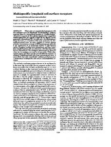
FIG. 1. Size of the cell surface proteoglycan from various tissues
and cells. Western blot of proteoglycans is as follows: uterus (lane UT), vagina (lane VG), duodenum (lane DU), gastric cutaneous (lane GA), mammary cells (lane MA), and keratinocytes (lane KC). Proteoglycans were purified by DEAE-cellulose chromatography and by immunoisolation using monoclonal antibody 281-2 (17) linked to Sepharose 4B beads (281-2 beads). Antibody 281-2 binds to the proteoglycan by way of an epitope on the core protein. Tissues and cells were extracted and then diluted 1:10 in a solution of 8 M urea, 50 mM sodium acetate (pH 4.5), and 0.15 M NaCl and applied to 5 ml of DEAE-Sephacel. Proteoglycans were eluted with 8 M urea and 50 mM sodium acetate (pH 4.5) containing 1 M NaCl and 0.1% Triton X-100. Pooled fractions were then diluted with Tris-buffered saline to a final concentration of 1 M urea and 0.15 M NaCl, adjusted to pH 7.4, and mixed with 281-2-linked beads. After incubation on a rocker overnight at 40C, beads containing bound proteoglycan were placed in a column and washed with 20 mM acetic acid (pH 3.1) to remove nonspecifically bound molecules, and proteoglycans were eluted with a solution of 50 mM triethylamine (pH 11.5) containing 0.1% Triton X-100, immediately neutralized with a 0.1 vol of 1 M Tris-HCl (pH 7.4), and quantitated by using 1251-labeled monoclonal antibody 281-2 in an immunodot assay as described (10). Proteoglycans were fractionated on a 3.8-20%o NaDodSO4/polyacrylamide [7.5% (wt/ wt) N,N'-methylene bisacrylamide] gel containing urea and boric acid, as described by Koda et al. (8), and transferred by the method of Towbin et al. (18) to Gene-Trans (Plasco, Woburn, MA), a cationic nylon membrane. After transfer, the membrane was fixed in 0.025% glutaraldehyde, washed, and probed with 1251-labeled monoclonal antibody 281-2. Forty nanograms of proteoglycan was applied to each lane except the duodenum where 80 ng was loaded because of the extensive heterogeniety in size. Experiments were carried out using a variety ofextraction buffers (Triton X-100 without urea, octyl glucoside, 4 M guanidine hydrochloride) and tissue harvest proce-
dures (homogenization of whole tissues, scraping, and extraction of eplthelium only). Despite these variations in procedures, the size difference in proteoglycans from simple and stratified epithelia remained the same (data riot included).
Proc. Natl. Acad. Sci. USA 85 (1988)
9563
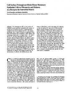
identical to that seen on simple epithelia in vivo. The keratinocytes can be induced to stratify in culture, and the proteoglycan on these cells is localized over the entire cell surface, in a pattern identical to that seen on stratified epithelia in vivo (unpublished data). Western blots of purified proteoglycans from both tissues and cultured cells show that the cell surface proteoglycans from simple epithelia are distinct in molecular size from the proteoglycans in stratified epithelia (Fig. 1). Proteoglycans of =92 kDa are in each source of stratified epithelia examined (vagina, gastric cutaneous, and keratinocyte) and proteoglycans of =160 kDa are in each source of simple epithelia (uterus, duodenum, and mammary cells). The duodenum also contains proteoglycans of a smaller size. Identical results were obtained when Western blots were probed with an affinity-purified serum antibody specific for the cell surface proteoglycan core protein (data not shown). The Proteoglycans from Epithelial Tissues Contain both Heparan Sulfate and Chondroitin Sulfate. The modal size of 35SO4-labeled proteoglycans from the uterus (=160 kDa) was larger than that from the vagina (=90 kDa) (Fig. 2), as was observed by Western blotting (cf., Fig. 1). Moreover, the size of the proteoglycans was reduced by removal of either heparan sulfate or chondroitin sulfate, indicating that at least a portion of the molecules contain both types of glycosaminoglycan chains. In the proteoglycan from vagina, the small shift in modal size after removal of chondroitin sulfate suggests that either a relatively small amount of chondroitin sulfate is present or a proportion of the molecules contain only heparan sulfate (see below). However, because Western blots indicate that no core protein bands are generated by removal of only chondroitin sulfate from either uterine or vaginal proteoglycans, the proteoglycans all contain heparan sulfate (data not shown). The Distinct Size Classes of Proteoglycans Contain Core Proteins of Identical Molecular Size and Immunoreactivity. The tissue-specific differences in size of the cell surface proteoglycan could be due to differences in core protein size and/or glycosaminoglycan chain number and size. To invesUTERUS VAGINA a
b c
a b c
kDa
200-
92.5
694630
FIG. 2. Hybrid nature of the cell surface proteoglycan extracted from tissues. Autoradiogram of radiosulfate-labeled proteoglycan from uterus and vagina (lanes a) and after removal of glycosaminoglycan chains with chondroitinase ABC (lanes b) or with heparitinase (lanes c). Removal of glycosaminoglycans was as described (7) except digestions were carried out on proteoglycan bound to 2812-linked beads. After digestion and washing of beads, remaining proteoglycan was eluted from the beads with PAGE sample buffer containing 2% (wt/vol) NaDodSO4, 5% (vol/vol) glycerol, 0.025% bromophenol blue, 140 mM Tris, and 60 mM boric acid (pH 8.0). Samples were fractionated by PAGE as described in Fig. 1.
9564
Cell Biology: Sanderson and Bernfield
Proc. Natl. Acad. Sci. USA 85
tigate core protein size, the proteoglycans extracted and purified as described above were treated to remove glycosaminoglycan chains and analyzed by Western blots. Regardless of the tissue (or cell) source, the core proteins migrate as bands at 69 kDa (Fig. 3), suggesting that the difference in proteoglycan size is due to the number and/or size of glycosaminoglycan chains on the core protein. The duodenum shows two size classes of proteoglycans and one major and two minor core protein bands. These distributions are attributable to proteoglycan from two types of cells in the duodenum that react with monoclonal antibody 281-2; the simple epithelial cells lining the lumen and the plasma cells in the lamina propria and any remaining Peyer's patches. A small (92 kDa) immunoreactive proteoglycan containing a predominant core protein band of 69 kDa and a minor core protein band of 76 kDa is known to be present in cells of the B-cell lineage (unpublished data). This suggests that the smaller proteoglycan in this tissue is derived from the plasma cells, and the larger proteoglycan is apparently derived from the duodenal epithelial cells. The smallest core protein band (50 kDa) is probably due to adventitious degradation because this band increases in relative intensity when the duodenum is extracted at room temperature (unpublished data). The Distinct Size Classes of Proteoglycans Contain Glycosaminoglycan Chains of Different Sizes. The heparan sulfate chains on the proteoglycan from the uterus (simple epithelium) are larger than the heparan sulfate on the vagina (stratified epithelium) as demonstrated by Sepharose CL-6B chromatography (Fig. 4). The size of chondroitin sulfate chains are also larger in uterus than in vagina. An analogous, but not directly comparable difference is seen in the proteoglycans from cultured cells. The heparan sulfate chains from the mammary cells (derived from simple epithelium) are larger than those from the keratinocytes (derived from stratified epithelium) and the size of chondroitin sulfate chains are also larger on the proteoglycan from mammary cells than the keratinocytes. The Distinct Size Classes of Proteoglycans Contain Different Proportions of Heparan Sulfate and Chondroitin Sulfate. The percent of heparan sulfate and chondroitin sulfate on 35S04labeled proteoglycans was determined by degrading these A
B UT
DU
VG +
-
+
MA
GA -
+
-+
KC -
+
kDa
200
Ia 92.5--.
69--do
a *
4630-
FIG. 3. Size of the core protein of the cell surface proteoglycan from various cells and tissues. Western blots of proteoglycans from tissues (A) and cells (B) before (-) and after (+) enzymatic removal of glycosaminoglycan chains. Proteoglycans bound to 281-2-linked beads were digested with both heparitinase and chondroitinase ABC (7), eluted from the beads, fractionated by PAGE, and Western blotted as in Fig. 1. Abbreviations are as in Fig. 1.
50 25
kDa 15
(1988)
5
10 A. -
8. c
HS chains
--- CS chains
6 4
*
Uterus
~
Vagina
0 0
Pe*
° 2I 0~ a.
ay ,-o
o 0
ct) 10 11
cV)
C
8
0
0
a
6.
Kav
FIG. 4. Size of the glycosaminoglycan chains of the cell surface proteoglycans. Chromatograms of 350S4-labeled glycosaminoglycans from proteoglycans that were isolated from tissues (A), uterus (o) and vagina (e), or cells (B), mammary (o) and keratinocyte (0). HS, heparan sulfate; CS, chondroitin sulfate. Radiosulfate-labeled proteoglycan bound to 281-2-linked beads was treated with nitrous acid or chondroitinase ABC (7), and digested glycosaminoglycan was washed from the beads and collected. Remaining glycosaminoglycan on the proteoglycan-containing beads was released with 0.1 M sodium hydroxide in 1 M potassium borohydride for 24 hr at 37°C. Glycosaminoglycan was fractionated on Sepharose CL-6B in a buffer of 2% (wt/vol) NaDodSO4, 0.15 M NaCI, 50 mM sodium acetate (pH 5.0), and 0.02% sodium azide. Size of the glycosaminoglycan chains was determined by comparison with a molecular size calibration curve generated with chondroitin sulfate standards (19).
chains selectively with nitrous acid or chondroitinase ABC (Table 1). The extent of sulfation of the heparan sulfate and chondroitin sulfate chains is equivalent in the proteoglycan from normal murine mammary gland cells (20). If the extent of sulfation of the two types of glycosaminoglycans is equivalent in the proteoglycans from tissues, the proportion of chondroitin sulfate is lower in the proteoglycans from vagina (stratified epithelium) than in the proteoglycans from uterus (simple epithelium). Thus the smaller proteoglycan from the stratified epithelium contains a higher proportion of heparan sulfate. Again, an analogous result is seen from proteoglycans of cultured cells.
DISCUSSION The cell surface proteoglycan in adult mouse tissues is localized differently on simple and stratified epithelial cell surfaces (12). Our current results indicate that the proteoglycan in mature tissues contains both heparan sulfate and chondroitin sulfate and exists in two distinct size classes: a 160-kDa proteoglycan in simple epithelia and a 92-kDa proteoglycan in stratified epithelia. The core proteins of these proteoglycans are identical in size and immunoreactivity, but they differ in the number and size of their glycosaminoglycan chains and in their proportion of heparan sulfate and chondroitin sulfate. The large proteoglycan is located at the basolateral surfaces of simple epithelial tissues and is richer
Cell
Biology: Sanderson and Bernfield
Proc. Natl. Acad. Sci. USA 85 (1988)
9565
Table 1. Relative amount and size of heparan and chondroitin sulfate glycosaminoglycan chains from various proteoglycans Heparan sulfate Chondroitin sulfate Chain mass, Chain mass, kDa % of total Chain(s), n % of total kDa Chain(s), n Epithelial tissues 21 2 57 21 2 Uterus 43 Vagina 71 16 1 29 11 1 Cultured cells 74 39 2 17 2 Mammary 26 7 92 21 1 8 1 Keratinocyte Size of glycosaminoglycan chains was determined by the modal Kay of the chains on Sepharose CL-6B columns (Fig. 4). Values for percent of heparan sulfate and chondroitin sulfate in the proteoglycans from uterus and vagina were determined by the amount of radiolabeled glycosaminoglycan released by nitrous acid or chondroitinase ABC. Numbers represent the mean of triplicate determinations. SEM: for uterus, 1.8, and for vagina, 3.2.
in chondroitin sulfate while the smaller proteoglycan is over the entire surface of stratified epithelial tissues and is richer in heparan sulfate. These results demonstrate a hitherto unsuspected tissue type-specific structural polymorphism of cell surface proteoglycans and suggest that the structural difference is related to the organization of the epithelial cells. However, the present study includes a limited number of epithelial tissues, each from adult mice. It is possible that not all simple and stratified epithelia produce proteoglycans of these distinct sizes and that developing tissues do not show this polymorphism. Models of the Proteoglycan Forms. Analyses of the proteoglycans extracted from uterus (simple epithelium) and vagina (stratified epithelium) were used to construct models of the polymorphic proteoglycans (Fig. 5). The number of glycosaminoglycan chains per proteoglycan was determined from the measured percent of heparan sulfate and chondroitin sulfate (Table 1), the molecular mass of these glycosaminoglycan chains (Fig. 4), and the size of the intact proteoglycan (Fig. 1). The typical uterine proteoglycan has two heparan sulfate and two chondroitin sulfate chains. Because each glycosaminoglycan chain is 21 kDa, the total glycosaminoglycan is 84 kDa (4 x 21 kDa). This value, plus a 69-kDa core protein, yields a total proteoglycan size of 153 kDa, close to the 160-kDa size of the uterine proteoglycan determined by
VAGINA
UTERUS
modal
size
(kDa) 160
modal size
(kDa)
PROTEOGLYCAN 69 CORE PROTEIN 21 * HEPARAN SULFATE * 21 O CHONDROITIN SULFATE O
92 69 16 11
FIG. 5. Model of cell surface proteoglycan from uterus (simple) and vagina (stratified) epithelium. Typical structures-for the proteoglycans from uterus and vagina were constructed from the molecular sizes of the proteoglycans (Fig. 1) and glycosaminoglycan chains (Fig. 4) and the proportions of radiosulfate-labeled heparan sulfate and chondroitin sulfate (Table 1). Location of the glycosaminoglycan chains relative to the core protein is speculative.
PAGE. This model proteoglycan contains nearly equal proportions of heparan sulfate and chondroitin sulfate, as was found experimentally (Table 1). The typical vaginal proteoglycan contains one heparan sulfate (16 kDa) and one chondroitin sulfate chain (11 kDa). Together with a 69-kDa core protein, the total proteoglycan size is 96 kDa, again close to the 92-kDa size determined by PAGE. However, the calculated percent of heparan (60%) and chondroitin sulfate (40%) in this model proteoglycan differs from that found experimentally (71% and 29%). This discrepancy may be due to the lack of a chondroitin sulfate chain on some proteoglycan molecules from the vagina. Indeed, such molecules would have a molecular weight of -85 kDa and can be seen on Western blots of the vaginal proteoglycan (cf., Fig. 1). Basis for the Molecular Polymorphism. Proteoglycans are heterogeneous molecules, consisting of a core protein, often with 0- and N-linked oligosaccharides, and covalently bound glycosaminoglycan chains (21). The cell surface proteoglycans of simple and stratified epithelia are heterogeneous, but occur in at least two distinct structural forms and thus are polymorphic. Part of the heterogeneity could be due to variability in the size of the carbohydrate substituents. However, a specific class of proteoglycan contains uniformly sized carbohydrate chains (see refs. 22-25), although the size of glycosaminoglycan chains may change with maturation (26, 27), rate of synthesis (28-31), intracellular concentration of core protein (32-34), or during cell culture (35, 36). The polymorphic proteoglycans described here have the same size core protein and these are equivalently immunoreactive on Western blots with either serum or monoclonal antibodies (Fig. 3). In addition, as seen by Northern analysis of RNA gel blots, both epithelial tissue types produce the same size and relative abundance of mRNA transcripts for the core protein (S. Saunders, M. Jalkanen, S. O'Farrell, and M.B., unpublished data). Although we cannot completely exclude differences in these core proteins until they are sequenced, these data suggest that the proteoglycan polymorphism is not due to differences in the core protein. Thus, polymorphism of the epithelial cell surface proteoglycan is due, in largest part, to differences in the number of glycosaminoglycan chains on a core protein. The mechanisms controlling glycosaminoglycan chain number are unclear, but the available data suggest that the number of possible glycosaminoglycan chain acceptor sequences in the core protein is not the sole determinant (37). The Cell Surface Proteoglycan in Tissues Is a Hybrid. The cell surface proteoglycan contains both heparan sulfate and chondroitin sulfate chains on the same core protein (Fig. 2 and ref. 7), as do proteoglycans from the Engelbreth-HolmSwarm (EHS) tumor (38), rat Sertoli cells (39), and human placenta (40), among others. The core proteins of these hybrid proteoglycans, unlike those of most other proteogly-
9566
Cell Biology: Sanderson and Bernfield
cans, must participate in both chondroitin sulfate and
heparan sulfate addition mechanisms. Analogously, an intracellular mast cell core protein apparently participates in either of these addition mechanisms in different cell types. The cDNA for the core protein (37) of a chondroitin sulfate proteoglycan from rat yolk sac (41) recognizes the same size mRNA in mucosal mast cells (42), which contain a chondroitin sulfate proteoglycan (43), as in serosal mast cells, which contain a heparan sulfate proteoglycan (44, 45). Consequences of the Proteoglycan Polymorphism for Epithelial Behavior. Epithelia are tissues composed entirely of contiguous cells whose fundamental property is extensive cell-cell adhesion. These cells are organized into simple and stratified epithelia; simple epithelia are adherent at their basolateral surfaces and nonadherent at their apical surfaces, whereas stratified epithelia are adherent over their entire surfaces. The polymorphism of the epithelial cell surface proteoglycan occurs in epithelial tissues that differ in cell organization, suggesting that the difference in proteoglycan structure is a solution to the distinct adhesive requirements of the two types of epithelial cell organization. On simple epithelia, the cell surface proteoglycan is known to be a matrix receptor (8, 9). Here, the proteoglycan is larger in size (160 kDa) and is localized solely to basolateral cell surfaces. On stratified epithelia, the proteoglycan is smaller in size (92 kDa), surrounds the entire cell, and may function in cell-cell adhesion. We thank Dr. Gerald R. Cunha (Department of Anatomy, University of California, San Francisco) for helpful advice and assistance. Discussions with Scott Saunders and Jim Brauker, critical reading of the manuscript by Susan O'Farrell, and excellent technical assistance by Margareta Svensson-Rosenberg are gratefully acknowledged. This work was supported by an Arthritis Foundation Postdoctoral Fellowship and Public Health Service Grant CA-09302 to R.D.S. and National Institutes of Health Grants HD-06763 and
CA-28735 to M.B. 1. Damskyj C. H., Knudsen, K. A. & Buck, C. A. (1984) in The Biology of Glycoproteins, ed. Ivatt, R. J. (Plenum, New York), pp. 1-64. 2. Edelman, G. M. (1986) Annu. Rev. Cell Biol. 2, 81-116. 3. Simons, K. & Fuller, S. D. (1985) Annu. Rev. Cell Biol. 1, 243288. 4. Obrink, B. (1986) Exp. Cell Res. 163, 1-21. 5. Hynes, R. 0. (1987) Cell 48, 549-554. 6. Rapraeger, A. C. & Bernfield, M. (1983) J. Biol. Chem. 258, 3632-3636. 7. Rapraeger, A., Jalkanen, M., Endo, E., Koda, J. E. & Bernfield, M. (1985) J. Biol. Chem. 260, 11046-11052. 8. Koda, J. E., Rapraeger, A. & Bernfield, M. (1985) J. Biol. Chem. 260, 8157-8162. 9. Saunders, S. & Bernfield, M. (1988) J. Cell Biol. 106, 423-430. 10. Jalkanen, M., Rapraeger, A., Saunders, S. & Bernfield, M. (1987) J. Cell Biol. 105, 3087-3096. 11. Rapraeger, A., Jalkanen, M. & Bernfield, A. (1986)J. Cell Biol. 103, 2683-2696. 12. Hayashi, K., Hayashi, M., Jalkanen, M., Firestone, J. H., Trelstad, R. L. & Bernfield, M. (1987) J. Histochem. Cytochem. 35, 1079-1088. 13. Sanderson, R., Hayashi, K., Cunha, G., Trelstad, R. & Bernfield, M. (1987) J. Cell Biol. 105, 132a (abstr.).
Proc. Natl. Aead. Sci. USA 85
(1988)
14. David, G. & Bernfield, M. (1979) Proc. Natl. Acad. Sci. USA 76, 786-790. 15. Hennings, H., Michael, D., Cheng, C., Steinert, P., Holbrook, K. & Yuspa, S. H. (1980) Cell 19, 245-254. 16. Owens, R. B., Smith, H. S. & Hackett, A. J. (1974) J. NatI. Cancer Inst. 53, 261-269. 17. Jalkanen, M., Nguyen, H., Rapraeger, A., Kurn, N. & Bernfield, M. (1985) J. Cell Biol. 101, 976-984. 18. Towbin, H., Staehelin, T. & Gordon, J. (1979) Proc. Natl. Acad. Sci. USA 76, 4350-4354. 19. Wasteson, A. (1971) Biochem. J. 122, 477-485. 20. David, G. & Van Den Berghe, H. (1983) J. Biol. Chem. 258, 7338-7344. 21. Heinegird, D. & Paulsson, M. (1984) in Extracellular Matrix Biochemistry, eds. Piez, K. A. & Reddi, A. H. (Elsevier, New York), pp. 277-328. 22. Hook, M., Kjelldn, L., Johansson, S. & Robinson, J. (1984) Annu. Rev. Biochem. 53, 847-869. 23. Poole, A. R. (1986) Biochem. J. 236, 1-14. 24. Gallagher, J. T., Lyon, M. & Steward, W. P. (1986) Biochem. J. 236, 313-325. 25. Hassell, J. R., Kimura, J. H. & Hascall, V. C. (1986) Annu. Rev. Biochem. 55, 539-567. 26. Thonar, E. J.-M.A., Buckwalter, J. A. & Kuettner, K. E. (1986) J. Biol. Chem. 261, 2467-2474. 27. Schick, B. P., Walsh,- C. J. & Jenkins-West, T. (1988) J. Biol. Chem. 263, 1052-1062. 28. Schwartz, N. B. (1977) J. Biol. Chem. 252, 6316-6321. 29. Kato, Y., Kimata, K., Ito, K., Karasawa, K. & Suzuki, S. (1978) J. Biol. Chem. 253, 2784-2789. 30. Schwartz, N. B. (1979) J. Biol. Chem. 254, 2271-2277. 31. Lohmander, S., Madsen, K. & Hinek, A. (1979) Arch. Biochem. Biophys. 192, 148-157. 32. Caputo, C. B., Kimura, J. H. & Hascall, V. C. (1984) Arch. Biochem. Biophys. 230, 594-604. 33. Kimura, J. H., Caputo, C. B. & Hascall, V. C. (1981) J. Biol. Chem. 256, 4368-4376. 34. Mitchell, D. & Hardingham, T. (1981) Biochem. J. 196, 521529. 35. Hassell, J. R., Robey, P. G., Barrach, H.-J., Wilczek, J., Rennard, S. I. & Martin, G. R. (1980) Proc. Natl. Acad. Sci. USA 77, 4494-4498. 36. Ledbetter, S. R., Tyree, B., Hassell, J. R. & Horigan, E. A. (1985) J. Biol. Chem. 260, 8106-8113. 37. Bourdon, M. A., Oldberg, A., Pierschbacher, M. & Ruoslahti, E. (1985) Proc. Natl. Acad. Sci. USA 82, 1321-1325. 38. Kato, M., Koike, Y., Ito, Y., Suzuki, S. & Kimata, K. (1987) J. Biol. Chem. 262, 7180-7188. 39. Skinner, M. K. & Fritz, I. B. (1985) J. Biol. Chem. 260, 1187411883. 40. Isemura, M., Sato, N., Yamaguchi, Y., Aikawa, J., Munakata, H., Hayashi, N., Yosizawa, Z., Nakamura, T., Kubota, A., Arakawa, M. & Hsu, C.-C. (1987) J. Biol. Chem. 262, 89268933. 41. Oldberg, A., Hayman, E. G. & Ruoslahti, E. (1981) J. Biol. Chem. 256, 10847-10852. 42. Tantravahi, R. V., Stevens, R. L., Austen, K. F. & Weis, J. H. (1986) Proc. Natl. Acad. Sci. USA 83, 9207-9210. 43. Stevens, R. L., Lee, T. D. G., Seldin, D. C., Austen, K. F., Befus, A. D. & Bienenstock, J. (1986) J. Immunol. 137, 291295. 44. Yurt, R. W., Leid, R. W., Jr., Austen, K. F. & Silbert, J. E. (1977) J. Biol. Chem. 252, 518-521. 45. Robinson, H. C., Homer, A. A., Hook, M., Ogren, S. & Lindahl, U. (1978) J. Biol. Chem. 253, 6687-6693.