suspeita de mastite, nos últimos três dias, sem melhora clÃnica. O exame fÃsico mostrou o seio esquerdo aumentado, um nódulo axilar e uma estrutura dolorosa ...
DOI: 10.1590/S0100-72032014000300008 Rita Mesquita Pinto1 Pedro Manso1 Cecília Urzal1 Joana Batista1 Carlos Aragão2
Mondor’s disease in puerperium: case report Doença de Mondor no puerpério: caso clínico
Francisco Cortez Vaz1
Case Report Abstract Keywords
Mondor’s disease is a rare entity characterized by sclerosing thrombophlebitis classically involving one or more of the subcutaneous veins of the breast and anterior chest wall. It is usually a self-limited, benign condition, despite of rare cases of association to cancer. We present the case of a 32 year-old female, breast-feeding, who went to emergency due to left mastalgia for the past week. She was taking antibiotics and non-steroidal anti-inflammatory drugs, previously prescribed for suspicious of mastitis, for three days, with no clinical improvement. Physical examination showed an enlarged left breast, an axillary lump and a painful cord-like structure in the upper outer quadrant of the same breast. Ultrasound scan showed a markedly dilated superficial vein in the upper outer quadrant of left breast. The patient was given a ventropic therapy and was kept in anti-inflammatory, with progressive pain improvement. Ultrasound control was performed after four weeks, showing reperfusion.
Thrombophlebitis/diagnosis Postpartum period Mastitis Case reports Palavras-chave Tromboflebite/diagnóstico Período pós-parto Mastite Relatos de casos
Resumo A doença de Mondor é entidade rara caracterizada por tromboflebite esclerosante envolvendo classicamente uma ou mais das veias subcutâneas da mama e da parede torácica anterior. Trata-se de condição benigna normalmente auto-limitada, apesar de raros casos de associação ao câncer. Descreve-se o caso de uma mulher lactante de 32 anos de idade, que procurou serviço de emergência devido a mastalgia do lado direito na última semana. A paciente estava tomando antibióticos, mas não medicamentos esteroides anti-inflamatórios, previamente prescritos devido a suspeita de mastite, nos últimos três dias, sem melhora clínica. O exame físico mostrou o seio esquerdo aumentado, um nódulo axilar e uma estrutura dolorosa em formato de corda no quadrante superior externo da mesma mama. A ultrassonografia revelou uma veia superficial acentuadamente dilatada no quadrante superior externo da mama esquerda. A paciente recebeu terapia venotrópica and manteve o tratamento com anti-inflamatórios, com melhora progressiva da dor. Controle ultrassonográfico foi realizado após quatro semana, mostrando reperfusão.
Correspondence Rita Mesquita Pinto Centro Hospitalar de Tondela-Viseu, Hospital S. Teotónio Avenida Rei D. Duarte, 3504-509 Viseu, Portugal Received 08/14/2013 Accepted with modifications 01/21/2014
Centro Hospitalar de Tondela-Viseu, Hospital S. Teotónio – Viseu, Portugal. Department of Gynecology and Obstetrics, Centro Hospitalar de Tondela-Viseu, Hospital S. Teotónio – Viseu, Portugal. 2 Department of Medical Imaging, Centro Hospitalar de Tondela-Viseu, Hospital S. Teotónio – Viseu, Portugal. Conflict of interests: none. 1
Pinto RM, Manso P, Urzal C, Batista J, Aragão C, Vaz FC
Introduction Mondor’s disease is a rare entity characterized by sclerosing thrombophebitis classically involving one or more of the subcutaneous veins of the breast and anterior chest wall (superior epigastric, thoracoepigastric and/or lateral thoracic vein). Rare cases have been reported in atypical sites (penis, groin, antecubital fossa and abdomen)1. Henri Mondor first described it in detail in 19392 and this condition is rarely reported, in part, due to the lack of awareness to identify it. This is a benign, s elf-limited condition. Diagnosis is usually clinical, by the presence of a cord-like structure, superficial in the thorax, and can be confirmed by imaging exams, such as ultrasound scan with Doppler study. The majority of reports describe thrombophlebitis on the thoracoabdominal wall as a sequela of breast surgery3,4. The authors report a case of Mondor’s disease during puerperium.
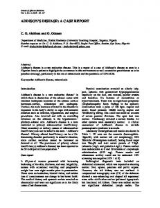
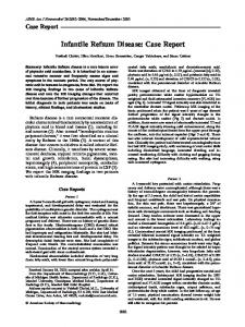
Case presentation Thirty-two year-old white, puerperal female, delivered vaginally 40 days before and no previous medical problems. She was still breastfeeding and went to Emergency due to left mastalgia for the past week, in the absence of previous trauma. She was taking antibiotics and no steroidal anti-inflammatory drugs, previously prescribed for suspicious of mastitis, for three days, with no clinical improvement. Physical examination showed an enlarged left breast, an axillary lump and a painful cord-like structure in the upper outer quadrant of the same breast (Figure 1). No fever and no inflammatory signs were registered. The clinical suspicious of Mondor’s disease was confirmed by ultrasound scan, showing a markedly dilated superficial vein in the upper outer quadrant of left breast (Figure 2). The patient was given a ventropic therapy (diosmin 500 mg twice a day) and was kept in anti-inflammatory (diclofenac 75 mg per day), for two weeks, with progressive pain improvement. Ultrasound control was performed after four weeks, showing reperfusion. Mammogram performed was normal.
Figure 1. Left breast with a lump in the upper outer quadrant.
140
Rev Bras Ginecol Obstet. 2014; 36(3):139-41
Figure 2. Ultrasound scan showing markedly dilated superficial vein (3.18 mm) in the upper outer quadrant of left breast, with a stop area of color Doppler (arrow).
Discussion Mondor’s disease affects mainly women (10:1), with the peak incidence at the age of 435. Fewer than 400 cases have been reported in the world literature6. However, its incidence after breast cancer surgery and aesthetic mammaplasties has been estimated at 1%7. The pathophysiology is not clear, but it has been explained as pressure on the vein affected with stagnation of blood or as direct trauma to the vein itself. In cases that do not show such evidence, the most reasonable explanation is on the basis of repeated movement of the breast along with the contracting and relaxing pectoral muscles, which causes stretching and relaxing of the veins8. The majority of patients reported in the literature had no identifiable cause9. However, trauma, including surgery, seems to play an important role in some cases, with the disease developing 5–7 weeks after surgery, possibly as a result of the injury to subcutaneous veins. Other reported possible aetiology factors include excessive physical activity, concomitant breast inflammation in the form of mastitis or abscess, pendulous breast, tightfitting brasserie, rheumatoid arthritis, febrile episodes and filariasis, as well as, associated with pregnancy, oral contraceptives and intravenous drug use10. In the present case, a previous mastitis could have been in the origin of the thrombophlebitis or there could have been a misdiagnosis, reason why it was not improving with the antibiotics prescribed. Clinically, it presents as a palpable subcutaneous cordlike structure. It is associated with sudden onset of breast pain, breast enlargement and skin retraction. The most common site is the upper outer quadrant; bilaterality is rare. There are some asymptomatic cases5. Ultrasound scan can be the first exam in younger patients and it is useful to confirm and monitor the evolution, however clinical suspicious is essential. Ultrasound scan
Mondor’s disease in puerperium: case report
shows initially a superficially located, long, tubular anechoic structure, with no flow on color Doppler study10. During evolution, the flow can return and the vein size decreases until normal caliber11. Mammogram should be performed to exclude cancer, even if it is rare11. The condition, though benign and self-limited, has been associated with breast cancer. The incidence of breast cancer was lower in older series (2.4%)12, but Catania et al.11 reported the highest incidence of breast cancer yet reported (12.7%), reason why attention should be paid even in young patients13. This relationship with breast cancer and risk factors suggests that routine mammogram is advisable6 and should always be performed, even when the results of physical examination are negative11. Once cancer is excluded, the expectant management is desired, reassuring the patient that it is a benign
condition. Most of the cases have a good prognosis, with spontaneous complete remission, requiring only symptomatic treatment (anti-inflammatory medication and local application of heat)5. The recent guidelines of the American College of Chest Physicians on superficial thrombophlebitis suggested prophylactic or intermediate doses of low molecular-weight heparin for at least 4 weeks (grade 2B evidence) without concomitant addition of non-steroidal anti-inflammatory drugs14. There is no clear recommendation on Mondor’s disease itself but considering the syndrome as a form of thrombophlebitis, which could have been an option. A six-month follow-up, with both mammogram and sonogram, is appropriated5. Very rarely, there is the need of surgical excision of the thrombosed vein.
References 1. Camargo Jr HSA, Camargo MMA, Teixeira SRC. Doença de Mondor: apresentação de três casos com características clínicas distintas. Rev Bras Mastologia. 2003;13(4):175-8. 2. Mondor H. Tronculite sous-cutanée subaigue de la paroi thoracique antéro-latérale. Mem Acad Chir. 1939;65(28):1271-8. 3. Niechajev I. Mondor’s subcutaneous banding after transaxillary breast augmentation: case report and the review of literature. Aesthetic Plast Surg. 2013;37(4):767-9. 4. Coscia J, Lance S, Wong M, Garcia J. Mondor’s thrombophlebitis 13 years after breast augmentation. Ann Plast Surg. 2012;68(4):336-7. 5. Faucz RA, Hidalgo RT, Faucz RS. Doença de Mondor: achados mamográficos e ultra-sonográficos. Radiol Bras. 2005;38(2):153-5. 6. Quéhé P, Saliou AH, Guias B, Bressollette L. [Mondor’s disease, report on three cases and literature review]. J Mal Vasc. 2009;34(1):54-60. 7. Khan UD. Mondor disease: a case report and review of the literature. Aesthet Surg J. 2009;29(3):209-12.
8. Hogan GF. Mondor’s disease. Arch Intern Med. 1964;113(6):881-5. 9. Shetty MK, Watson AB. Mondor’s disease of the breast: sonographic and mammographic findings. AJR Am J Roentgenol. 2001;177(4):893-6. 10. Shousha S, Chun J. Ulcerated Mondor’s disease of the breast. Histopathology. 2008;52(3):395-6. 11. Catania S, Zurrida S, Veronesi P, Galimberti V, Bono A, Pluchinotta A. Mondor’s disease and breast cancer. Cancer. 1992;69(9):2267-70. 12. Hou MF, Huang CJ, Huang YS, Hsieh JS, Chan HM, Wang JY, et al. Mondor’s disease in the breast. Kaohsiung J Med Sci. 1999;15(11):632-9. 13. Barata S, Santos P, Valério O, Simões D, Calhaz-Jorge C. Doença de Mondor e carcinoma da mama – caso clínico. Acta Obstet Ginecol Port. 2009;3(1):60-2. 14. Kearon C, Kahn SR, Agnelli G, Goldhaber S, Raskob GE, Comerota AJ, et al. Antithrombotic therapy for venous thromboembolic disease: American College of Chest Physicians evidence-based clinical practice guidelines (8th edition). Chest. 2008;133(6 Suppl):454S-545S.
Rev Bras Ginecol Obstet. 2014; 36(3):139-41
141